Laboratory diagnosis of (hiv)
•
15 likes•4,002 views

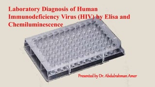
Laboratory Diagnosis of Human Immunodeficiency Virus (HIV) by Elisa and Chemiluminescence Presented by Dr. Abdulrahman Amer

Recommended
More Related Content
What's hot
What's hot (20)
Similar to Laboratory diagnosis of (hiv)
Similar to Laboratory diagnosis of (hiv) (20)
Recently uploaded
Recently uploaded (20)
Laboratory diagnosis of (hiv)
- 1. Presentedby Dr. Abdulrahman Amer Laboratory Diagnosis of Human Immunodeficiency Virus (HIV) by Elisa and Chemiluminescence
- 3. HIV-Structure (Viral Antigens) 1- Gag = Group Antigen: ( (Capsid), P17 (Matrix), & P7 (Nucleocapsid). 2- Pol= polymerase and associated enzymes: P66 (reverse transcriptase -RT), P32 (integrase) , P9 (protease). 2- Env= Envelope glycoproteins.
- 6. Why AIDS is more important today Apart from Men, there is a increasing burden of Infections in Women. Implication on Mother to Child Transmission Female Adolescents are infected 3 – 6 times higher than male counterparts. AIDS has turned out to be Social Problem.
- 7. Implication of AIDS Being diagnosed as having HIV/AIDS has life time repercussions. Every body understands AIDS as a life threatening condition. A casual way of dealing as being HIV + will have Moral, Legal, and Social consequences.
- 8. Importance of precise Diagnosis of AIDS All patients should be informed about consequences of testing for HIV infection, Legality of respective National Laws should be followed. A pretest and posttest counseling is Growing importance, even in Developing countries. Today’s debate on testing for HIV status as matter of Health protocol ?
- 9. Purpose of Testing for HIV Infection A matter of great importance as in Blood donors to prevent infected blood being transfused. To diagnose the patients infected with HIV virus. For surveillance purpose. Persons with high risk behavior. In Pregnant women to prevent Mother to Child transmission. Patient's presenting with opportunistic infections.
- 10. Diagnosis of HIV/ AIDS Basic Tests. 1. We can detect Antigen or Antibody 2. We can detect Antigen and Antibody. 3. Majority of Laboratories depend on Commercial Kits. 4. Devloped Nations going for Molecular and more precise Methods
- 11. Screening Tests for HIV/AIDS Detection A screening test posses high Sensitivity. We rarely miss the Diagnosis in Infected patients. Done as mass screening procedure. Economical. Developing countries depend on these tests even for Diagnosis.
- 12. Confirmatory Tests in AIDS It is important to confirm all screening tests with Confirmatory tests, or we brand some one without infection as infected. Confirmatory tests differentiates false reactive tests. and identifies truly infected or not.
- 13. Screening test High degree of Sensitivity Few false negatives Confirmatory test High degree of Specificity Few / No false positive results. Differences of Screening and Confirmatory Tests.
- 14. Why patients should we tested with Screening and Confirmatory tests. Before you declare a person infected with HIV / AIDS you should perform both methods. Faulty testing methods can lead to catastrophic consequences, and legal litigations.
- 15. Spectrum of HIV Tests HIV diagnosis (Antibody/Antigen testing) Enzyme Immunoassays (EIAs) Rapid tests Western blot (WB) Early diagnosis in infants p24 Initiation and monitoring of ART CD4 Viral Load
- 16. Challenges of HIV Testing Early detection of seroconversion Early detection in infants born to HIV positive mothers Effect of HIV subtypes on test performance Impact of other health conditions on test performance Product specific equipment Technical skill
- 17. Diagnostic lab test for HIV infection A- Tests to diagnose HIV infections: 1- Screening 2- Confirmatory B- Tests for follow up the disease: 1- Viral load by Quantitative PCR 2- Th-cells count (CD4+) C- Tests for the complication of the disease: 1- TB infection 2- HBV 3- HCV 4- Toxoplasma 5- Liver function tests 6- UTIs 7- Others.
- 18. A- Tests to diagnose HIV infections 1- Screening tests These test are rapid in giving results however, they are not diagnostic and need confirmation by the confirmatory tests. The Screening tests include: - ELISA tests (combined Ag-Ab Immunoassays) for detecting HIV-1 & HIV-2 antibodies and P24 Ag of HIV-1. chemiluminescent (immunoassay testing). - Rapid tests
- 19. ELISA or chemiluminescent (immunoassay testing) Were previously for detection of HIV Antibodies Now detection of P24 Antigen of HIV is included (Combo ELISA). Even though, need confirmation Done upon serum sample If initially reactive, repeat in triplicate, if positive send for WB or PCR.
- 20. ELISA METHOD Universally accepted test ,most popular even in the developing nations. Useful in large scale screening. A common method used in Blood banks in mass screening of Human blood.
- 21. ELISA plateHIV EIAs •Based on color change/fluorescence •Change compared with standardized cut-off •Result positive or negative •No specific antibody reaction information •Multiple samples run with traditional EIA 96-Well Microtiter Plate EIA Interpretation of EIAs
- 22. Sequence of Events in ELISA test
- 24. ELISA (Enzyme-linked immuno-sorbent assay) is one of immunoassay method used to detection of 1-Antibodies 2-Proteins 3-Peptides 4-Biomolecules ELISA
- 25. Immunoassay The term “immunoassay” is a combined term of “immuno”(= immunological, practically immunochemical antigen-antibody-reaction) and “assay” means the determination of the purity of a substance or the amount of any constituent of a mixture.
- 26. 1. Antigen/antibody of interest is absorbed on to plastic surface (sorbent). 2. Antigen is recognised by specific antibody (immuno). 3. This antibody is recognised by second antibody (immuno) which has enzyme attached (enzyme-linked). 4. Substrate reacts with enzyme to produce product, usually coloured. Enzyme Linked Immunosorbent Assay
- 27. Radioimmunoassay was first described in a scientific paper by Rosalyn Sussman Yalow and Solomon Berson published in 1960. In 1971, Peter Perlman and Eva Ingval at the University of Stockholm in Sweden, Anton Schuurs and Bauke van Weemen in the Netherlands published research papers that synthesized this knowledge into methods to perform EIA/ELISA.
- 28. 1-Antibody (Antiserum) 2-Antigen 3-Labeling materials Important components in immunoassay
- 29. 1-Antibody (antiserum) Antibody: proteins produced by the immune system which help defend against antigens SYMBOL FOR ANTIBODY The variable regions are though to be the place for recognition and binding with the antigen.
- 30. Any molecule that induces production of antibodies when introduced in the body is called antigen. e.g. bacteria, viruses, (or their parts), pollen, etc. SYMBOL FOR ANTIGEN 2-Antigen
- 31. 3-Labeling materials In immunoassay, it is necessary to use any marker to know the antigen-antibody binding. For such purpose, we label either antigen or antibody with some materials that do not interefere with the binding.
- 32. ELISA READER
- 34. Components of Kit Pre-Coated, Stabilized 96-well Microtiter Plate. Sample Diluent Standards and controls Conjugated Detection Antibody 10X Wash Solution Substrate Stop Solution
- 37. Advantages of ELISA • Reagents are relatively cheap & have a long shelf life • ELISA is highly specific and sensitive • No radiation hazards occur during labelling or disposal of waste. • Easy to perform and quick procedures • Equipment can be inexpensive and widely available. • ELISA can be used to a variety of infections.
- 38. Disadvantages of ELISA Measurement of enzyme activity can be more complex than measurement of activity of some type of radioisotopes. Enzyme activity may be affected by plasma constituents. Kits are commercially available, but not cheap Very specific to a particular antigen. Won’t recognize any other antigen False positives/negatives possible, especially with mutated/altered antigen
- 40. 1-Direct ELISA 2-Indirect ELISA 3-Sandwich ELISA 4-Competitive ELISA 5-Ogives ELISA Types Of Elisa
- 41. The direct detection method uses a labeled primary antibody that reacts directly with the antigen. Direct detection can be performed with antigen that is directly immobilized on the assay plate . Direct detection is not widely used in ELISA but is quite common for immunohistochemical staining of tissues and cells.
- 42. The indirect ELISA utilizes an unlabeled primary antibody in conjunction with a labeled secondary antibody. The secondary antibody has specificity for the primary antibody
- 43. Direct and Indirect ELISA
- 44. The sandwich measures the amount of antigen between two layers of antibodies. Sandwich are especially useful if the concentration of antigens is low or they are contained in a mix of high concentrations of contaminating protein To utilize this assay, one antibody (capture) is bound to a microtiter plate well. Antigen is then added and bound to the antibody. Unbound products are then removed, and 2ry antibody is added (detection), then add the 3rd labeled antibody to complete the sandwich Major advantages of this technique are that the antigen does not need to be purified prior to use, due to its high specificity. Sandwich ELISA
- 46. In this Unlabeled antibody is incubated in the presence of its antigen. These bound antibody/antigen complexes are then added to an antigen coated well. The plate is washed unbound antibody is removed. The secondary antibody, specific to the primary antibody is added. This second antibody is coupled to the enzyme. A substrate is added, and remaining enzymes elicit a chromogenic or fluorescent signal. For competitive ELISA, the higher the original antigen concentration, the weaker the eventual signal. Competitive ELISA
- 48. A newer technique uses an solid phase made up of an immuno-sorbent polystyrene rod with 8-12 protruding ogives. The entire device is immersed in a test tube containing the collected sample and the following steps (washing, incubation in conjugate and incubation in chromogenous ) are carried out by dipping the ogives in microwells of standard microplates pre-filled with reagents Multiple and portable ELISA
- 51. 1. Coating of Wells with Antibody Add 25µL of the sample diluent IG4 to all the wells Add 100 μl of the anti-HIV Negative control in well A1,B1 and C1 Add 100 μl of the anti-HIV Positive control in well D1. Add 100 μl of the HIV P24 Antigen Positive control in well E1. 2- Add 100 μl of the first sample in the well E1, second sample in the well G1 and so on. 3- Incubation with Test Samples Cover the wells with strip sealers &block cover, incubate for 60 minuts at 37 °C. Remove the sealers
- 52. 5. Incubation with enzyme- Conjugated Antibody. . Add 50 μL Assay buffer IG4 and 50μL conjugate G4 to all the wells Cover the wells with strip sealers and black cover, 30 minutes at 37 °C. The enzyme-conjugated antibody should be directed against the antigen to be determined The conjugatec antibody must be specific for the antigen of interest 4. Washing 6 cycles 350 μl/well and 30 seconds soak time with diluted wash buffer(1:20). For 500mL wash buffer, mix 25mL concentrare in 475 mL distilled water
- 53. 6. Wash as described 6 cycles (350 μL/well and 30 seconds soak time with diluted wash buffer(1:20)). 7. Colour Development Add 50μL color reagent to each well(chromogenic substrate ). Incubution 30 minutes at room temperature (20-30 °C) in dark. The plate should preferably be protected against light during this incubation. 8. Stopping the Colour Development Add 100μL stop solution to each well to stop the reaction . 9. Reading of Results Read results directly through the bottom of the micro well plate using an automated (ELISA-reader). The subtraction of the absorbance at a reference wavelength (between 620 and 650 nm) is recommended.
- 55. Add mach. photo
- 56. HOW TO READ
- 57. Important points in performing ELISA and improvement of assay performance 1-Sample treatment. 3-Stability of assay samples. 2-Infleunce of humidity and air stream.
- 58. Important points in performing ELISA and improvement of assay performance Sampling and treatments of samples Serum or plasma
- 59. In general, we recommend using Use of fluoride must be avoided because fluoride ion is a potent inhibitor of peroxidase. sampling When getting heparin is most often used as an anti-coagulant
- 60. An important phenomenon with frozen plasma is that an insoluble substance (fibrin) will be formed when thawed. In this case, the sample must be mixed and centrifuged, then the insoluble cluster flowing in the plasma should be taken out by a thin wire needle sharply bent at an end. If such fibrin remains in the sample, it may clog the tip of a pipette and influences assay variability
- 62. Serum or plasma, when fresh, shows pH near neutral, however, it very quickly goes to alkaline more than pH 8 by losing CO2. In alkaline pH, the antigen-antibody reaction is interfered. resulting in cancellation of the assay or giving inaccurate assay values. So, in such case, before hand dilution of serum or heparinized plasma with assay buffer will be helpful. pH Of the sample
- 63. Sample storage temperature is better to be lower than -35 C. Ultra- low temperature such as -80 C is recommended for a long-term storage. Repeated freezing and thawing is also harmful to the protein, and may cause inactivation. Storage temperature and freezing-thawing.
- 64. When samples are taken out from the freezer and thawed, never forget to mix these samples because the solution after thawing is not homogeneous, and the bottom area contains more solute DON’T FORGET
- 65. Stability of assay samples. In assay, the problem of sample stability, i.e. how long the substance to be measured can keep its immunoreactivity, in serum or plasma, is very important. Blood samples also contain enzymes to destroy peptides or proteins, and stability against those enzymes differs from substance to substance. Freezer of –20C is not trustable for the constancy of temperature but use of a freezer of –35 C or lower temperature is recommended.
- 66. Avoid sunlight Avoid air fans
- 67. Influence of humidity and air stream During all the incubation process, the well-plate should be covered using the attached plate cover. Plate cover is effective only under the most suitable condition, i.e. room temperature, humidity more than 50%, and air stream of less than 0.2m/sec. It is recommend to get a small semi-transparent plastic box, and put moistened paper towel on the bottom .
- 68. Trouble shooting in ELISA
- 69. Poor or no coloration after the last step 1) The standard or samples might not be added. 2) Reagents necessary for coloration might not be added. 3) Wrong reagents related to coloration might have been added. 4) Influence of the temperature under which the kits had been stored. 5) Excessive hard washing of the well plate.
- 70. 2)The standard curve obtained was not smooth. There might be some mistake in the serial dilution of the original standard solution. 3)Flat standard curve. Standard solutions are not added.
- 71. 4-Big variation between two wells in duplicated assay was observed. 1) Scratching the bottom of the well by aspirator tip during aspiration of washing buffer. 3)Assay might be started while the well-plate was still cooler than room temperature. 2) Scratching the bottom of the well by pipette tip during addition of standards, samples, or reagents.
- 72. 4) Air stream, warmer or cooler than room temperature 5) Air stream from air conditioner or other instruments might dry wells. 6) Insufficient removal of washing buffer from the wells might dilute reagent solution added in the following step of the procedure. 7)Big variation would be obtained if the sample is not homogeneous.
- 73. Chemiluminescence Chemiluminescence is a chemical reaction that emits energy in the form of light. When used in combination with immunoassay technology, the light produced by the reaction indicates the amount of analyte in a sample. Direct chemiluminescent reactions directly measure the light energy without the use of added steps or amplifying molecules. The ADVIA Centaur XP assays use acridinium ester (AE) as the chemiluminescent label, because AE does not require the addition of a catalyst or substrate.
- 74. Direct Chemiluminescence It is easy to automate direct chemiluminescence using AE and provides many benefits, such as long reagent shelf life, fast reaction time, and assay sensitivity. The ADVIA Centaur XP assays use the dimethyl form of AE because its stability allows long reagent shelf life.
- 75. Assay Reaction Formats The system uses a variety of formats to detect antigens as well as antibodies. 1 2 3 Sandwich format Competitive format Antibody- capture format
- 88. SAMPLE HANDLING
- 91. Reagent Handling
- 92. Reagent Cooling • 30-position reagent tray cooled 4°C to 8°C Reagent Integrity Control • Barcode reagent identification, automatic inventory tracking and flagging, calibration validity tracked and flagged, reagent on board stability tracked and flagged, reagent expired/ reagent low flagging Reagent Mixing • Ready Packs automatically rocked onboard
- 93. Quality Control Advanced QC package includes Levy-Jennings plots and Westgard rules; 65,000 control results can be stored Results within ± 2SD accepted
- 94. TYPES OF ASSAYS
- 97. • AFP • BR • CA 15-3 • CA 19-9 • CA 125II • CEA • Complexed PSA • Free PSA • PSA • Serum HER-2/neu Tumor Markers/Onc ology
- 98. ToRCH and Special ID • CMV IgG • CMV IgM • Rubella IgG • Rubella IgM • Syphilis • Toxo IgG • Toxo IgM
- 99. • BNP • CKMB II • D-Dimer • Myoglobin • Tnl-Ultra • C-Peptide (serum, urine) • Insulin
- 100. • Anti-HBe • Anti-HBs • Anti-HBs-2 • eHIV 1/O/2† • HAV IgM • HAV Total • HBc Total • HBc IgM • HBe Ag • HBs Ag • HBs Ag Confirmatory • HBs AgII • HCV† • HIV Combo (CHIV)†
- 102. • HA (ELF Marker) • TIMP (ELF Marker) • PIIINP (ELF Marker) Liver Fibrosis • Cortisol (serum, urine) • Homocysteine Metabolic
- 103. • AFP • DHEAS • Estradiol-6 • Estradiol-6 III • FSH • LH • Progesterone • Prolactin • SHBG • Testosterone • Total hCG Reproductive Endocrinology
- 104. • Cyclosporine • Tacrolimus* Immunosuppresants • Procalcitonin Sepsis
- 105. Thyroid Function • Anti-TG Ab • Anti-TPO Ab • Free T3 • Free T4 • Intact PTH • TSH • Third Generation TSH • T Uptake • Total T3 • Total T4 • TSH 3-UL
- 106. Solid-phase EIA with immobilized viral antigens to detect antibodies to specific HIV proteins. Western Blot (Immunoblotting)
- 107. AIDS is caused by at least 2 etiological agents HIV-1 & HIV-2 Inactivated and denatured protein of HIV-1 are fractioned by polyacrylamide gel electrophoresis Protein bands are transferred into nitrocellulose strips HIV-1 sample diluted with buffer are then incubated with the strip Principle
- 108. Conjugate peroxidase labeled anti human IgG is added It will bind to the antibodies already bound to the strip Chromogen is then added forming color reaction Reaction is then stopped by aspiration and reaction
- 109. Serum sample Maximum 8 days Stored 2o C – 8oC or frozen at – 25oC Lipemic sample must be centrifuged well Avoid heating Sample requirement:
- 110. Creating Western Blot Strips Proteins are transferred (blotted) onto the surface of a membrane Strips are incubated with patient serum and antihuman IgG conjugated with an enzyme (and chromagen) HIV lysate proteins are separated by size using gel electrophoresis The membrane is cut into strips
- 111. HIV Western Blot Banding Pattern env gp160 gp120 gp 41 gag p55 p18 p24 pol p65 p51 p31
- 112. Interpretation of Results (General Consensus) Negative: No bands present Positive: 2 ENV band present (WHO Guidelines) Indeterminate Any bands present but do not meet criteria for positive
- 113. * * *
- 114. When should WB be used? Western Blot assay should not be used as a screening test. WB should be viewed as a supplemental test which can be used to confirm positive results obtained from EIA. HOWEVER: Specificity is less than that of EIA A significant number of indeterminate blots are seen in low risk populations
- 115. Advantages Specific interaction of antibody and antigen can be directly visualized. Disadvantages Technically demanding Expensive Subject to interpretation Presence or absence of bands Intensity of those bands
- 116. HIV-1 Western Blot Antigens p = protein gp = glycoprotein Number = molecular weight
- 117. Components Used in HIV-1 Western Blot Human HIV Antibodies (from patient serum) Y YY Y HIV Western blot Strip YY HIV Antigens (on Western blot) YY Y Antihuman IgG Antibodies Enzyme Detector Color Reagent
- 118. Sample HIV-1 Western Blot YY Y YY Y Y Y YY Y Y Antibodies to gp120 Anti-human IgG Enzyme Detector HIV gp120 antigen Color Reagent Antibodies to p24 Enzyme Detector HIV p24 antigen Color Reagent Anti-human IgG Test Completed gp120 & p24 bands Visible
- 119. HIV-1 Gene Products & Western Blot
- 120. HIV-1 and HIV-2 Gene Products & Western Blot
- 121. Interpretive Criteria for HIV-1 Western Blot Source: CDC. MMWR. 1989:38(S-7):1-7. Positive Control
- 122. Interpretive Criteria for HIV-1 Western Blot Source: CDC. MMWR. 1989:38(S-7):1-7. Negative No bands:
- 123. Interpretive Criteria for HIV-1 Western Blot Source: CDC. MMWR. 1989:38(S-7):1-7. Positive At least two of the following bands: p24 gp41 gp120/160
- 124. Interpretive Criteria for HIV-1 Western Blot Source: CDC. MMWR. 1989:38(S-7):1-7. Indeterminate One or more bands present Not meeting positive criteria Examples Most common bands seen with indeterminate Western blot (IWB) p17, p24, p55
- 125. THANK YOU