
Recommended
More Related Content
Similar to Diaphragmatic hernia.pptx
Similar to Diaphragmatic hernia.pptx (20)
Recently uploaded
Recently uploaded (20)
Diaphragmatic hernia.pptx
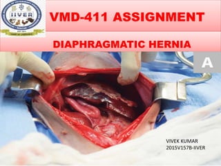
- 1. VMD-411 ASSIGNMENT DIAPHRAGMATIC HERNIA VIVEK KUMAR 2015V157B-IIVER
- 2. INTRODUCTION • The diaphragm is the muscular separation between the chest and abdominal cavities that functions as a barrier and aids in respiration • A break in the continuity of the diaphragm allows protrusion of abdominal viscera into the thorax. OR • Diaphragmatic hernia is the disruption of the diaphragm which allows abdominal organs to migrate into the chest cavity.
- 3. ETIOLOGY • In small animals, automobile-related trauma is a common cause of diaphragmatic hernia. • Congenital defects of the diaphragm may also result in herniation (eg, peritoneopericardial hernia). • In horses, diaphragmatic hernia may occur, less commonly, congenitally or after trauma, dystocia, or recent strenuous activity. • Diaphragmatic hernias are extremely rare in cattle.
- 4. PATHOGENESIS • Achalasia of reticulo-omasal sphincture or impairment of Oesophageal groove • Food get into Rumen but cannot pass to Abomasum • Hypermotility due to over distention of Rumen • FROTHY BLOAT • Respiratory problems arise due to displacement & compression of heart.
- 5. TYPES • Two types of diaphragmatic hernias occur in dogs and cats: • Traumatic ̶ caused by an event that tears the diaphragm • Congenital ̶ pets have these at birth (The most common type of this subcategory is the peritoneal- pericardial diaphragmatic hernia (PPDH).
- 6. Peritoneal-Pericardial Diaphragmatic Hernia (PPDH). • This is the picture of PPDH in cats, necropsy from right side of the cat, to the left Abdomen, Diaphragm in middle, Thorax at the right.
- 7. Congenital diaphragmatic hernia in an 8-month-old Rottweiler. Most of the intestine and stomach are lodged in the thoracic cavity as well as the spleen and part of the liver. On the right, a clamp is holding the fibrous and regular border of the diaphragmatic defect.
- 8. CLINICAL SIGNS • A diaphragmatic hernia can cause significant respiratory difficulty. • The trauma that caused the hernia may also result in rib fractures, lung lacerations, and lung bruising. • These injuries may lead to : • pneumothorax (air in the chest outside the lungs), • hemothorax (blood in the chest cavity)
- 9. ACUTE DIAPHRAGMATIC HERNIA • If abdominal contents have entered the chest cavity, this can further compromise the ability to expand the lungs. • Abdominal organs, displaced through a diaphragmatic hernia, may experience compromise to their blood supply. • Signs associated with an acute diaphragmatic hernia are: • Difficulty in breathing • Rapid, shallow breathing pattern • Abnormal breathing posture with extended head and neck
- 10. Clinical Findings: Acute & Chronic • Dogs and cats are characteristically dyspneic in the acute case. • If the stomach is herniated, it may bloat and the animal may deteriorate rapidly. • In chronic cases, systemic signs such as weight loss may be more prominent than respiratory signs. • Congenital peritoneopericardial hernia is most frequently an incidental finding.
- 11. Clinical findings in Horse & Cattle • Horses most frequently present with acute, severe colic secondary to displaced intestines, or with respiratory signs and dyspnea. • In cattle and water buffalo, diaphragmatic hernias may be associated with traumatic reticulitis and herniation of the reticulum.
- 12. • Herniation of reticulum through diaphragm Fig 2. Nails and small stones recovered from reticulum
- 13. DIAGNOSIS • Careful physical examination, including auscultation and percussion, usually suggests the presence of thoracic disease. • The definitive diagnosis is most frequently made from Radiographs. • Loss of diaphragmatic contour, abdominal viscera in the thorax, and displacement of viscera from the abdomen may be apparent. • Radiographic contrast studies may be necessary to make the diagnosis.
- 14. Additional imaging tests • Additional imaging tests to include: • Abdominal and thoracic Ultrasound • Contrast radiography (taking radiographs after placing a contrast medium in gastrointestinal system) • Computed tomography (CT scan)
- 15. Normal chest x-ray of a dog. The dog’s head is to the left. The diaphragm is not distinctly visible but is the curved surface separating the white abdominal contents on the right from the grey of the air-filled lungs. The oval structure in the center of the lung field is the heart. • Chest x-ray of a dog with a diaphragmatic hernia, showing loss of the normal diaphragmatic line. Note that the white abdominal organs overlap the heart and the curve of the diaphragm is no longer visibly distinct.
- 16. • Normal chest x-rays of a dog on its back. The dog’s head is off the top of the image. The diaphragm is the curved dome separating the white abdominal contents at the bottom from the dark grey lungs. The oval structure in the center of the lung field is the heart. • Chest x-ray of a dog on their back with a diaphragmatic hernia, showing loss of the normal diaphragmatic line. Note that the white abdominal organs overlap the heart and the curve of the diaphragm is no longer visibly distinct along its entire length.
- 17. TREATMENT • The only treatment to repair the diaphragmatic hernia is Surgery. • Surgery is performed on an emergency basis if the stomach is herniated into the chest cavity and becomes distended with gas. • This can prevent lung expansion and cause respiratory distress. A needle can be passed through the chest wall into the stomach to decompress the stomach, and then surgery can be performed. • Surgical repair of the diaphragmatic hernia is typically performed by entering the abdominal cavity along the ventral midline, retracting the abdominal organs back into the abdomen, and suturing the tear in the diaphragm.
- 18. SURGICAL PROCEDURE • Surgical repair of the diaphragmatic hernia is typically performed by entering the abdominal cavity along the ventral midline, retracting the abdominal organs back into the abdomen, and suturing the tear in the diaphragm. • If the diaphragmatic tear is chronic, it is necessary to be especially careful with anesthesia, because reexpansion pulmonary edema is likely fatal.
- 19. Aftercare & Prognosis • A hospital stay will be required after surgery. • It is important to encourage rest and avoid activity during the post-operative period. • The prognosis for animals presenting with a traumatic diaphragmatic hernia is variable depending on other injuries incurred. It is estimated that roughly 15% of animals suffering traumatic diaphragmatic hernia will die before presentation. • Successful treatment of shock prior to surgery results in the best survival rates.
- 20. THANK YOU