
Recommended
Recommended
More Related Content
Similar to malaria - lab. diagnosis.pptx
Recently uploaded
Recently uploaded (20)
malaria - lab. diagnosis.pptx
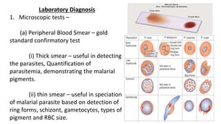
- 1. Laboratory Diagnosis 1. Microscopic tests – (a) Peripheral Blood Smear – gold standard confirmatory test (i) Thick smear – useful in detecting the parasites, Quantification of parasitemia, demonstrating the malarial pigments. (ii) thin smear – useful in speciation of malarial parasite based on detection of ring forms, schizont, gametocytes, types of pigment and RBC size.
- 3. (b) Fluorescence Microscopy – Kawamoto technique Steps - Blood smears are prepared – stained with acridine orange – examined under a fluorescence microscope. Nuclear DNA is stained green. (c) Quantitative Buffy Coat examination –
- 4. Advantages – faster, more sensitive and quantification is possible. Disadvantages – expensive, less specific and speciation is difficult. 2. Non – Microscopic Test – (a) Antigen Detection by RDT – Interpretation is based on immobilization of malarial antigens at test lines 1 and/or 2 forming colored bands. 1. If band is formed at only test line 1: indicates P. falciparum infection 2. If band is formed at only test line 2: indicates Plasmodium species other than P. falciparum infection
- 5. 3. If bands are formed at both test lines 1 and 2: indicates P. falciparum or mixed infection. Note: The band at control line must come to validate the test; if it does not come, test is considered invalid. Control line is coated with antibody against polyclonal malarial antibody present in buffer. Ab detection tests – banned Automated systems – Parasite F digital cytometry, automated microscopy methods Non – specific tests – normocytic hemolytic anemia, leukopenia, raised ESR, metabolic acidosis, hypoglycemia.