1. Hippocampal long-term potentiation in SUMO 1-3
knockdown mice in vivo
Titus John1,3, Huaxin Sheng2, Robert Pearlstein3
1Department of Biomedical Engineering, North Carolina State University, 2Department of Anesthesiology, Duke University, and 3Department of
Surgery/Division of Neurosurgery, Duke University
SUMO 1-3 Knockdown Mice
• Small Ubiquitin-like Modifiers(SUMO) are proteins shown
to play role in signal transduction pathways.
• Evidence suggest that SUMO expression plays role in
synaptic plasticity (indicated by motor and memory
function)
• Prior study found mice had impaired novel object
recognition and fear conditioning
• Current results have so far been based on in vitro
approaches
• Two Group Study 5 WT 5 SUMO 1-3 Knockdown
Surgical Preparation
• Animals anesthetized with Urethane (1.5 g/kg i.p.)
• Procaine applied to both inner ear and scalp
• Animal body temperature maintained 36.5-37 C on DC
heating pad via feedback from rectal probe
• Hair was removed from the surgical area
• Midline incision was made along the scalp to expose skull
bilaterally
Electrode Insertion Method
• Two trephine holes were made in order to insert
stimulating and recording electrodes sterotatically
• Locations CA 1 (P. 2.2 L 1.3 H 1.3-1.5 mm) , CA3 (P 2.2
L 2.5 H2.3-2.5 mm) with respect to bregma
• Monopolar recording electrode positioned in CA1
stratum radium for recording field potentials
Optimizing Recording Condition
• Electrode depth was adjusted to evoke largest response
• Voltage/Current characterization was tested in order to ensure
stimulation current did not evoke population spike
• Baseline recording takes place for 20 minutes to establish stable
baseline
• 100 Hz tetanus frequency was delivered in order to induce LTP
• Potentiated EPSP was followed for 2 hours after tetanus
frequency delivered
Figure 2: The stimulating and recording electrodes were
dipped in florescent dye (DiI) to confirm location.
• SUMO 1-3 mice have previously been tested for
behavioral abnormalities and found to have
impaired memory consolidation.
• Current observation are consistent with a role of
Hippocampal synaptic plasticity as effected by
SUMO expression.
Results
Figure 1: Mouse surgical preparation, two screws inserted
into skull to serve as ground for monopolar stimulation and
reference screw for recording electrode
In vivo noise reduction methods
• Mice were positioned on stereotactic frame to allow chest
movement from respiration in the ventral direction
• EKG leads were attached to the left anterior and right
posterior paws to determine heart rate frequency
• Respiration and EKG frequency can then be compared to
continuous neural signal to identify source of periodic
noise interference
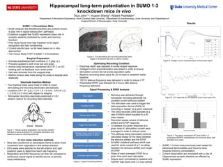
Figure 5: Voltage current characterization of stimulated signal
Figure 6: LTP response 20 min baseline and 2 hours post HFS
Figure 7: Two group comparison WT and SUMO 1-3
knockdown fEPSP% characterized at 2 mins and 2hour with
standard error mean.
Raw Signal
Amplified 1000X
Filtered 30Hz-10kHz
Trigger from stimulator
Digitized signal using NI-DAQ at
sampling frequency of 40kHz
Recoded 50 msec sweep of signal
10 sweeps signal averaged
Signal Processing & EPSP Analysis
• Stimulus was delivered through
monopolar recording electrode via
constant current stimulus isolator
• The stimulator was used to trigger the
data acquisition device (DAQ) for
recording a “sweep” of a given response
• The DAQ recorded 2000 samples at a
rate of 40kHz which equated to a 50
msec sweep
• Recorded sweep consist of stimulus
artifact and EPSP response
• 10 sweeps of a given response at a
given level of stimulation current were
averaged in order to reduce noise
• The pathway being stimulated could be
calculated based on the delay between
the stimulus artifact and the EPSP
• Schaffer collateral pathway which was
used for study consist of a 7 ms delay
between the stimulus artifact and trough
of EPSP
• Slope of each averaged EPSP for a
given time point was calculated
• Slopes were normalized to baseline and
%EPSP was found over 2.5 hour periodFigure 3: Signal Processing Method