2. Eur J Appl Physiol
1 3
(Bailey et al. 2010a, b; Vanhatalo et al. 2011; Wylie et al.
2013). Much of the recent literature has focussed on
increasing the bioavailability of NO using dietary nitrate
supplementation and this has been shown to reduce the
oxygen cost of submaximal exercise (Lansley et al. 2011;
Vanhatalo et al. 2010; Kuennen et al. 2015), lower arterial
pressure (Vanhatalo et al. 2010; Keen et al. 2015; Levitt
et al. 2015), increase muscle blood flow and increase cuta-
neous vascular conductance (CVC) (Ferguson et al. 2013;
Keen et al. 2015; Levitt et al. 2015). Dietary nitrate sup-
plementation is one of the two pathways in which NO bio-
availability can be elevated, the other is the nitric oxide
synthase (NOS) -dependent l-arginine (L-ARG) pathway
(Palmer et al. 1987). Beneficial physiological effects of
L-ARG supplementation have regularly been observed
in some clinical populations (Adams et al. 1997; Clark-
son et al. 1996; Rector et al. 1996); however, the effect of
L-ARG supplementation in healthy, non-diseased partici-
pants is equivocal with beneficial physiological and perfor-
mance effects reported in some (Bailey et al. 2010b; Bode-
Boger et al. 1998; Koppo et al. 2009; Schaefer et al. 2002),
but not all (Bescos et al. 2009; Bode-Boger et al. 1998;
Koppo et al. 2009; Schaefer et al. 2002), studies.
Cutaneous blood flow is regulated by two branches of
the sympathetic nervous system- the noradrenergic vaso-
constrictor nerves and the cholinergic active vasodila-
tor nerves (Kellogg et al. 1995). While resting in temper-
ate ambient conditions and during dynamic exercise, the
cutaneous vasculature is predominantly controlled by the
noradrenergic vasoconstrictor system and endothelial NOS
(eNOS) (Kellogg et al. 1999; McNamara et al. 2014); how-
ever, increases in body temperature lead to sympathetic
cholinergic mediation of skin blood flow regulated by neu-
ronal NOS (nNOS) (Kellogg et al. 1995, 1999). The cho-
linergic active vasodilator system accounts for 80–95 % of
the increased skin blood flow observed during passive heat
stress (Johnson and Kellogg 2010) and in situations where
the magnitude of skin blood flow increases is naturally
reduced (e.g., ageing), the skin blood flow response to pas-
sive heat stress has been shown to be augmented by intra-
dermal L-ARG administration (Holowatz et al. 2006). Hol-
owatz et al. (2006) suggested that L-ARG supplementation
may enhance the vasodilatory response in older populations
because of a reduced endogenous L-ARG concentrations,
and therefore lower NO bioavailability, while younger indi-
viduals may have sufficient L-ARG concentrations to meet
the vasodilatory stimulus provided by passive heating and
so do not benefit from supplementation (Holowatz et al.
2006).
Passive and active heating protocols both place the body
under thermal stress and elevate cutaneous blood flow;
however, the mechanisms which control this increase in
peripheral blood flow differ. Previous investigations have
used passive heating protocols (Holowatz et al. 2006; Lev-
itt et al. 2015) during which the increase in skin blood flow
appears to be dependent on nNOS (Kellogg et al. 2009),
whereas skin blood flow increases during exercise appear
dependent on eNOS (McNamara et al. 2014). Recent data
have shown that dietary nitrate supplementation increases
rectal temperature without altering skin temperature during
marching in hot conditions (Kuennen et al. 2015). No skin
blood flow data were recorded and so the authors used the
lack of effect on skin temperature to suggest that there was
a lack of effect on skin blood flow. Unlike the nitrate-NO
pathway (Govoni et al. 2008), the L-ARG-NO pathway is
NOS-dependent (Moncada and Higgs 1993) and so when
a greater strain is placed upon this system, such as during
exercise and possibly during recovery from exercise in the
heat, it seems prudent to suggest that L-ARG may offer a
hyperaemia-induced thermoregulatory benefit to healthy
individuals, but this is yet to be investigated. Recently, it
has been reported that nitrate supplementation increases
rectal temperature by ~11 % despite a ~6 % reduction in
the oxygen cost of submaximal treadmill exercise in a
hot environment (Kuennen et al. 2015) but effects of oral
L-ARG supplementation on whole-body thermoregulatory
and cardiovascular responses to active and passive hyper-
thermia are also unknown.
An intervention which enhances heat dissipation and
reduces thermal strain would be of interest to a range of
researchers, athletes, and coaches and elevating NO bio-
availability by systemic L-ARG supplementation may be
such an intervention. The aim of this study was to inves-
tigate the physiological responses to an acute oral dose of
L-ARG during rest, exercise and recovery in the heat, in
recreationally active, healthy males. We hypothesised that
acute L-ARG supplementation would offer no hyperaemia-
induced thermoregulatory benefit to healthy individuals
during passive heat exposure but that it might augment heat
loss responses during active heat stress, when the cutane-
ous vasculature is predominantly controlled by eNOS
rather than nNOS.
Materials and methods
Participants
Nine healthy, recreationally active, non-heat acclimated,
non-smoking, Caucasian males volunteered for the study;
however, one of the participants experienced gastrointes-
tinal discomfort following ingestion of the L-ARG and
withdrew from the study. The mean (±standard devia-
tion) age, stature, body mass and maximum power output
in the heat (Wmax) of the 8 participants were 27 ± 6 years,
176 ± 6 cm, 76 ± 4 kg, and 237 ± 39 W, respectively. It
3. Eur J Appl Physiol
1 3
was calculated that a sample size of eight participants
would provide sufficient statistical power (0.8; β = 0.20)
to detect a difference in skin blood flow using an esti-
mated effect size of d = 0.95 [the estimated effect size of
10 g intravenous L-ARG on skin blood flow was d = 1.40
(Giugliano et al. 1997); however, the bioavailability from
oral dosages is ~70 % that of infusion (Bode-Boger et al.
1998) so the estimated effect size was reduced proportion-
ately] and an alpha level of 0.05. None of the participants
were users of dietary supplements or taking any medica-
tion. The participants were blinded to the main aim of the
investigation but were informed of any associated risks and
discomforts before giving their oral and written informed
consent to participate. Once consent was given the partici-
pants completed a health screen questionnaire (American
College of Sports Medicine Position Stand and American
Heart Association 1998) and this screening procedure was
repeated prior to each laboratory visit to assess the health
status of the individual. Following the completion of the
trials, each participant was provided with a post-participa-
tion information sheet, where full details of the study were
provided and participants were asked for their consent to
allow the use of their data in the final analysis. All 8 par-
ticipants provided their consent at this stage. The study was
approved by the University of Roehampton’s Ethical Advi-
sory Committee.
Pre‑trial (visit 1)
Prior to the main trials, participants completed an incremen-
tal, cycle ergometer (Monark 874E, Monark, Sweden) test
to determine Wmax (Kuipers et al. 1985) in a walk-in envi-
ronmental chamber (Design Environmental, Wales, UK)
set to 35 °C and 50 % relative humidity (rh). To determine
Wmax, participants initially cycled for 5 min at 100 W after
which the workload was increased by 50 W every 2.5 min
until a heart rate of 160 b min−1
was reached. Upon attain-
ing a heart rate of 160 b min−1
the work was increased by
21 W every 2.5 min until volitional exhaustion. The maxi-
mum workload was calculated using the following equation:
Wmax = Wcom + ((t/150) × �W) (Kuipers et al. 1985)
where Wcom is the last workload completed, t is the time in
seconds that the final, uncompleted, stage was sustained for
and ΔW is the final load increment (21 W).
Following the Wmax test, participants undertook a partial
familiarisation where they sat for 15 min and cycled for
10 min to be familiarised with the ambient conditions and
the power output required during the experimental trials
(60 % Wmax). Participants abstained from alcohol, caffeine,
strenuous exercise, and completed a food record for the
24 h period prior to the initial pre-test. They adopted the
same diet and abstained from strenuous exercise, caffeine,
and alcohol for 24 h prior to each subsequent trial.
Trials (visits 2 and 3)
Following the preliminary test, participants visited the lab-
oratory twice at the same time of the day (±1 h) separated
by 7–9 days. Participants arrived at the laboratory ~45 min
before the commencement of the trial and ~2 h postpran-
dial. On arrival, nude body mass was recorded (Seca,
Birmingham, UK), a rectal probe (Rectal 401, Varioham
Eurosensor ltd, UK) was self-inserted ~10 cm past the anal
sphincter for measurement of rectal temperature (Trectal),
and a heart rate (HR) monitor (Polar, UK) was attached.
Four skin thermistors (Varioham Eurosensor ltd, UK) were
placed on the sternal notch, forearm, thigh, and calf, and
connected to a digital reader (Thermistor Thermometer
5831, DigiTec Corp, USA) for the subsequent calcula-
tion of mean-weighted skin temperature (Tskin) (Ramana-
than 1964). All thermistors were attached via a transparent
dressing (Tagaderm, 3 M Health Care, USA) and water-
proof tape (Transpore, 3 M Health Care, USA). A blood
pressure cuff (Digital Blood Pressure Monitor UA-767,
AD Instruments LTD, Japan) was placed on the upper,
right arm and a laser-Doppler local heater probe (PeriF-
lux 5000, Perimed AB, Stockholm, Sweden) was attached
to the left forearm and maintained at 35 °C to clamp local
skin temperature to environmental conditions. CVC was
calculated as laser-Doppler flux divided by mean arterial
pressure (MAP) and was standardised to %CVCmax. CVC-
max was calculated as the mean of a 5 min plateau in laser-
Doppler flux observed during a bout of local skin heating to
43 °C (~35 min of heating).
Upon arrival at the laboratory, participants consumed
one of two drinks. Both drinks were made up of 200 ml
blackcurrant cordial diluted in 300 ml of water- one con-
tained 10 g of dissolved commercially available L-ARG
powder (NOW Foods, USA) whereas the other did not
(PLA). The drinks were prepared by an impartial techni-
cian and administered in a double-blind, crossover man-
ner. A single, 10 g oral dose of L-ARG was selected based
upon previous data suggesting that it is the largest tolerable
dose and one that would definitely increase plasma L-ARG
concentrations (Tang et al. 2011). The data were not un-
blinded until the completion of data analysis.
Following the ingestion of the L-ARG or PLA beverage,
participants rested in a seated position for 30 min in ambi-
ent, laboratory conditions before entering the environmen-
tal chamber (35 °C, 50 % rh) for a total duration of 90 min.
During the 90 min, participants initially rested in a seated
position for 30 min, then cycled in a seated position at
60 % Wmax (143 ± 22 W) for 30 min and finally undertook
30 min of passive, seated, recovery. The time-periods were
chosen to maximise the L-ARG concentrations during the
trial because previous data have shown that plasma L-ARG
concentrations peak ~60 min after ingestion (i.e., after the
4. Eur J Appl Physiol
1 3
30 min rest in the heat) and remain elevated for ~120 min
(i.e., the end of test) (Bode-Boger et al. 1998). Participants
consumed 100 ml of water at the beginning of each 30 min
section to reduce participant discomfort associated with
perceptions of thirst.
HR, Tskin and Trectal were recorded every 5 min during
the 90 min trials, whereas oxygen consumption (VO2) was
measured every 10 min using the Douglas bag method.
CVC and arterial pressure were recorded at the start, end
and at 5 min intervals of the rest and recovery periods but
no measurements were taken during the exercise period due
to issues with limb movement. MAP was calculated using
the formula ((DAP × 2) + SAP)/3 (where DAP = dias-
tolic arterial pressure and SAP = systolic arterial pressure).
After the completion of each trial, participants towel-dried
and recorded a dry post-exercise nude body mass (Seca
813, Seca Birmingham, UK; ±0.1 kg) from which sweat
loss was calculated, taking into account the pre-trial body
mass and the 100 ml of water consumed at the beginning of
each 30 min section.
Blood analyses
Capillary blood samples were taken at upon arrival to the
laboratory prior to any ingestion (PRE) and at 0, 30, 60,
and 90 min during the protocol. Whole-blood was col-
lected into EDTA-treated micro-cuvettes and aliquots were
immediately centrifuged in a micro-centrifuge for 15 min
at 1000×g after which the supernatant was removed and
frozen at −80 °C until analysis. Plasma concentrations
of l-arginine were determined in duplicate via enzyme-
linked immunosorbent assay (Human l-Arginine ELISA
#MBS728648, My Biosource Incorporated, USA). Analy-
sis was completed in one freeze–thaw cycle. The intra-
assay coefficient of variation was 5.5 %.
Statistical analyses
One-way repeated-measures ANOVA tests were conducted
to evaluate differences between sweat loss, whereas two-
way repeated-measures ANOVA (trial x time) tests were
performed to evaluate differences between trials for HR,
SAP, DAP, MAP, %CVCmax, Tskin, Trectal, and VO2. Analy-
ses were conducted separately for rest, exercise, and recov-
ery bouts. Due to equipment issues forearm skin blood flow
data were collected from only 5 participants. Greenhouse-
Geisser corrections were made where appropriate and Bon-
ferroni corrections were made for multiple comparisons.
Descriptive data are reported as mean ± SD unless other-
wise stated. SPSS (version 20; SPSS Inc., Chicago, IL) was
used to analyse data. Statistical significance was set at the
P ≤ 0.05 level.
Results
The effect of l‑arginine supplementation on plasma
l‑arginine concentrations and participant tolerance
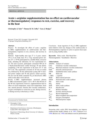
The 10 g dose used resulted in a relative dose of
0.13 ± 0.01 g kg−1
and elevated plasma L-ARG concen-
trations compared to PLA (main trial effect, p 0.001;
Fig. 1). Plasma concentrations increased in L-ARG but not
PLA (main effect time, p 0.001; interaction p 0.001)
and peaked at 60 min. Concentrations were 1.3 ± 5.3 %
higher in PLA and 246.7 ± 44.1 % higher in L-ARG com-
pared to baseline at 60 min. The L-ARG supplementation
caused gastrointestinal upset and nausea in one participant
which resulted in the participant failing to complete the
experiment reducing the sample size to 8.
The effect of l‑arginine supplementation
on thermoregulatory variables
L-ARG supplementation had no effect on Trectal or Tskin at
rest, during exercise or recovery (Fig. 2). There was no dif-
ference between the mean body mass lost during L-ARG
(1.1 ± 0.1 kg) and PLA (1.1 ± 0.2 kg) trials (p 0.99).
The effect of l‑arginine supplementation
on cardiovascular variables
Heart rate, DAP, SAP, MAP, and %CVCmax were altered
by the exercise and heat stress but were largely unaffected
by the L-ARG supplementation (Fig. 2; Table 1). At rest,
HR and VO2 remained stable (p 0.05), DAP, SAP, and
0
100
200
300
400
500
600
Pre 0 30 60 90
Plasmal-arginineconcentration(µmol.L-1)
Time (min)
Fig. 1 Mean ± SD plasma l-arginine concentrations for l-argi-
nine (dashed line, open squares) and placebo (solid line, filled cir-
cles) trials at rest and immediately before and after consecutive
30 min bouts of rest, submaximal exercise and recovery in the heat.
0–30 min = seated rest; 30–60 min = submaximal cycling exercise;
60–90 min = seated recovery. Main effect for trial, time and trial ×
time interaction (p 0.001). N = 8 at each time-point
5. Eur J Appl Physiol
1 3
MAP declined (p 0.001–0.02), and % CVCmax increased
(p 0.001) whereas during exercise HR and VO2 increased
(p 0.001). During the recovery bout all cardiovascu-
lar variables reduced (p 0.001–0.02). For MAP, there
was a trial × time interaction at rest but not during recov-
ery suggesting that the decline was more pronounced and
prolonged in L-ARG. The magnitude of decline in MAP
was greater during the 30 min rest period following sup-
plementation (8.5 ± 5.6 vs. 4.6 ± 4.9 mm Hg; p = 0.045,
d = 0.79). Supplementation had no effect on the mag-
nitude of the decline during recovery (15.8 ± 10.5 vs.
17.0 ± 11.8 mmHg, p = 0.85, d = 0.12 (negligible effect)).
The exercise had a similar effect on DAP (p = 0.47;
d = 0.39), SAP (p = 45; d = 0.40), MAP (p = 0.45;
d = 0.42) and %CVCmax (p = 0.61; d = 0.26) in each trial
(Table 1). Mean VO2 was also affected by the exercise and
heat stress but unaffected by the L-ARG supplementation
at rest (0.4 ± 0.1 vs. 0.4 ± 0.1 L min−1
; p = 0.11, d = 0.4),
during exercise (3.6 ± 0.6 vs. 3.6 ± 0.5 L min−1
; p = 0.34,
d = 0.1) and during recovery (0.6 ± 0.1 vs. 0.6 ± 0.1
L min−1
; p = 0.62, d = 0.1).
Discussion
The main finding of this investigation was that despite
increasing plasma L-ARG concentrations by ~250 %, acute
L-ARG supplementation had no effect on cardiovascular or
thermoregulatory responses to rest, exercise or recovery in
hot conditions in healthy, male participants.
The effects of l‑arginine supplementation on peripheral
blood flow and the thermoregulatory responses to rest,
exercise, and recovery in hot ambient conditions
Direct administration of L-ARG via intradermal microdi-
alysis can improve skin blood flow response during pas-
sive heat stress in older populations (Holowatz et al. 2006).
While the lack of physiological changes with oral L-ARG
supplementation during passive hyperthermia in young
individuals in the present study is congruent with data pre-
viously reported by Holowatz et al. (2006), the lack of an
increase in the exercise-induced cutaneous hyperaemia
following L-ARG ingestion is noteworthy, as exercise-
induced increases in skin blood flow are eNOS-dependent
(McNamara et al. 2014) while the skin blood flow response
to passive heating are nNOS-dependent (Kellogg et al.
2009). The present study suggests that L-ARG supple-
mentation does not augment skin blood flow during either
active nor passive hyperthermia in young, healthy partici-
pants. Two recent studies examining nitrate supplementa-
tion via beetroot juice reported increases in CVC (Keen
et al. 2015; Levitt et al. 2015); however, the authors sug-
gested that the observed increases in CVC during local skin
heating (eNOS-dependent) (Keen et al. 2015) and passive
heat stress (nNOS-dependent) (Levitt et al. 2015) were
due to reductions in arterial pressure as laser-Doppler flux
was unchanged pre- and post-nitrate supplementation. It
is important to note, that the vascular responses to dietary
nitrate supplementation are NOS-independent whereas the
responses to L-ARG supplementation are NOS-dependent.
MAP was unaffected by acute L-ARG supplementation
in the current study. Ultimately, the data from the current
study and that from Holowatz et al. (2006) strongly suggest
that skin blood flow responses to passive and active heat
stress are unaffected by acute L-ARG supplementation in
healthy, young individuals.
36.5
37.0
37.5
38.0
38.5
Rest
0
5
10
15
20
25
30
35
40
45
50
55
60
65
70
75
80
85
90
Rectaltemperature(°C)
31.5
32.5
33.5
34.5
35.5
36.5
37.5
Rest 5 15 25 35 45 55 65 75 85
Meanskintemperature(°C)
50
70
90
110
130
150
170
190
Rest 5 15 25 35 45 55 65 75 85
HR(b.min-1)
R 0 10 20 30 40 50 60 70 80 90
Time (min)
a
b
c
Fig. 2 Mean ± SD rectal temperature (a), mean skin temperature (b)
and heart rate (c) for l-arginine (dashed line, open squares) and pla-
cebo (solid line, filled circles) trials at rest and throughout consecu-
tive 30 min bouts of rest, submaximal exercise and recovery in the
heat. 0–30 min = seated rest; 30–60 min = submaximal cycling exer-
cise; 60–90 min = seated recovery. N = 8 at each time-point
7. Eur J Appl Physiol
1 3
The L-ARG dose used in the present study had no effect
on peripheral vasodilation and so it is unsurprising that it
had no effect on any of the whole-body thermoregulatory
variables measured. During active and passive heat stress,
core temperature increases and there is a redistribution of
blood to the periphery caused by sympathetic cholinergic
mediated NO-dependent vasodilation (Kellogg et al. 2003)
to facilitate heat loss. While increases in core and skin tem-
peratures were observed in the present study, acute L-ARG
supplementation had no effect on either. This observation is
congruent with previous passive heating data showing that
peripheral blood flow responses to elevations in body tem-
perature are unaffected by acute L-ARG supplementation in
young, humans (Holowatz et al. 2006). Interestingly, rectal
temperature during submaximal treadmill walking in the
heat was elevated by nitrate supplementation (Kuennen et al.
2015), despite reductions in the oxygen cost of the exercise
and no change in skin temperature (skin blood flow was not
measured). There was also no effect on sweat loss despite
the role that NOS plays in its initiation (Kellogg et al. 2009;
Mills et al. 1997). Data show that the sweat response can be
augmented by large doses of L-ARG in horses (Mills et al.
1997); however, in the current study, there was no effect of
L-ARG on sweat response suggesting that the increase in
NO that occurs due to hyperthermia (Kellogg et al. 2003) is
sufficient to initiate the appropriate recruitment of the sweat
glands during rest and submaximal exercise in the heat in
healthy, humans. The lack of effect on markers of dehydra-
tion and sweating were also observed following nitrate sup-
plementation in the heat (Kuennen et al. 2015).
The effects of l‑arginine supplementation
on cardiovascular responses to rest, exercise,
and recovery in hot ambient conditions
The lack of effect on the oxygen cost of moderate exer-
cise observed in the current study is similar to most (Bai-
ley et al. 2015; Bescos et al. 2009; Forbes et al. 2013;
Koppo et al. 2009), but not all (Bailey et al. 2010b) data
previously reported. Bailey et al. (2010b) reported reduc-
tions in the oxygen cost of submaximal exercise following
L-ARG supplementation; however, other studies reported
no effect on oxygen cost of submaximal exercise (Bescos
et al. 2009; Forbes et al. 2013; Koppo et al. 2009) using
similar or greater dosages to the present study. Bailey et al.
(2010b) suggested that the contrasting data may have been
because the other two studies failed to alter markers of
NO synthesis; however, more recently, Bailey et al. (2015)
reported that 7 days of L-ARG supplementation (6 g d−1
)
had no effect despite elevating plasma nitrite (NO2) con-
centrations. Although NO2 was not measured in the current
study, the elevations in plasma L-ARG were greater than
those observed by Bailey et al. (2010b) (~250 vs. 108 %),
yet SAP was unaffected unlike in the study by Bailey et al.
(2010b). Previous studies investigating the effect of acute,
oral L-ARG supplementation in healthy participants have
also reported no differences in SAP or DAP (Bailey et al.
2015; Bode-Boger et al. 1998; Tang et al. 2011); although
higher dosages (Bode-Boger et al. 1998; Giugliano et al.
1997) can have hypotensive effects. The reasons for the
differences between the arterial pressure data from the pre-
sent study, and others (Bailey et al. 2015; Bode-Boger et al.
1998; Tang et al. 2011), and that from Bailey et al. (2010b)
are unclear, but it is of note that resting SAP in the placebo
trial reported by Bailey et al. (2010b) was higher than in
the present study and higher than those reported in simi-
lar studies (Forbes et al. 2013; Koppo et al. 2009). L-ARG
supplementation appears to be more effective in clinical
populations than healthy ones (McConell 2007). Although
the participants in Bailey et al. (2010b) are not a clinical
population, the higher blood pressures at rest in the placebo
group may help to explain the reduction seen following
supplementation in that study. In the present study, we also
observed no effect of L-ARG on HR which is in line with
similar previous investigations (Bescos et al. 2009; Bode-
Boger et al. 1998; Forbes et al. 2013; Koppo et al. 2009).
The increases in plasma L-ARG observed in the present
study were higher than those recently reported by Forbes
et al. (2013) who used a lower dose of L-ARG (~250
vs. ~150 %), but comparable to those reported by Tang
et al. (2011) who also used a 10 g dose (300 % increase)
and Bailey et al. (2015) who used 7 days of 6 g (265 %
increase). Intravenous supplementation of large (30 g)
doses of L-ARG elevated plasma L-ARG concentrations
to a greater extent than smaller doses (6 g) administered
orally or intravenously, and only the intravenously admin-
istered high dose had any effect on physiological responses
in healthy males (Bode-Boger et al. 1998). Despite eleva-
tions in plasma L-ARG concentrations, neither Forbes
et al. (2013) nor Tang et al. (2011) reported elevations in
markers of nitric oxide production, which is consistent
with data reported by Alvares et al. (2012), but in contrast
to data reported elsewhere (Bailey et al. 2010b, 2015). The
data from the current study in combination with that from
Alvares et al. (2012), Bailey et al. (2015), Bode-Boger
et al. (1998), Forbes et al. (2013) and Tang et al. (2011)
suggest that tolerable, oral doses of L-ARG have no effect
on the physiological responses to rest or exercise in normal
or elevated ambient temperatures in young, healthy males;
and that elevations in plasma L-ARG may not represent
increases in markers of NO production.
Limitations
A potential limitation of this study is that no measurements
of NO or its markers were taken. L-ARG supplementation
8. Eur J Appl Physiol
1 3
consistently increases plasma concentrations of L-ARG,
but rarely increases markers of NO production. Resting
intracellular concentrations of L-ARG (Baydoun et al.
1990) exceed the Km of endothelial NOS for L-ARG (Closs
et al. 2000); however, paradoxically, Bailey et al. (2010b)
reported that L-ARG supplementation may increase
endothelial NO production. Most investigations have not
observed this effect (Alvares et al. 2012; Forbes et al. 2013;
Tang et al. 2011) and it appears that an increase in plasma
L-ARG concentrations is only of physiological importance
if NO concentrations are also increased (Bode-Boger et al.
1998). We cannot confirm that NO concentrations were
unaffected in the present study, but the lack of physiologi-
cal changes, despite marked elevations in plasma L-ARG,
suggests that this was the case.
Further research
It seems apparent that oral L-ARG supplementation does
not improve the NOS-dependent vasodilatory response
to heat exposure of the cutaneous circulation, nor does
it improve other thermoregulatory and cardiovascular
responses in healthy populations (Holowatz et al. 2006;
Keen et al. 2015; Levitt et al. 2015). It would be of inter-
est to directly compare the efficacy of oral L-ARG sup-
plementation and intradermal L-ARG administration in
healthy and unhealthy, young and old, populations and
this should be done using both active and passive heating
models in order to isolate different mechanisms of cuta-
neous blood flow. A key component of any further studies
examining physiological responses to L-ARG supplemen-
tation, particularly in healthy populations, should include
measures of NO, or NO synthesis biomarkers, as part of
the analysis. This will help establish the effect, if any, that
L-ARG supplementation is having on endogenous NO
levels.
Conclusions
The results of the current study do not support the use of
L-ARG for improving whole-body responses to active and
passive heat exposure in young, healthy males. Acute, oral
supplementation with 10 g of L-ARG had no effect on ther-
moregulatory or physiological responses to rest, exercise or
recovery in the heat. Although indirect markers of NO were
not measured, it seems prudent to suggest that the lack of
effect may be due to the L-ARG supplementation regimen
failing to increase NO bioavailability.
Acknowledgments The authors would like to thank the participants
for their time and effort, Tom Reeve (University of Roehampton, UK)
for his technical support and assistance, and Nottingham Trent Uni-
versity for the loan of equipment.
Compliance with ethical standards
Conflict of interest No conflicts of interest, financial or otherwise,
are declared by the authors.
Ethical approval All procedures performed in studies involving
human participants were in accordance with the ethical standards of
the institutional and/or national research committee and with the 1964
Helsinki declaration and its later amendments or comparable ethical
standards.
References
Adams MR, McCredie R, Jessup W, Robinson J, Sullivan D, Celer-
majer DS (1997) Oral l-arginine improves endothelium-depend-
ent dilatation and reduces monocyte adhesion to endothelial
cells in young men with coronary artery disease. Atherosclerosis
129:261–269
Alvares TS, Conte-Junior CA, Silva JT, Paschoalin VM (2012) Acute
l-arginine supplementation does not increase nitric oxide pro-
duction in healthy subjects. Nutr Metab (Lond) 9:54
American College of Sports Medicine Position Stand and American
Heart Association (1998) Recommendations for cardiovascular
screening, staffing, and emergency policies at health/fitness facil-
ities. Med Sci Sports Exerc 30:1009–1018
Bailey SJ, Fulford J, Vanhatalo A, Winyard PG, Blackwell JR,
DiMenna FJ, Wilkerson DP, Benjamin N, Jones AM (2010a)
Dietary nitrate supplementation enhances muscle contractile effi-
ciency during knee-extensor exercise in humans. J Appl Physiol
(1985) 109:135–148
Bailey SJ, Winyard PG, Vanhatalo A, Blackwell JR, DiMenna FJ,
Wilkerson DP, Jones AM (2010b) Acute l-arginine supple-
mentation reduces the O2 cost of moderate-intensity exercise
and enhances high-intensity exercise tolerance. J Appl Physiol
(1985) 109:1394–1403
Bailey SJ, Blackwell JR, Lord T, Vanhatalo A, Winyard PG, Jones
AM (2015) L-citrulline supplementation improves O2 uptake
kinetics and high-intensity exercise performance in humans. J
Appl Physiol (1985) jap
Baydoun AR, Emery PW, Pearson JD, Mann GE (1990) Substrate-
dependent regulation of intracellular amino acid concentrations
in cultured bovine aortic endothelial cells. Biochem Biophys Res
Commun 173:940–948
Bescos R, Gonzalez-Haro C, Pujol P, Drobnic F, Alonso E, Santo-
laria ML, Ruiz O, Esteve M, Galilea P (2009) Effects of dietary
l-arginine intake on cardiorespiratory and metabolic adaptation
in athletes. Int J Sport Nutr Exerc Metab 19:355–365
Bode-Boger SM, Boger RH, Galland A, Tsikas D, Frolich JC (1998)
l-Arginine-induced vasodilation in healthy humans: pharma-
cokinetic-pharmacodynamic relationship. Br J Clin Pharmacol
46:489–497
Clarkson P, Adams MR, Powe AJ, Donald AE, McCredie R, Robinson
J, McCarthy SN, Keech A, Celermajer DS, Deanfield JE (1996)
Oral l-arginine improves endothelium-dependent dilation in
hypercholesterolemic young adults. J Clin Invest 97:1989–1994
Closs EI, Scheld JS, Sharafi M, Forstermann U (2000) Substrate sup-
ply for nitric-oxide synthase in macrophages and endothelial
cells: role of cationic amino acid transporters. Mol Pharmacol
57:68–74
Ferguson SK, Hirai DM, Copp SW, Holdsworth CT, Allen JD, Jones
AM, Musch TI, Poole DC (2013) Effects of nitrate supplementa-
tion via beetroot juice on contracting rat skeletal muscle micro-
vascular oxygen pressure dynamics. Respir Physiol Neurobiol
187:250–255
9. Eur J Appl Physiol
1 3
Forbes SC, Harber V, Bell GJ (2013) The acute effects of l-arginine
on hormonal and metabolic responses during submaximal exer-
cise in trained cyclists. Int J Sport Nutr Exerc Metab 23:369–377
Giugliano D, Marfella R, Verrazzo G, Acampora R, Coppola L, Coz-
zolino D, D’Onofrio F (1997) The vascular effects of l-argi-
nine in humans. The role of endogenous insulin. J Clin Invest
99:433–438
Govoni M, Jansson EA, Weitzberg E, Lundberg JO (2008) The
increase in plasma nitrite after a dietary nitrate load is mark-
edly attenuated by an antibacterial mouthwash. Nitric Oxide
19:333–337
Holowatz LA, Thompson CS, Kenney WL (2006) l-Arginine sup-
plementation or arginase inhibition augments reflex cutaneous
vasodilatation in aged human skin. J Physiol 574:573–581
Johnson JM, Kellogg DL Jr (2010) Thermoregulatory and thermal
control in the human cutaneous circulation. Front Biosci (Schol
Ed) 2:825–853
Keen JT, Levitt EL, Hodges GJ, Wong BJ (2015) Short-term dietary
nitrate supplementation augments cutaneous vasodilatation and
reduces mean arterial pressure in healthy humans. Microvasc
Res 98:48–53
Kellogg DL Jr, Pergola PE, Piest KL, Kosiba WA, Crandall CG,
Grossmann M, Johnson JM (1995) Cutaneous active vasodila-
tion in humans is mediated by cholinergic nerve cotransmission.
Circ Res 77:1222–1228
Kellogg DL Jr, Liu Y, Kosiba IF, O’Donnell D (1999) Role of nitric
oxide in the vascular effects of local warming of the skin in
humans. J Appl Physiol (1985) 86:1185–1190
Kellogg DL Jr, Zhao JL, Friel C, Roman LJ (2003) Nitric oxide con-
centration increases in the cutaneous interstitial space during
heat stress in humans. J Appl Physiol 94:1971–1977
Kellogg DL Jr, Zhao JL, Wu Y (2009) Roles of nitric oxide synthase
isoforms in cutaneous vasodilation induced by local warming of
the skin and whole body heat stress in humans. J Appl Physiol
(1985) 107:1438–1444
Koppo K, Taes YE, Pottier A, Boone J, Bouckaert J, Derave W (2009)
Dietary arginine supplementation speeds pulmonary VO2 kinet-
ics during cycle exercise. Med Sci Sports Exerc 41:1626–1632
Kuennen M, Jansen L, Gillum T, Granados J, Castillo W, Nabiyar A,
Christmas K (2015) Dietary nitrate reduces the O cost of desert
marching but elevates the rise in core temperature. Eur J Appl
Physiol
Kuipers H, Verstappen FT, Keizer HA, Geurten P, van Kranenburg G
(1985) Variability of aerobic performance in the laboratory and
its physiologic correlates. Int J Sports Med 6:197–201
Lansley KE, Winyard PG, Fulford J, Vanhatalo A, Bailey SJ, Black-
well JR, DiMenna FJ, Gilchrist M, Benjamin N, Jones AM
(2011) Dietary nitrate supplementation reduces the O2 cost of
walking and running: a placebo-controlled study. J Appl Physiol
(1985) 110:591–600
Levitt EL, Keen JT, Wong BJ (2015) Augmented reflex cutaneous
vasodilatation following short-term dietary nitrate supplementa-
tion in humans. Exp Physiol
McConell GK (2007) Effects of l-arginine supplementation on exer-
cise metabolism. Curr Opin Clin Nutr Metab Care 10:46–51
McNamara TC, Keen JT, Simmons GH, Alexander LM, Wong BJ
(2014) Endothelial nitric oxide synthase mediates the nitric
oxide component of reflex cutaneous vasodilatation during
dynamic exercise in humans. J Physiol 592:5317–5326
Mills PC, Marlin DJ, Scott CM, Smith NC (1997) Nitric oxide and
thermoregulation during exercise in the horse. J Appl Physiol
(1985) 82:1035–1039
Moncada S, Higgs A (1993) The l-arginine-nitric oxide pathway. N
Engl J Med 329:2002–2012
Palmer RM, Ferrige AG, Moncada S (1987) Nitric oxide release
accounts for the biological activity of endothelium-derived relax-
ing factor. Nature 327:524–526
Ramanathan NL (1964) A new weighting system for mean surface
temperature of the human body. J Appl Physiol 19:531–533
Rector TS, Bank AJ, Mullen KA, Tschumperlin LK, Sih R, Pillai K,
Kubo SH (1996) Randomized, double-blind, placebo-controlled
study of supplemental oral l-arginine in patients with heart fail-
ure. Circulation 93:2135–2141
Schaefer A, Piquard F, Geny B, Doutreleau S, Lampert E, Mettauer B,
Lonsdorfer J (2002) l-arginine reduces exercise-induced increase
in plasma lactate and ammonia. Int J Sports Med 23:403–407
Tang JE, Lysecki PJ, Manolakos JJ, MacDonald MJ, Tarnopolsky
MA, Phillips SM (2011) Bolus arginine supplementation affects
neither muscle blood flow nor muscle protein synthesis in young
men at rest or after resistance exercise. J Nutr 141:195–200
Vanhatalo A, Bailey SJ, Blackwell JR, DiMenna FJ, Pavey TG, Wilk-
erson DP, Benjamin N, Winyard PG, Jones AM (2010) Acute and
chronic effects of dietary nitrate supplementation on blood pres-
sure and the physiological responses to moderate-intensity and
incremental exercise. Am J Physiol Regul Integr Comp Physiol
299:R1121–R1131
Vanhatalo A, Fulford J, Bailey SJ, Blackwell JR, Winyard PG, Jones
AM (2011) Dietary nitrate reduces muscle metabolic pertur-
bation and improves exercise tolerance in hypoxia. J Physiol
589:5517–5528
Wylie LJ, Mohr M, Krustrup P, Jackman SR, Ermiotadis G, Kelly
J, Black MI, Bailey SJ, Vanhatalo A, Jones AM (2013) Die-
tary nitrate supplementation improves team sport-specific
intense intermittent exercise performance. Eur J Appl Physiol
113:1673–1684