
Recommended
More Related Content
Similar to SAF_RECURRENT PATELLA DISLOCATION (1).pptx
Similar to SAF_RECURRENT PATELLA DISLOCATION (1).pptx (20)
Recently uploaded
Recently uploaded (20)
SAF_RECURRENT PATELLA DISLOCATION (1).pptx
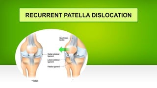
- 2. ANATOMY
- 3. ANATOMY
- 4. ANATOMY
- 5. ANATOMY
- 6. ANATOMY
- 7. EPIDEMIOLOGY & RISK FACTOR Most commonly occurs in 2nd-3rd decades of life Risk factors • previous patellar instability event • increased Q angle • femoral anteversion • genu valgum • external tibial torsion / pronated feet
- 11. Anatomical factors BONE : • Patella alta --> not articulate with sulcus, losing its constraint effects • Trochlear dysplasia • Excessive lateral patellar tilt • Lateral femoral condyle hypoplasia
- 15. Anatomical factors MUSCLE • Dysplastic vastus medialis oblique (VMO) muscle • overpull of lateral structures • Iliotibial band • Vastus lateralis
- 16. Passive stability • Medial patellofemoral ligament (MPFL)--> avulsion of MPFL • femoral origin-insertion is between medial epicondyle and adductor tubercle • patellar-femoral bony structures account for stability in deeper knee flexion • Trochlear groove morphology, patella height, patellar tracking Dynamic stability • Provided by vastus medialis (attaches to MPFL)
- 17. SYMPTOMS and EXAMINATION Symptoms • Complaints of instability • Anterior knee pain PE • Acute dislocation usually associated with a large hemarthrosis • Medial sided tenderness (over MPFL) • Patellar apprehension (+) • Increased Q angle • J sign
- 18. IMAGING • X-RAY : – AP views • best to evaluate overall lower extremity alignment and version – Lateral views • best to assess for trochlear dysplasia • Evaluate for patellar height (patella alta vs. baja) • Sunrise/Merchant views – best to assess for lateral patellar tilt CT scan MRI
- 19. Treatment • No study addresses specifically recurrent patellar dislocation in the young child • Conservative – Physical therapy – Knee bracing • Surgical
- 21. PATELLAR INSTABILITY TREATMENT Faktor soft tissue • Medial longgar : 1. Mpfl defisiensi : Mpfl Reconstruction 2. VMO weakness : VMO Strenghtening 3. Medial kapsul/retinakulum laxity : Medial Patellar Retinaculum Plasty • Lateral tightness: 1. ITB kontaktur : lateral release 2. Lateral kapsul/retinakulum : lateral release
- 23. PATELLAR INSTABILITY TREATMENT • Fibrosis/kontrakturbony factor 1. Valgus knee : DFO (Distal Femur Osteotomy) 2. External rotasi tibia : Derotation Tibial Osteotomy 3. Internal rotasi femur : Derotation Femoral Osteotomy 4. Patellar facet displacia : Patellar osteotomy 5. Trochlear displacia : Trocheoplasty 6. Patella alta : Tibial tubercle distalization
- 24. DFO
- 25. DFO
- 26. DFO
- 28. Trochlear Dysplasia Surgery Procedure Crossing sign : radiographic line of trochlear groove cross the projection of femoral condyle Trochlear bump : Trochlear line extend to anterior femoral cortex
- 29. Sulcus Deepening Tracheoplasty (Lyon/Dejour)