
Recommended
More Related Content
What's hot
What's hot (20)
Similar to Infectious uveitis
Similar to Infectious uveitis (20)
Recently uploaded
Recently uploaded (20)
Infectious uveitis
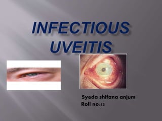
- 1. Syeda shifana anjum Roll no:43
- 2. Uvea =uveal layeruveal tractvascular layer It constitutes the midle vascular coat of eye ball{grape shaped} from anterior to posterior iris ciliary body coroid
- 4. Functions; Nutrition and gas exchange Light absorption=improves contrast of retinal images by reducing reflected light within eye{analogue to black paint inside camera}
- 5. Uveitis; inflammation of uveal tract and its adjacent structures such as retina, vetrious ,sclera and cornea.
- 6. Anatomical anterior •Iris to pars plicata of cilaiary body •Iritis,iridocyclitis intermediate •Pars plana and peripheral part of cilaiary body posterior •Involves choroid with or without retinal inflammation •Choroiditis,corioretinitis,neuroretinitis
- 7. clinical acute •Sudden symptomatic onset •Lasts for 6weeks to 3 months chronic • insidious and asymptomatic onset •3months to years recurrent •Repeated episodes seperated by inactive episodes of >3months with no treatement
- 9. etiological infectious • Bacterial • Fungal • Viral • Parasitic • rickettsial Non infectious • Immune related • Traumatic • Toxic • idiopathic masquerade • Non neoplastic • neoplastic
- 10. Inflammation of uveal tissue is induced by invasion of micro organisms. It may be , Exogenous:infection directly gain entry into eyes from outside.occurs following penetrating injuries,perforation of corneal ulcer and postoperatively .this causes suppurative irido cyclitis which turns to endophthalmitis Secondary:from the neighbouring structures Example:keratitis,retinitis Endogenous:through the blood stream.this plays important role in inflammation of uvea
- 11. I. Suppurative:by direct invasion of pathogen into uveal tissue II. Non suppurative:involves uveitogenic processes such as, Due to hypersensitivity reation:to the protein and toxin causes antigen antibody reaction. Microbial induced autoimmune uveitis: Molecular micmicry;cross rxn of Microbial antigens Stimulation of innate immunity:autoimmunity following the stimulation with microbial products.eg:bacterial DNA,viral RNA
- 12. Uveitis induced by bacterial exotoxins and other secretory products:hemolysin,alpha toxin,collagenases. Microbial cellwall stimulated alternate pathway for inflammation:complementary system,C reactive protein
- 13. Clinical features: Symptoms:pain,photophobia,redness,lacrimati on,decreased vision,severe inflammation,choroiditis,circumcorneal congestion,vitreous haze,involvement of retina,[patches]. Signs:lid edema corneal signs:corneal edema,KPs,posterior corneal opacities AC signs:aqueous cells,aqueous flare,hypopyon.
- 15. Differential diagnosis:red eye condition Scleritis Episcleritis Lens induced glaucoma Conjunctivitis
- 16. Routine investigation of uveitis: o Carefull history taking,detailed ocular examination,intraocular pressure,slit lamp examination,fundus examination after dilating pupil. o Haematological investigation o Urine examination o Stool examination o Radiological investigation o Skin tests
- 18. Types: Uveitis in chronic systemic bacterial infections: Tubercular Uveitis:chronic granulomatous infection by bovine or human tubercle bacilli Accounts for 1%of uveitis Clinical presentation: Tubercular anterior uveitis:ocurs as multiple miliary tubercles on choroid appears as round yellow white nodules 1/6-1/2 disc in size.associated with tubercular meningitis. Douse or multifocal choroiditis Choroidal granuloma Vasculitis[eales disease]
- 20. Diagnosis: postive skin test Associated systemic tuberculosis findings Positive response to isoniazid test[300mg OD for 3 days] Treatement:chemoterapy with rifamicin and isoniazid for 12 months Treatement for uveitis
- 21. leprotic uveitis:hansen disease Clinical types: o Acute iritis by ag-ab deposition,leads to severe exudation reaction o Chronic granulomatous iritis due to direct organism invasion,charecterised by the presence of iris pearl near pupillary margins in neclace form,coalesce of small pearls to form larger once. Treatement:dapsone 50-100mg+local theray of iridocyclitis
- 22. Spirochetetial uveitis: >Acquired syphylitic uveitis; chronic veneral infection caused by TREPONEMA PALLIDUM{spirochaete}.affects both anterior and posterior uvea. o syphilitic anterior uveitis:occurs as acute plastic iritis or granulomatous iritis Acute plastic iritis occurs as secondary stage of syphilis also as herxheimer rxn 24-48 hrs of penicillin dose. Gummatous anterior uveitis late in secondary or tertiary stage of syphilis .forms yellowish red highly vascularised multiple nodules arranged near the pupillary border.
- 24. o Syphilitic posterior uveitis; occurs as disseminated peripheral or diffuse choroiditis. Clinical diagnosis:confirmed by FTA-ABSblood test,more specific and sensitive than TPI and VDRL Treatement:systemic penicillin or other anti syphilitic drugs Local therapy of uveitis
- 25. Parasitic uveitis : toxoplasmosis:protozoan infestation caused by TOXOPLASMA GONDII .primarily affects CNS.systemic lesion are more marked in immunocompromised patient. Most common cause of posterior uveitis accounts for 90% of focal necrotising retinitis. Clinical presntation: 3 forms Congenial toxoplasmosis :more common ,acquired by the fetus through transplacental route during pregnancy.49%are born with either active or inactive form.most of them born with inactive disease,characterised by bilateral healed punched out heavily pigmented chorioretinal scars in macular area.discoverd when child is brought for defective vision or squint.
- 27. Acquired toxoplasmosis:infestation is acquired by eating the under cooked meat of intermediate host containing cyst from the parasite.lesions are similar to congenital one added with punctate outer retinal toxoplasmosis.
- 28. Recurrent toxoplasmic retinochoroiditis: Pathogenesis: Parasite in fetus through transplacental route involving retina and brain Excite ab formation. after healing of lesion,parasite remain encysted in inactive form After 10-40 yrs cyst rupture and release hundreds of parasite,adjacent to old inactive pigmented scar
- 29. Features:whitish yellow slightly raised area of infiltration located near margins of old punched out scarred lesion in macula ,radial pigmentation with necrosis in the centre with severe vitritis. Diagnosis by IFA,HA,ELISA. treatement by topical and systemic steroids +spiramycin/clindamycin
- 30. Toxocariasis:caused by TOXOCARA CANIS Ocular toxocariasis is always unilateral and presents as, 1. Toxocara chronic endophthalmitis with leucocoria due to marked vitrious clouding.mimics retinoblastoma and seen in 2-10 yrs. 2. Posterior pole granuloma:yellow white solitary raised nodule affecting unilateral vision 3. Peripheral granuloma:assosiated with vitreous band formation. Daignosis by ELISA BLOOD TEST.treatement consiss of periocular injection of steroids and systemic.pars plana vitrectomy is also done.
- 32. Fungal uveitis: o Presumed ocular histoplasmosis syndrome:caused by HISTOPLASMA CAPSULATUM. Features: 1) histo spots,atrophic spots scattered in mid retinal periphery.roundish yellowish white lesion .begins to appear in early childhood. 2) Macular lesions starts as atrophic macular scar, a hole in bruchs membrane sub retinal choroidal neovascularisation leakage of fluid serous detachment complicated by haemorrhages repeatedly later forms fibrous disciform scar. Daignosis by +ve histoplasmin skin test and CFT.FA inearly diagnosis.treatement by laser photo coagulation of subretinal neovascular membrane.
- 33. o Candidiasis:CANDIDA ALBICANS in immunocompromised patients. Ocular candidiasis: 1) Anterior uveitis with hypopyon 2) Multifocal chorioretinitis:roth’s spots 3) Candida endophthalmitis:puff/cotton ball,string of pearls Treatement :topical cycloplegics+antifungals systemic antifungals pars plana vitrectomy
- 35. Viral uveitis: Herpeszooster ophthalmicus:involvement of fifth nerve by VZV,causes non granulomatous iridocyclitis.complicated by iris atrophy and secondary glaucoma. Herpes simplex uveitis:forming dendritic or geographical corneal ulcers with disciform keratitis. Treated by topical steroids,cycloplegics,systemic acyclovir
- 36. CMV retinitis:occurs only en CD4 count,50 cells/mm3. Features: Symptoms.oten asymptomatic,some presents with scotoma/floaters/decresed vision. Signs:anterior segment usually rare. Posterior include, haemorrhagic retinitis with necrosis{pizza pie appearance}. Granular retinitis Complications including RD, retinal atrophy,optic nerve disease. Treated bt HAART+anti-CMV drugs.
- 38. Acute retinal necrosis:caused by VZV{commom},HSV1and 2,CMV Features: Symptoms.pain,photophobia,floaters,ocular discomfort,decresed visual acuity. Signs:ant segment may or may not show inflammation in AC Marked vitritis Retinal lesions Treatement:,systemic antivirals{lifelong},Systemic steriods,aspirin,barrier laser photocoagulation,vitrectmy with silicon oil injection.
- 39. Progressive outer retinal necrosis:rare devastating retinal necrosis caused by VZV in immunocompromised persons. Features: Painless loss of vision Retinal lesions with rapid coalese white areas. RD common complication. Treatement:IV gancyclovir/foscarnet+intra vitreal gancyclovir.