1. Invited Review
Acute phase proteins in dogs
and cats: current knowledge
and future perspectives
´ ´
Jose Joaquın Ceron, Peter David Eckersall, Silvia Martınez-Subiela
´ ´
Abstract: The acute phase response is a nonspecific inflammatory reaction of the host that occurs shortly after any tissue injury.
The response includes changes in the concentration of plasma proteins called acute phase proteins (APPs), some of which
decrease in concentration (negative APPs), such as albumin or transferrin, and others of which increase in concentration (positive
APPs), such as C-reactive protein, serum amyloid A, haptoglobin, alpha-1-acid glycoprotein, and ceruloplasmin. Most positive
APPs are glycoproteins synthesized mainly by hepatocytes upon stimulation by proinflammatory cytokines and released into the
bloodstream. The acute phase response and clinical application of monitoring APPs in dogs and cats are reviewed in this article,
including biochemical characteristics, assays developed for each individual APP, and preanalytic and analytic factors influencing
APP results that should be taken into account for proper and adequate clinical interpretation. In addition, the diagnostic use of
APPs and their possible application in monitoring treatment, which can be considered one of the most interesting and promising
practical applications of these proteins, will be discussed. Finally, challenges and future developments of APPs in dogs and cats
will be considered, because it is expected that new and cheaper automated assays for determination of the main APPs in small
animals will contribute to a wider use of these proteins as biomarkers of infection and inflammatory lesions. (Vet Clin Pathol.
2005;34:85–99)
Ó2005 American Society for Veterinary Clinical Pathology
u
I. General Concepts of the Acute Phase Response . . . . . . . . . . . . . . 85 1. Infectious disease . . . . . . . . . . . . . . . . . . . . . . . . . . . . . . . . . . . . . . . 93
II. Biochemical Properties and Methods of Measurement . . . . . . . 87 2. Surgery. . . . . . . . . . . . . . . . . . . . . . . . . . . . . . . . . . . . . . . . . . . . . . . . . . 94
A. C-reactive protein . . . . . . . . . . . . . . . . . . . . . . . . . . . . . . . . . . . . . . . . . 87 3. Gastrointestinal disease . . . . . . . . . . . . . . . . . . . . . . . . . . . . . . . . . 94
B. Serum amyloid A. . . . . . . . . . . . . . . . . . . . . . . . . . . . . . . . . . . . . . . . . . 87 4. Autoimmune disease . . . . . . . . . . . . . . . . . . . . . . . . . . . . . . . . . . . 94
C. Haptoglobin . . . . . . . . . . . . . . . . . . . . . . . . . . . . . . . . . . . . . . . . . . . . . . . 88 5. Endocrine disease . . . . . . . . . . . . . . . . . . . . . . . . . . . . . . . . . . . . . . . 94
D. Alpha-1-acid glycoprotein . . . . . . . . . . . . . . . . . . . . . . . . . . . . . . . . 88 6. Neoplasia . . . . . . . . . . . . . . . . . . . . . . . . . . . . . . . . . . . . . . . . . . . . . . . 94
E. Ceruloplasmin . . . . . . . . . . . . . . . . . . . . . . . . . . . . . . . . . . . . . . . . . . . . . 89 7. Other (including monitoring of new
F. Fibrinogen . . . . . . . . . . . . . . . . . . . . . . . . . . . . . . . . . . . . . . . . . . . . . . . . . 89 drugs and vaccines). . . . . . . . . . . . . . . . . . . . . . . . . . . . . . . . . . . . . 94
G. Negative acute phase proteins: albumin D. Limitations in monitoring treatment . . . . . . . . . . . . . . . . . . . . . . 95
and transferrin . . . . . . . . . . . . . . . . . . . . . . . . . . . . . . . . . . . . . . . . . . . . 89 VI. Challenges and Future Developments . . . . . . . . . . . . . . . . . . . . . . . . 95
III. Preanalytic Factors Influencing Results . . . . . . . . . . . . . . . . . . . . . . . 89
VII. References . . . . . . . . . . . . . . . . . . . . . . . . . . . . . . . . . . . . . . . . . . . . . . . . . . . . . 96
A. Storage . . . . . . . . . . . . . . . . . . . . . . . . . . . . . . . . . . . . . . . . . . . . . . . . . . . . 89
B. Anticoagulants . . . . . . . . . . . . . . . . . . . . . . . . . . . . . . . . . . . . . . . . . . . . 89
C. Interfering substances (hemolysis, lipemia, General Concepts of the Acute Phase Response
and bilirubinemia). . . . . . . . . . . . . . . . . . . . . . . . . . . . . . . . . . . . . . . . . 89
IV. Values in Healthy Animals and the Influence The acute phase response refers to a nonspecific and complex
of Biological Factors . . . . . . . . . . . . . . . . . . . . . . . . . . . . . . . . . . . . . . . . . . . 90 reaction of an animal that occurs shortly after any tissue injury.
A. Age and breed . . . . . . . . . . . . . . . . . . . . . . . . . . . . . . . . . . . . . . . . . . . . 90 The origin of the response can be attributable to infectious,
B. Sex and pregnancy . . . . . . . . . . . . . . . . . . . . . . . . . . . . . . . . . . . . . . . . 90
immunologic, neoplastic, traumatic, or other causes, and the
C. Circadian rhythms and day-to-day variation . . . . . . . . . . . . . 91
D. Environment and housing . . . . . . . . . . . . . . . . . . . . . . . . . . . . . . . . 91 purpose of the response is to restore homeostasis and to
E. Drug treatment . . . . . . . . . . . . . . . . . . . . . . . . . . . . . . . . . . . . . . . . . . . . 91 remove the cause of its disturbance.1,2 The acute phase re-
V. Clinical Value of Acute Phase Protein Determination. . . . . . . . . 91 sponse is considered a part of the innate host defense system,
A. Magnitude and time course of the acute which is responsible for the survival of the host during the
phase response . . . . . . . . . . . . . . . . . . . . . . . . . . . . . . . . . . . . . . . . . . . . 91 critical early stages of attack, and in evolutionary terms, it
B. Comparison with other markers of predates the acquired immune response.3
inflammation . . . . . . . . . . . . . . . . . . . . . . . . . . . . . . . . . . . . . . . . . . . . . . 92 The acute phase response is characterized by a number of
C. Diagnosis and monitoring of disease . . . . . . . . . . . . . . . . . . . . . 93 different systemic effects, including fever, leukocytosis, in-
´
From the Department of Animal Medicine and Surgery (Ceron, Martı´nez-Subiela), Faculty of Veterinary Medicine, University of Murcia, 30100, Espinardo, Murcia, Spain; the Division of
Animal Production and Public Health (Eckersall), Institute of Comparative Medicine, Faculty of Veterinary Medicine, University of Glasgow, Glasgow, G61 1QH, UK. Corresponding
author: Jose J Ceron (jjceron@um.es). This article has been peer-reviewed. ª2005 American Society for Veterinary Clinical Pathology
´ ´
Vol. 34 / No. 2 / 2005 Veterinary Clinical Pathology Page 85
2. Acute Phase Proteins in Dogs and Cats
has been postulated that production of APPs at the site of the
initial acute phase reaction in addition to the liver may
contribute to maintaining homeostasis by reducing tissue
damage associated with the inflammatory process.11
Excellent detailed reviews about the physiopathologic
mechanisms involved in the acute phase response can be
found in the literature.4,17–19 In view of its great complexity
involving a large number of physiologic, immunologic, and
biochemical alterations and reactions, it would be useful to
highlight 3 major characteristics of the acute phase response
that have practical application from a clinical point of view:
The acute phase response is a very fast response,
developing before stimulation of the specific immune
response and in many cases before the onset of clinical
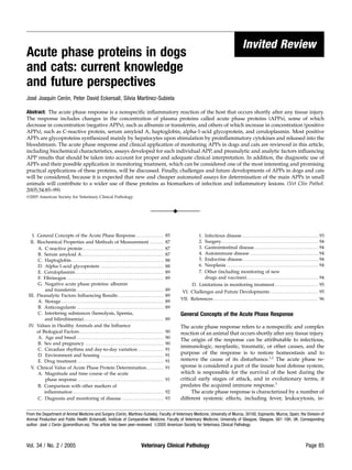
Figure 1. Mechanisms of APP production by hepatocytes, indicating 4 signs. Therefore, it can be considered as one of the
components that can be differentiated: triggering factors, local reaction, earliest markers for any pathologic process or disease.
mediators, and systemic reaction. Adapted from Heinrich6; Ebersole and
Capelli,1 and Martınez Subiela et al.7 IFN-c indicates interferon-c; TNF-a,
´ The acute phase response is highly nonspecific because it
tumor necrosis factor-a. develops secondary to numerous conditions that can
produce tissue injury (infectious, immunologic, neo-
plastic, traumatic, etc).
creased blood cortisol and decreased thyroxine concentra-
tions, metabolic changes (ie, lipolysis, gluconeogenesis, Production and response of APPs varies depending on
muscle catabolism), and decreased serum iron and zinc the species. For example, in the dog, a strong response
concentrations. The response also includes changes in the occurs with CRP; however, in cats, significant increases
concentrations of plasma proteins, called acute phase proteins of CRP have not been detected after an inflammatory
(APPs),4,5 some of which decrease in concentration (negative stimulus.20
APPs; eg, albumin or transferrin) and others of which increase
in concentration (positive APPs; eg, C-reactive protein [CRP], Biological functions of acute phase proteins recently have
serum amyloid A [SAA], haptoglobin [Hp], alpha-1-acid been widely reviewed by Murata et al.19 Although their
glycoprotein [AGP], ceruloplasmin (Cp), and fibrinogen). physiologic role still is not well understood, it is apparent that
Most positive APPs are glycoproteins synthesized mainly by APPs are involved in regulation of the immune response,
hepatocytes upon stimulation by proinflammatory cytokines inflammation, and protection against infection, and in the
and released into the bloodstream (Figure 1). repair and recovery of damaged tissue (Table 1). The same
The main proinflammatory cytokines are interleukin individual APP can have both a pro- and anti-inflammatory
(IL)-6, IL-1, and tumor necrosis factor (TNF)-a. In humans, effect, with a delicate balance between the 2 functions.23
IL-6 is also considered as one of the most important cytokines Interestingly, some proteins such as AGP and albumin
able to induce synthesis of different APPs either directly, such function to bind drugs. This can have important pharmaco-
as Hp, or combined with IL-1, as in the case of CRP and SAA,4 kinetic implications in clinical therapy because variations in
and serum levels of IL-6 markedly increase during an acute the plasma levels of APPs during inflammation can alter the
phase response in dogs.8 Cytokine assays could be used for free plasma concentration of drugs.11,26,27
quantifying the induced systemic response to infection or The acute phase response, by definition, only lasts a few
inflammation; however, the assay of APPs has been proposed days and, as was described above, seems to play a positive role
as a robust alternative for this purpose.9 in the innate host defense mechanisms. However, increases in
While APPs conventionally have been thought to be APPs also have been described in chronic inflammation.28,29 In
hepatocyte derived, there is growing evidence that they also these cases, an aberrant continuation of some aspects of the
can be produced in other tissues. Lymphocytes have been acute phase response may contribute to the underlying tissue
shown to produce AGP,10 which could explain its high serum damage which accompanies the disease and also may lead to
levels in dogs and cats with lymphoma. Extrahepatic pro- further complications, for example, protein deposition, such as
duction of AGP has also been described in other organs in reactive amyloidosis, or cardiovascular disease in humans.30,31
humans, such as kidney, intestine, and heart, and in different The main purpose of this review is to update the
types of white blood cells.11 In addition, AGP can originate knowledge about APPs in dogs and cats and present some
from the prostate gland and appear in seminal fluid.12 SAA ideas about the challenges and future developments of APPs
production has been demonstrated in tissues such as intestine, in these species, especially regarding methods for determina-
kidney, bone marrow, adipocytes (in cases of hyperglycemia), tion and clinical applicability. We will focus on APPs such as
and mammary gland (in cases of mastitis) in different animal CRP, SAA, Hp, AGP, and Cp. These APPs have not been
species13,14 Lung, adipose tissue, spleen, and kidney can exhaustively studied in the past in dogs and cats and they
produce Hp.1,15 Kidney can produce CRP in humans, and CRP could, in the future, provide valuable clinical information.
can be used as an indicator of renal transplant rejection.16 It However, it should be noted that many well-known proteins
Page 86 Veterinary Clinical Pathology Vol. 34 / No. 2 / 2005
3. ´ ´
Ceron, Eckersall, Martınez-Subiela
are also negative (albumin, transferrin) or positive (fibrinogen, Table 1. Biological functions of the main acute phase proteins.1,11,15,19,21–25
ferritin) APPs, and brief information also will be provided
about these classical APPs, especially as methods of measure- Protein Main Biological Functions
ment for them are readily available in most clinical pathology C-reactive protein When bound to bacteria, promotes the binding
laboratories for use in routine practice. Most information in of complement, which facilitates bacterial
this review will be related to dogs because APPs have been uptake by phagocytes; it has been
much more widely studied in this species than in cats. considered as a primitive form of antibody
However, data about APPs in the feline species will be specifically interacting with cell membrane
included where it is available. components of microorganisms
Induction of cytokines
Inhibition of chemotaxis and modulation
Biochemical Properties and Methods of Measurement of neutrophil function
Serum amyloid-A Chemotactic recruitment of inflammatory cells
Biochemical properties and different methods of analysis for to sites of inflammation
each individual APP will be described as, in many cases, Downregulation of the inflammatory process
biochemical characteristics influence methods of measure- (inhibition of myeloperoxidase release and
ment. For laboratories that are currently unable to set up inhibition of lymphocyte proliferation)
specific methods for APP measurement, it can be useful to Involvement in lipid metabolism and transport
note that most APPs migrate to the a- and b-globulin areas, so Haptoglobin Involvement in host defense responses to
routine electrophoresis on agarose or cellulose acetate gels infection and inflammation; acts as a natural
can be used to identify overall increases in APP concentra- antagonist for receptor-ligand activation of
tions in inflammation.5 However, this method is much less the immune system
sensitive than the individual APP assays.29 Binding of free hemoglobin (a toxic and
proinflammatory product resulting from
C-reactive protein hemolysis)
Bactericidal effect in infected wounds by binding
Canine CRP has a molecular weight of 100 kD, which consists hemoglobin and limiting the availability of
of 5 subunits of 20 kD each. This protein was the first APP to hemoglobin iron for bacterial growth
Inhibition of granulocyte chemotaxis and
be described. Originally named for its ability to bind the
phagocytosis
C-polysaccharide of Pneumococcus pneumoniae, CRP has been
defined in humans as an exquisitely sensitive systemic marker Alpha-1-acid glycoprotein Antiinflammatory and immunomodulatory agent
with antineutrophil and anticomplement
of inflammation and tissue damage.32,33 Examined by electron
activity; increases the secretion of
microscopy, canine CRP resembles human CRP; the main interleukin-1 receptor antagonist by
difference between the proteins is that 2 of the 5 subunits of macrophages
canine CRP are glycosylated,34,35 which could explain in part Drug binding to numerous basic and neutral
the difficulties of using antibodies raised against human CRP lipophilic drugs and also acidic drugs, such
for canine measurements.36 as phenobarbital
Measurement of serum CRP is generally by immuno- Ceruloplasmin Transport of copper needed for wound healing,
assays using specific canine CRP antibodies, and several collagen formation, and maturation
formats have been developed and described for this purpose, Protection of cells and tissues against oxidant
such as an immunoturbidimetric assay adapted for automated compounds generated by phagocytes in
biochemical analyzers,37 ELISA,38,39 or slide/capillary reverse the course of clearing microorganisms
passive latex agglutination tests.40,41 New methods based on or tissue debris
Reduction in the number of neutrophils attaching
time-resolved fluorometry (TRFIA) have been recently de-
to endothelium
veloped for CRP assays in canine whole blood,42 saliva,43 and
effusions.44
Currently, a commercial ELISA kit is available that is about the biochemical properties and methods of measure-
specific for canine CRP, although technical improvements are ments of CRP in cats is scarce because CRP does not seem to be
needed to decrease between-run imprecision.45–47 A commer- involved in the acute phase response in this species.20
cially available automated turbidometric immunoassay for
human serum CRP has been found to be valid for measuring Serum amyloid A
canine serum CRP concentration48; however, in other inves-
tigations very weak or negligible cross-reactivity of canine SAA is a small serum protein with a molecular weight of
CRP with different antihuman CRP antibodies has been 15 kD. It is thought to be the precursor of amyloid protein A,
found.36,49,50 Recently, a rapid assay giving qualitative results the major protein of a-amyloid, so it is potentially involved in
by sample dilution followed by immunochromatography on the pathogenesis of amyloidosis and other chronic inflamma-
a prepared test strip has been produced for canine CRP51 tory diseases such as rheumatoid arthritis.21 In humans, this
which could differentiate between samples with CRP concen- protein shows a sensitivity, response speed, and dynamic
trations of ,5mg/L and .5mg/L. False positive results are range comparable with those of CRP.52 However, its use in
a major limitation of this test. To our knowledge, information animals has, until recently, been limited due to difficulties in
Vol. 34 / No. 2 / 2005 Veterinary Clinical Pathology Page 87
4. Acute Phase Proteins in Dogs and Cats
purification and quantification, probably because it is a hy- eases.67,68 Further studies of these changes could reveal more
drophobic apolipoprotein that is complexed within serum details about the possible application and use of Hp gly-
high-density lipoproteins.28,53 cosylation patterns for differentiating pathologic processes
Amyloid protein A from dogs and humans shows and monitoring disease activity.1
considerable homology,54 although the primary structure of Assays for serum Hp concentration can be divided into 2
canine SAA has an additional peptide of 8 amino acids.55 It main groups: a) spectrophotometric assays and b) immuno-
seems the sequence and inductive capacity of SAAs are highly assays. Different spectrophotometric manual assays have been
conserved across evolutionarily-distinct vertebrate species.21,56 based on the ability of Hp to bind hemoglobin (Hb), forming
Specific sandwich ELISAs using anti-canine53 and anti- Hp–Hb complexes that either alter the absorbance character-
feline57 SAA antibodies for canine and feline SAA measure- istic of Hb in proportion to the concentration of Hp in a serum
ments, respectively, have been developed, although technical sample,22,69 or preserve peroxidase activity at an acidic pH,
difficulties in the preparation of antiserum to canine SAA have which then can be detected and quantified.70 In addition, an
been reported.53 Monoclonal antiserum against human SAA automated spectrophotometric multispecies assay based on
has been successfully used in a sandwich ELISA to measure the peroxidase activity of Hp–Hb complexes, in which in-
canine SAA,58 and polyclonal antiserum against canine SAA terference by serum albumin is eliminated, has been de-
has been used to detect feline SAA.59 In addition, a commer- scribed71 and validated at different laboratories for use with
cially available ELISA for SAA determination in veterinary canine serum, giving satisfactory results.46,71 This methodol-
species using monoclonal antiserum against human SAA has ogy has been developed into a commercially available kit for
been proven to be useful for canine46 and feline60 SAA routine analysis and is also being used for Hp measurements
quantification; however, technical improvements are needed in cats.60 Nephelometric immunoassays, in which the rate of
to reduce between-run imprecision.46 Recently, an automated precipitation of the antibody–antigen complex is measured,
commercially available human SAA turbidimetric immuno- have been validated for estimation of Hp in dogs.66 These
assay has been validated for feline SAA determination.61 assays depend on the cross-reactivity of antiserum raised to
Several recommendations have been made about stand- human Hp with canine Hp and must be properly validated
ards and calibration to improve SAA assays, including deter- before use.
mining the concentration of SAA in the standard by sequence It is important to note that canine serum specimens must
analysis and amino acid composition. Because several proteins be diluted in many cases when Hp assays developed for other
copurify with canine SAA,58 use of SAA-enriched high-density species are used, as the concentrations of Hp in health and
lipoprotein as an assay standard,62 or use of serum with a high disease are significantly higher in dogs than in other species,
concentration of SAA as a calibration standard instead of the such as ruminants or humans. For example, the mean (6 SD)
purified protein also are recommended.53 serum Hp concentration in healthy dairy cows was undetect-
able (,0.02 mg/mL) and in cows with mastitis was 1.26 6
0.66 mg/mL, whereas the mean serum Hp concentration in
Haptoglobin healthy dogs was 0.60 6 0.55 mg/mL and in dogs with
polyarthritis was 11.6 6 3.2 mg/mL.71 Development of assays
Dogs have only 1 subtype of Hp compared with humans, who for canine and feline Hp with species-specific standards that
have 3 subtypes (Hp 1-1, Hp 2-1, and Hp 2-2). However, allow measurement of a wider range of concentrations would
canine Hp closely resembles human Hp 1-1 with respect to be desirable to avoid sample dilution.
amino acid content, molecular weight (81 kD), electrophoretic
pattern on starch gel, and the existence of a and b subunits in
a tetra-chain arrangement (b-a-a-b).63 Compared with human Alpha-1-acid glycoprotein
Hp 1-1, canine Hp has 2 structural differences:64 the 2 ab
chains are joined by a noncovalent interaction rather than by The main biochemical characteristic of AGP is that it is
a disulfide bridge (this noncovalent linkage also exists in feline a highly glycosylated protein and is the main protein com-
Hp), and the a-chain has an oligosaccharide-binding sequence ponent in seromucoid, the fraction of plasma that is most
and is glycosylated, whereas the human a-chain is non- resistant to acid precipitation.3 Canine AGP has been purified
glycosylated. These structural particularities may be respon- and biochemically characterized and, similar to AGP in hu-
sible for the divergence of findings in the recognition of canine mans, has been described as a very unusual protein of 43 kD
and feline Hp by antibodies directed against human Hp. For with a very low pI of 2.8–3.8 and a very high carbohydrate
example, it has been reported that a monoclonal antibody content of 45%.11,72
against human Hp did not recognize canine and feline Hp, Although AGP can be estimated by precipitation of the
but this antibody was directed to the disulfide bond linking majority of serum proteins by perchloric acid and quantifica-
the Hp chains that is not present in dogs and cats.65 How- tion of the remaining soluble proteins,9 this protein usually is
ever, other authors found that human antiserum can recognize measured by single radial immunodiffusion on agarose gel
dog Hp and have demonstrated that human methods based impregnated with anti-species AGP rabbit serum, using
on antigen–antibody reaction can be applied to the measure- different commercial kits. These tests are species-specific and
ment of canine Hp.66 are currently available for dogs and cats; however, they have
The glycosylation pattern of Hp can vary in dogs with the disadvantage of requiring 24 or 48 hours for diffusion to be
various inflammatory, autoimmune, and neoplastic dis- complete.9 Immunoturbidimetric assays for the measurement
Page 88 Veterinary Clinical Pathology Vol. 34 / No. 2 / 2005
5. ´ ´
Ceron, Eckersall, Martınez-Subiela
of canine and feline AGP have been developed,73,74 offering polypeptide chain of about 700 amino acids. Transferrin binds
the advantages of being rapid and adaptable to biochemical iron as a ferric ion in 2 binding sites at a neutral pH, but the ion
analyzers. is dissociated when the pH falls below 5.5.86 Although total
Changes in glycosylation of AGP have been described in transferrin can be measured by immunoassay, it is commonly
acute inflammatory conditions in humans11,23,75,76 and also in assessed by measuring the total iron-binding capacity (TIBC)
cats with feline infectious peritonitis (FIP),77 in which de- of serum, though in dogs and cats this test has been used
creased sialylation77 and variations in monosaccharides were largely for the assessment of iron metabolism and homeosta-
expressed on AGP.78 sis.86,87
In humans, there has been debate on the balance between
nutritional and disease effects on the production of negative
Ceruloplasmin APPs.88,89 A recent hypothesis is that the acute phase response
has a stronger effect than the nutritional plane on concen-
Ceruloplasmin is an a2-glycoprotein and one of the positive trations of transferrin, albumin, and other negative APPs.90
APPs in dogs.7 Although there are no reports about the Thus, a decreased concentration of transferrin may be more
structure of canine or feline Cp, studies of human Cp have indicative of an acute phase response than of poor diet.
shown that it is a blue protein with a molecular weight of 151
kD containing about 0.34% copper, which corresponds to 8
atoms of copper per molecule. Additionally, it is a glycoprotein Preanalytic Factors Influencing Results
containing hexosamine, hexose, and neuraminic acid.79
Many quantitative methods based on different principles Storage
have been used for Cp measurement in plasma or serum.79 Canine CRP has been reported to be stable at À108C for at least
Assays based on oxidation of different compounds such as 3 months.91 Dillman and Coles92 reported that CRP in dogs was
p-phenylenediamine (PPD) or its N-dimethyl derivative and stable when stored at À208C for 2 months and was inactivated
o-dianisidine dihydrochloride have been used most often in when left at 708C for 30 minutes; however, their results should
veterinary medicine. Manual80,81 and automated82 methods be taken with caution because they used a human anti-CRP
based on PPD-oxidase activity have been reported for antibody test for measurements. In addition, Hp has been
measuring Cp in canine serum. reported to be more stable in serum than in a purified
One of the main problems with Cp assays is the lack of preparation.93 However, a decrease in Hp concentration in
commercially available reference materials to standardize Cp canine serum stored at À208C has been described and À708C
concentrations, such that different arbitrary units based on the has been suggested for prolonged storage.66
increase of absorbance per unit of time have been used and
expressed as oxidase units81 or UI/L.80
Anticoagulants
Fibrinogen In a study in which different anticoagulants (heparin, EDTA,
and citrate) were compared with serum, CRP values were
Fibrinogen is a b-globulin present in the plasma of all significantly lower in samples with citrate, while SAA results
vertebrates. It is composed of 3 nonidentical polypeptide were significantly higher in all plasma samples, and Cp
chains linked by disulfide bridges and a glycoprotein that concentrations were significantly higher with heparin and
contains 3–5% carbohydrate.83 Assays have been largely lower with EDTA.94 Increases in Hp concentration have been
dependent on fibrinogen’s biological activity based on the found when heparin was used as an anticoagulant.71 Overall,
rate of formation of insoluble fibrin in the presence of excess the effects of anticoagulants on APP concentrations seem to be
thrombin or on its precipitation following mild heat treat- small and without influence on clinical interpretation.
ment.83
Interfering substances (hemolysis, lipemia, and bilirubinemia)
Negative acute phase proteins: albumin and transferrin
The effects of hemolysis, lipemia, and bilirubinemia in assays
Albumin is the most abundant protein in blood, constituting for different APPs in canine serum are tabulated (Table 2).
35–50% of protein in the plasma of healthy dogs and cats; it is These results should be interpreted with caution because they
the major band observed in serum protein electrophoreto- have been obtained with specific kits and methods, and
grams. Albumin is responsible for about 75% of the osmotic significant variation could be found when other reagents or
pressure of plasma and is a major source of amino acids that analyzers are used; for example, use of TRFIA avoids the effect
can be utilized by the animal’s body when necessary. Albumin of hemolysis.42
usually is measured in routine practice by spectrophotometric The magnitude of differences caused by interfering
methods, such as the bromcresol green assay; however, substances is small, and unlikely to have a significant impact
overestimation of albumin can occur in heparinized plasma on the clinical interpretation of the test result with the
samples assayed by this method.84,85 exception of the decreases found in Hp and CRP concen-
Transferrin is a plasma glycoprotein that is responsible for trations as measured with immunoturbidimetric assays in
the transport of iron in the circulation; it has a single samples with hemolysis. It is postulated that the causes of
Vol. 34 / No. 2 / 2005 Veterinary Clinical Pathology Page 89
6. Acute Phase Proteins in Dogs and Cats
Table 2. Effects of interfering substances in some methods for analysis of acute phase proteins.37,71,66,95
Acute Phase Protein Method Hemolysis Lipemia Bilirubinemia
C-reactive protein (CRP) ELISA Increased* Increased* Decreased*
Immunoturbidimetric Decreased by 90% when Hb 5 80 mg/dL Not evaluated Not evaluated
Serum amyloid A ELISA No effect No effect No effect
Haptoglobin (Hp) Spectrophotometric Decreased by 25–30% maximum when No effect No effect
Hb 5 0.3–10 mg /mL
Immunoturbidimetric Decreased by 70–80% when Hb .25 mg/dL Increased when triglycerides .100 mg/dL Not evaluated
Ceruloplasmin Spectrophotometric Increased when Hb ,2.5 mg/dL; Increased* Increased*
decreased when Hb .2.5 mg/dL*
*No significant impact on clinical interpretation.
decreased Hp concentration can be in vivo or in vitro. In vivo As with other biochemical analytes, these values should be
decreases occur during episodes of intravascular hemolysis, interpreted with caution, as they can be greatly influenced by
when Hb is bound by Hp and the Hb–Hp complexes are analytic conditions, and it is recommended that each
removed from the circulation by macrophages, mostly in the laboratory should establish and use its own values. This is
spleen and liver, causing a transient decrease in plasma Hp particularly important for APPs due to the lack of reference
concentration.22 In vitro decreases may occur in immunotur- materials for international harmonization of assays in small
bidimetric methods because of Hb binding to the b-chain of animals.9
the Hp molecule, masking some of its antigenic determinants For diagnostic use, concentrations of APPs in an animal
and decreasing Hp/anti-Hp lattice formation.63,66 In hemo- are compared with a population-based reference interval
lyzed samples, large background absorption caused by Hb, derived from an observed distribution of measurements of the
which can be minimized by sample dilution, could be the APP in an appropriate group of animals and containing the
responsible for decreases in CRP concentration when using central 95% of the distribution. The distribution of serum
immunoturbidimetric assays.37 concentrations is non-Gaussian for many APPs, with only the
upper level of the reference interval reported. Other ways of
assessment, such as the critical difference, in which a patient
Values in Healthy Animals and the Influence of serves as its own reference by comparing analytic results
Biological Factors obtained from serial samples at appropriate intervals, also
could be used.104,105 A number of biological factors have been
Acute phase protein concentrations in adult, healthy dogs as examined that could influence the values of APPs.
determined by different investigators are tabulated (Table 3).
Age and breed
Table 3. Serum acute phase protein concentrations in healthy animals.
No significant age-related differences in serum APPs in
Acute Phase Protein Dogs Cats healthy animals have been found.40,101 However, it seems that
C-reactive protein ,5 mg/L8,96 —
APP responses in inflammatory conditions are higher in adult
,10 mg/L39 animals because, after experimental inoculation with turpen-
0.22–4.04 mg/L97 tine oil or Staphylococcus aureus, significantly greater peak CRP
0.8–16.4 mg/L98 concentrations in 3- and 18-month-old dogs were found
8.4 6 4.6 mg/L40 compared with those in 1-month-old dogs.106
0.48 6 0.17 mg/L99 Clinically healthy Yorkshire Terriers and Dachshunds
Serum amyloid A Nondetectable–2.19 mg/L97 10.21 6 8.32 mg/L60 were reported to have lower levels of AGP compared with
Nondetectable–69.6 U/mL53 breeds such as Poodle, Cocker Spaniel, Labrador Retriever, or
1.15 6 2.53 mg/L58 German Shepherd. This observation could contribute to the
Haptoglobin 0–3 g/L71 0.04–3.84 g/L100 wide range of AGP values (40–1070 mg/L) that can be found
0.3–1.8 g/L97 1.30 6 0.64 g/L60 in healthy dogs.107 To our knowledge, no other investigations
Alpha-1-acid glycoprotein 322 6 202 lg/mL101 0.1–0.48 g/L100 have examined interbreed differences in APP concentrations.
509 6 117 lg/mL102 1.20 6 0.62 g/L60
302 6 74 lg/mL103
,380 lg/mL38 Sex and pregnancy
480 6 149 lg/mL58
No significant sex-related differences have been found in CRP
Ceruloplasmin ,20 UI/L80 —
,0.4 oxidase units81
and AGP levels in healthy Beagle dogs40,78,101 or in SAA, AGP,
,4.93 mg/dL29 and Hp levels in healthy cats.20 However, an increase in APPs
occurs in pregnancy in the bitch during embryonic implanta-
Page 90 Veterinary Clinical Pathology Vol. 34 / No. 2 / 2005
7. ´ ´
Ceron, Eckersall, Martınez-Subiela
tion and placental development due to an inflammatory Table 4. Classification of acute phase proteins based on the magnitude
reaction induced by embryonic endometrial invasion. Based of response.
on this fact, serum APP profile determination has been
proposed as an early pregnancy test (from the third week of Species Major Moderate Negative
gestation) in bitches. Due to the low specificity of APPs, Dog C-reactive protein Ceruloplasmin Albumin
however, knowledge of mating dates and a complete evalu- Serum amyloid A Haptoglobin
ation of the animal’s health and condition should be Alpha-1-acid glycoprotein
performed to ensure there is no evidence of other inflamma- Cat Serum amyloid A Haptoglobin Albumin
tory processes and to avoid false-positive results.73,101,108,109 Alpha-1-acid glycoprotein
Circadian rhythms and day-to-day variation
As in humans, there appears to be no circadian rhythm in CRP clinical value in diagnosing and monitoring inflammation. For
concentration in adult dogs, as no significant variations were example, a decreased albumin/globulin (A/G) ratio provides
detected in serum CRP levels in samples collected from the an estimate of the acute phase reaction in infection or in-
same animal at different times within 24 hours. In addition, flammation in dogs and cats,116 because of decreased albumin
no significant variations in serum CRP were detected when concentration (as a negative APP) and increased globulins
the same animal was sampled on different days over a period concentration. However, the sensitivity and specificity of the
of 3 weeks.98 ratio for detecting clinical or subclinical disease are not as high
as those of positive APPs such as CRP.29 Similarly, in cats with
Environment and housing FIP, the A/G ratio has been a valuable diagnostic test for this
viral infection for some time, but the use of a positive APP,
Healthy dogs kept in private households showed higher AGP, has been shown to have superior diagnostic efficiency.100
serum CRP values (8.4 6 4.6 mg/L) than dogs kept in very Although the pathophysiologic reaction of fibrinogen to
clean animal facilities (0.48 6 0.17 mg/L), possibly due to infection and inflammation has been known for many years
exposure to environmental factors that stimulate the immune and occurs in dogs and cats, as in other mammalian species,116
system and to which animals in highly protected facilities are fibrinogen has not been routinely measured in veterinary
not exposed.40,99 Environmental effects also could be re- diagnostic laboratories as an acute phase protein. Indeed,
sponsible for the high intraindividual variation in serum most of the interest in measurement of fibrinogen in canine
CRP values found in healthy dogs.98 medicine has been for the diagnosis of disseminated in-
travascular coagulation and hyperfibrinolysis, in which the
concentration of fibrinogen falls.117 In a study of APPs in 161
Drug treatment dogs with inflammation, the predictive value of a positive or
negative test for Hp and Cp was comparable to or better than
A significant increase in serum Hp concentration has been that of fibrinogen.80 Thus, the use of fibrinogen as an APP in
demonstrated after administration of different doses and dogs and cats appears to largely have been superseded by the
protocols of glucocorticoid treatment in dogs; however, growing availability of specific assays for APPs that show
concentrations of CRP, Cp, and SAA were not affected. The greater and more rapid responses on stimulation than the 2- to
magnitude of increase in Hp concentration after the admin- 4-fold increase observed in the concentration of fibrinogen.
istration of glucocorticoids can be similar to the increase
produced by an inflammatory stimulus.97 In addition, other
drugs, such antihelmintics, produced an increase in serum
Hp,110 and phenobarbital, in a therapeutic dosage regime, Magnitude and time course of the acute phase response
induced a significant increase in canine AGP concentration.111
Acute phase proteins in dogs and cats can be classified by the
However, in other studies with dogs, doxorubicin adminis-
magnitude of their response to stimuli (Table 4) as major (10-
tration did not increase concentrations of AGP112 and
to 100-fold increase), moderate (2- to 10-fold increase), or
prostaglandin inhibitors did not affect CRP concentration in
negative (decrease) reactants. For major APPs, especially in
Escherichia coli sepsis.113
cats, upper limits of increase are lower than those de-
There is evidence that the concentrations of APPs in dogs
scribed for humans, who experience increases as high as
do not change significantly as a result of either the continuous
1000-fold.
handling or venous blood sampling of animals,104,105,92 sub-
In dogs, differences in the magnitude and also the time
cutaneous injections,114 or the administration of saline or other
course of response have been detected between major and
placebos.38,115
moderate APPs (Table 5). Major APPs (CRP and SAA) usually
have an early and high rise in concentration and a very rapid
Clinical Value of Acute Phase Protein Determination decline. The CRP increase in dogs is indeed more rapid than in
humans because, in the latter species, levels do not increase
Classical APPs, such as albumin and fibrinogen, although until after 6 hours. Although no data were given about the
currently easier and cheaper to measure, seem to have a lower half-life of CRP and SAA in dogs and cats, it appears that the
Vol. 34 / No. 2 / 2005 Veterinary Clinical Pathology Page 91
8. Acute Phase Proteins in Dogs and Cats
Table 5. Magnitude and time of response of different acute phase proteins in dogs following different stimuli.8,58,81,118
Acute Phase Protein Cause of Inflammation Magnitude of Time of Time of
Response Detecting a Maximum
(fold increase) Significant Peak
Increase Concentration
C-reactive protein Surgical trauma 953 4 hours 24 hours
Turpentine oil injection 40–503 — 48 hours
Intravenous dose of Escherichia coli 233 4 hours —
lipopolysaccharide
Serum amyloid A Experimentally induced parvovirus infection 30–8003 5 days 7 days
Intravenous dose of E coli lipopolysaccharide 5253 2 hours —
Haptoglobin Surgical trauma 2–33 24 hours 3–4 days
Alpha-1-acid glycoprotein Experimentally induced parvovirus infection 2–53 5 days 7 days
Turpentine oil injection 2–33 — 3 days
Ceruloplasmin Surgical trauma 2–33 24 hours 4 days
Albumin Turpentine oil injection Decrease of ;50% 5 days
half-life of canine CRP is short.81 The rapid production and as Trypanosoma brucei or Ehrlichia canis, the peak appeared at
clearance of CRP make it a very useful test to indicate the 4–10 days postinfection.38 Increases of 95-fold were found in
clinical situation of an animal at the time of sample CRP concentrations after surgical trauma compared with
collection.34 In humans, SAA is catabolized in the liver and 40- to 50-fold increases after turpentine oil injection. Five-fold
has a half-life of 1 day, although in acute or chronic increases have been found in Hp and Cp concentrations in
inflammation, the capacity of the liver to degrade SAA dogs with leishmaniosis,29 compared with the 2- to 3-fold
decreases.119 increases found with surgical trauma.81
On the other hand, moderate APPs (Hp, AGP, and Cp) In cats, CRP does not seem to be affected or has a very
need more time to increase and return to normal values, with small increase in inflammatory conditions (Table 6). Sasaki
a more gradual decline. Increases in Cp concentration in dogs et al57 found increases of up to 55-fold in SAA in different
are higher and evidently earlier than in humans, peaking at infectious and inflammatory diseases, and Duthie et al100
about double normal values on the fourth day following found increases of .10-fold in AGP concentrations in cats
surgery.81 Interestingly, the moderate APPs, which increase with FIP, so that SAA and AGP could be considered as major
less in terms of fold-increase, are in higher concentration than APPs in the cat. SAA is the APP that increases earliest after
CRP and SAA in the serum of healthy animals, and the total a stimulus in cats.20
amount of protein produced during the acute phase response
is usually higher.
The peak time and magnitude of an APP response can
Comparison with other markers of inflammation
vary depending on the type of stimulus. In dogs that had
undergone surgery or were infected with Bordetella bronchi-
Some relationship, although weak, exists between APPs and
septica, peak CRP responses were observed within 1 day;
WBC and neutrophil counts. A significant difference in serum
however, with infection by intracellular microorganisms, such
CRP concentrations between clinically normal dogs and dogs
with high neutrophil counts was detected, and a significant
positive correlation, although with low correlation coeffi-
cients, was found between the concentrations of different
Table 6. Response of different acute phase proteins in cats following
positive APPs (CRP, Hp, and Cp) and WBC and segmented
injection of lipopolysaccharide or turpentine oil.20
and band neutrophils counts.80,104 However, there are some
advantages of APPs compared with leukocyte counts as
Acute Phase Protein Magnitude Time of Time of
markers of inflammation:
of Response Detecting a Maximum
(fold increase) Significant Peak APPs have higher diagnostic sensitivity. Ceruloplasmin
Increase (h) Concentration and Hp are up to 6 times more sensitive than leukocyte
(h) counts in detecting inflammation, and are increased in
Serum amyloid A 4.23 8 24–48 cases in which total and differential WBC counts showed
Alpha-1-acid glycoprotein 5.73 24 48 no changes.80 In addition, APPs concentrations are of
Haptoglobin 2.93 24 48 value for detecting inflammation in animals in which the
C-reactive protein No significant change bone marrow cannot respond normally to an inflamma-
tory stimulus, such as those with myelosuppresion
Page 92 Veterinary Clinical Pathology Vol. 34 / No. 2 / 2005
9. ´ ´
Ceron, Eckersall, Martınez-Subiela
attributable to treatment with chemotherapeutic agents Table 7. Acute phase proteins found to be increased in different infectious
or those with leukemia.104,120 diseases in dogs.*
Longer analyte stability. Acute phase proteins are more
Disease Acute Phase Magnitude of Reference
stable than the cellular components of blood, and assays
Protein Increase
can be performed on frozen serum or plasma samples.80
Babesiosis CRP Not specified Matijatko et al122
Theoretically, APPs exhibit a faster response than AGP 3–53 Lobetti et al102
changes in WBC counts, especially in inflammation,
Bordetella bronchiseptica CRP 953 Yamamoto et al53
where new WBCs must be generated in the bone
SAA 203 Yamamoto et al115
marrow. However, significant increases in CRP concen-
tration after surgical trauma do not appear earlier than Ehrlichia canis CRP 3.3–6.53 Rikihisa et al38
alterations in CBC values.104 AGP 1.9–8.63
Escherichia coli sepsis CRP 3.5–43 Hulton et al113
Leishmaniosis CRP 25–303 Martinez-Subiela
Hp 4–53 et al29
Diagnosis and monitoring of disease Cp 3–53
SAA 80–903y Martinez-Subiela
It is important to recognize that APP concentrations are and Ceron123
elevated in animals with many different diseases, having very Leptospirosis CRP 16–803 Caspi et al96
poor diagnostic specificity in detecting the cause, so they (experimentally
cannot be used as the primary diagnostic test for a particular induced)
disease. However, they have very high sensitivity in detecting CRP 30X (naturally Yamamoto et al124
many conditions that alter the health of the animal and in occurring)
providing evidence that an animal has subclinical inflamma- Parvovirosis CRP 203 Yamamoto et al124
tion or infection. As summarized by Kent,121 APPs quickly and
SAA 30–8003 Yule et al58
precisely demonstrate the presence of infectious and in- AGP 2–53
flammatory conditions but not, unfortunately, the cause. A
Trypanosomiasis CRP at least 203z Ndungu et al125
very interesting characteristic of APPs is the possibility of Hp 33
detecting subclinical disease and predicting possible future
clinical disease in the animal. For example, measurement of *CRP indicates C-reactive protein; AGP, alpha-1-acid glycoprotein; SAA, serum
AGP in a health screen was useful for identifying dogs with amyloid A; Hp, haptoglobin; and Cp, ceruloplasmin
subclinical disease that, after 2 weeks, had clinical signs or ySome animals with leishmaniosis had SAA concentrations within the reference
even died of different causes such as parvovirus infection.103 interval.
zUpper range of measurement of the method was 200 mg/L, which was reached in
Infectious disease. To our knowledge, most experimentally many affected animals.
induced or naturally occurring infectious diseases studied
produce an increase in positive serum APP concentrations in concentration is not pathognomonic for the disease because
dogs, with CRP being the protein that shows the highest high levels were also found in cats infected with feline
response (Table 7). SAA has also been shown to increase very immunodeficiency virus.100 Increases in other APPs, such as
significantly in dogs infected with B bronchiseptica and Hp and SAA, have been detected in FIP-infected cats.60,126 In
parvovirus40,58; however, concentrations of SAA were less addition, increases in Hp concentration have been described in
sensitive for detecting animals infected with Leishmania cats with abscesses and upper respiratory tract infections;
infantum compared with others.123 Concentrations of other however, Hp concentration decreased after hemolysis associ-
APPs, such as Hp, AGP, and Cp, also have been shown to ated with hemobartonellosis (mycoplasmosis).126
increase in dogs with different infectious diseases.29,38,58,102,125 It is likely that measuring APPs is going to be especially
Increases in all of these APPs have been detected both in acute helpful in the care of animals diagnosed with infectious and
and chronic infections.38 inflammatory conditions to monitor clinical evolution and
In addition, a correlation has been described between APP assess response to treatment and its withdrawal. In human
levels and the severity of disease, with serum APP concen- medicine, CRP concentration has been found to rapidly
trations being higher in severe or complicated cases38,40,58,122 or decrease following effective therapy127 and was useful in
in animals with clinical signs of disease compared with those evaluating and assessing the duration of antibiotic therapy in
without clinical signs.29 No correlation has been found neonatal septicemia.128,129 Examples of the use of APPs in
between antibody titers and APP concentrations in different monitoring different canine infectious and inflammatory
infectious diseases29,38; an explanation for this fact could be diseases are:
that the APP reaction corresponds with the early innate
response, whereas antibody titers correspond with the later In dogs with Trypanosoma brucei infection, high levels of
acquired immune response. serum CRP appeared as long as the parasite persisted,
In cats, quantification of AGP has been demonstrated to be and a decrease in this protein was detected with
a reliable aid in the diagnosis of FIP; however, increased AGP elimination of the parasite by chemotherapy. Further-
Vol. 34 / No. 2 / 2005 Veterinary Clinical Pathology Page 93
10. Acute Phase Proteins in Dogs and Cats
more, relapsed infections resulted in a quick return of useful in monitoring clinical progression and response to
CRP to high concentrations.125 therapy in dogs with acute pancreatitis.133
CRP and AGP concentrations declined in dogs with E
canis infection, in which, although the parasite persisted, Autoimmune disease. Caspi et al96 found increases of CRP in
clinical signs resolved, suggesting healing of the initial canine rheumatoid arthritis and nonerosive polyarthritis.
tissue damage.38 In both processes, dogs with inactive disease had no increase
in CRP concentration. These authors also found increases of
Concentrations of CRP and Cp decreased significantly in CRP in dogs with autoimmune hemolytic anemia; however, as
dogs with leishmaniosis during short-term therapy with expected, low levels of Hp were associated with this disease22
2 different treatment protocols. In the dogs in which SAA due to the mechanism previously described.
concentrations were high before treatment, a significant
decrease was observed in this protein at the end of Endocrine disease. Hp has been particularly studied in ca-
therapy.114 nine hyperadrenocorticism, where moderate increases have
been detected, probably due to endogenous glucocorticoid-
Surgery. An increase in serum CRP concentration has been mediated stimulation of Hp production. Hp concentration
described after different surgical procedures in dogs; the also was increased in dogs with diabetes mellitus and diabetic
magnitude of increase was generally related to the intensity ketoacidosis, for which it was postulated that chronic in-
of the surgical trauma, being larger when more severe tissue flammation may have a role in this disease,135 as has been
injury was produced, such as in orthopedic surgery.96,124 In all shown in humans.13 Additionally, increased SAA concentra-
surgical procedures in dogs without clinical complications, tions have been found in cats with diabetes.57
and although WBC count was still increased, CRP concen-
tration was markedly decreased by the time of suture Neoplasia. In dogs with mammary tumors, CRP values
removal and disappearance of clinical signs. Therefore, seemed higher when there was disseminated disease and
serum CRP determination has been suggested as a more complications compared with localized primary malignancy
useful tool for monitoring postsurgical progress than WBC or benign lesions.96 In a different study, dogs with mammary
counts.86,124 In addition, in humans with signs of an acute tumors and various other neoplasms did not have higher AGP
phase response preoperatively, it was found that the patient’s levels in serum compared with healthy animals, although it
response to an operation and infection during the post- was postulated that the AGP molecule may have a higher sialic
operative period may be adversely affected.130 In cats, acid concentration in neoplasia.107 Increases in the concentra-
concentrations of SAA, AGP, and Hp increased significantly tion of this protein have been described in dogs with
1 day after surgery.114 lymphoma, various carcinomas (except mammary carcinoma),
and sarcomas.112 In cats, AGP concentrations were significant-
Gastrointestinal disease. Increases in CRP concentration have ly higher in tumor-bearing cats compared with healthy cats.136
been found in canine serum following experimentally induced Serum AGP measurement was useful for monitoring dogs
acute gastric mucosal injury with different compounds such as with lymphoma treated with doxorubicin because the
acetylsalicylic acid, indomethacin, and sodium chloride.131 concentration of AGP significantly decreased with complete
Moderate increases in CRP have been found in dogs with remission after doxorubicin treatment112 and increased again, 3
inflammatory bowel disease (IBD) (range 10–15 lg/mL), and weeks prior to the time of lymphoma relapse.137 However,
increases were strongly correlated with disease activity. serum AGP concentrations provided no useful information
Indeed, serum levels of CRP have been proposed as an regarding treatment response or survival in cats with
additional criteria to categorize and assess disease activity in lymphoma.138
canine IBD and were a suitable marker for laboratory
evaluation of the effect of therapy, with concordance between Other (including monitoring of new drugs and vaccines). In
the improvement of clinical signs and decreased levels of this dogs, increases in APP concentrations have been described in
APP.130 In this study, no significant differences in serum AGP different inflammatory processes, such as pyometra124 and
and Hp were found between healthy and IBD-affected dogs. pneumonia.40According to reports by Sevelius and Ander-
Animals with other enteric diseases, such as bacterial sson,139 APPs can be considered as a test of liver function
enteritis and intestinal obstruction, also had increased levels because, in terminal liver cirrhosis, there is a decrease in APP
of CRP caused by the infection or trauma of the obstruc- production; however, increased levels have been found in
tive process.5 However, in cases of intestinal malabsorption various types of hepatitis and could indicate a favorable
caused by exocrine pancreatic insufficiency, wheat-sensitive prognosis. In a recent report, quantification of CRP concen-
enteropathy, or anaerobic bacterial overgrowth, no consistent trations in canine effusions was useful to differentiate between
increase in CRP concentration was found in affected dogs.96 transudates and exudates.44
Serum CRP concentrations were increased in dogs with Anemia of inflammatory disease has been considered as
spontaneously occurring acute pancreatitis133 and were a consequence of the acute phase response,8 and decreases in
significantly higher in dogs with severe forms (pancreatic transferrin (ie, TIBC) and increases in ferritin concentrations
necrosis) compared with mild forms (interstitial) of pancrea- have been found in this process.140 Although there have been
titis.134 It has been suggested that CRP concentrations may be few definitive studies in dogs or cats, it is known that
Page 94 Veterinary Clinical Pathology Vol. 34 / No. 2 / 2005
11. ´ ´
Ceron, Eckersall, Martınez-Subiela
transferrin is a negative APP whose concentration falls during reports published in the last few years. Clinical application of
the acute phase response141 and that the acute phase response APPs has not been extensive in routine small animal practice,
to infection or inflammation leads to marked anemia.142,143 The however, due to practical limitations associated with analysis
rationale for the depletion of available iron in the circulation and interpretation. There are insufficient studies on the
during these conditions is that it aids in prevention of bacterial sensitivity of APPs for the diagnosis of many diseases. In
colonization by limiting iron availability to the bacteria in addition, few commercial kits for APP analysis are available
a response that may be mediated by lipocalin.144 for small animal species, and those available are relatively
In cats, the mean SAA concentration in Abyssinian cats with expensive. Although use of species-specific antibodies is
amyloidosis was significantly higher than in healthy Abyssi- highly recommended for immunologic methods, assays based
nian cats without clinical evidence of amyloidosis and in on antiserum raised against protein from different species
hospitalized non-Abyssinian cats.145 SAA concentrations were (mainly humans) has been described and for some APPs may
also increased in cats with renal failure and different feline provide a cheaper alternative for dogs and cats. These assays
urinary tract diseases.57 need to be validated for each use in alternative species,
APPs have been used in experimental studies for the however, and the validation procedure should be repeated for
assessment of new drugs or therapies, such as the effects of each new batch of antiserum.71 In addition, it is recommended
statin on atrial fibrillation in a canine sterile pericarditis that standards (purified protein or pools of acute phase serum)
model146 and evaluation of a diet rich in polyunsaturated n-3 and control samples be from the species under investigation.5
fatty acids in dogs with lymphoma.147 In addition, SAA and Calibration of assay methods ideally should be harmonized to
AGP concentrations proved to be useful in monitoring the ensure that results obtained in different laboratories are
efficacy of a parvovirus vaccine.58 It is expected that, in the universally comparable and of consistent quality. An in-
future, APPs will be widely included as an additional analyte ternational standardization project for APPs in farm animals
in biochemical profiles used to assess drug and vaccine has been developed,153 and similar efforts for companion
effectiveness. animals are needed.
Despite these challenges, a number of new developments
regarding the use of APPs in small animals show promise for
Limitations in monitoring treatment the future. Because APPs vary in their response to inflamma-
tion and tissue damage, measurement of several APPs in
Some cautions should be taken when APPs are used for combination or a serum APP profile may be more meaningful
monitoring treatment: than making observations on a single protein.5,120 Use of APP
Due the low specificity of APPs, different pathologic profiles involving at least 1 major (CRP or SAA), 1 moderate
processes and conditions, such as surgery, trauma, (Hp, AGP, or Cp), and 1 negative APP are likely to become
diverse infectious and parasitic diseases, endocrine more widely used instead of the determination of individual
disorders, and neoplasia, can increase and maintain high APPs. For example, the combination of a fast APP, such as CRP,
concentrations of APPs and impede a decrease of these and a slow one, such as Hp, has been recommended to
proteins in response to defined treatment against an differentiate between pathologic states150 and to provide more
established disease. information about the temporal evolution of the disease.
Additional comparative studies involving the simultaneous
In some cases, the concentration of selected APPs can be determination of various APPs would be of great help in
within the reference interval even with disease, in which deciding which major, moderate, and negative APPs are the
case, these APPs would not be useful in monitoring most applicable for clinical use in specific diseases. These kinds
response to treatment.114 In these situations, the critical of studies would facilitate the use of different analytic APP
difference derived from data on biological variation, profiles and also allow wider use of APP indexes, calculations
with the individual acting as their own reference, could based on the simultaneous analysis of various positive and
be of practical use. negative APPs that may improve the sensitivity of individual
CRP analysis is not useful for evaluating the short-term assays, as has been shown for canine leishmaniosis.151
response of dogs to treatment with nonsteroidal anti- Development and use of novel drugs that specifically
inflammatory drugs (NSAIDs). One of the reasons could block the in vivo proinflammatory effects of APPs, such as
be that NSAIDs do not directly block production of IL-6, CRP or SAA, have shown promise in initial investigations.152
which is the main inducer of CRP production.148 Such drugs could be used to avoid complications of the acute
phase response, such as amyloidosis.
Serum Hp levels can increase in dogs treated with Small animals also can serve as important models for
glucocorticoids, so use of a different APP for monitoring studying the role of the acute phase response in different
treatment would be recommended in these cases.97,149 human diseases, such as cardiovascular disease, atherosclero-
sis, or myocardial infarction.32 CRP, for example, binds to low-
and very low-density lipoproteins, and SAA is associated with
Challenges and Future Developments high-density lipoproteins. Assessing the behavior and poten-
tial value of lipoproteins in the acute phase response in
Practical uses and advantages of APP assays have been animals would be valuable and have potential application to
described and demonstrated in a large number of scientific human disease. Preliminary studies of CRP in renal trans-
Vol. 34 / No. 2 / 2005 Veterinary Clinical Pathology Page 95
12. Acute Phase Proteins in Dogs and Cats
plantation in dogs154 suggest that dogs may serve as a good 16. Jabs WJ, Logering BA, Gerke P, et al. The kidney as a second site of
model for investigating markers of transplant rejection in human C-reactive protein formation in vivo. Eur J Immunol. 2003;33:
152–161.
people.
It is expected that, in the near future, with the de- 17. Colten HR. Tissue-specific regulation of inflammation. J Appl Physiol.
1992;72:1–7.
velopment of new and cheaper tests and the harmonization of
assays, APPs could be more widely used in routine small 18. Petersen HH, Nielsen JP, Heegaard PM. Application of acute phase
protein measurements in veterinary clinical chemistry. Vet Res.
animal practice. APPs have proven to be very useful in the 2004;35:163–187.
early detection of subclinical diseases or alterations of the
19. Murata H, Shimada N, Yoshioka M. Current research on acute
health status of an animal, with predictive information phase proteins in veterinary diagnosis: an overview. Vet J. 2004;168:
regarding the development of disease in the future. Changes 28–40.
in serum APP concentrations indicate the need for a more 20. Kajikawa T, Furuta A, Onishi T, et al. Changes in concentrations of
detailed clinical evaluation of a patient. In addition, APPs can serum amyloid A protein, alpha-1-acid glycoprotein, haptoglobin,
be a powerful tool in patient management and the monitoring and C-reactive protein in feline sera due to induced inflammation and
surgery. Vet Immunol Immunopathol. 1999;68:91–98.
of treatment. As has been pointed out recently, in the future,
any health check regime that omits evaluation of the APP 21. Uhlar CM, Whitehead A. Serum amyloid A, the major vertebrate
acute phase reactant. Eur J Biochem. 1999;265:501–523.
response could be considered less than optimal.155
22. Harvey JW, West CL. Prednisone-induced increases in serum alpha-
2-globulin and haptoglobin concentrations in dogs. Vet Pathol.
1987;24:90–92.
References 23. Hochepied T, Berger FG, Baumann H, et al. a1-Acid glycoprotein: an
1. Ebersole J, Cappelli D. Acute phase reactants in infectious and in- acute phase protein with inflammatory and immunomodulating
flammatory diseases. Periodontology. 2000;23:19–49. properties. Cytokine Growth Factor Rev. 2003;14:25–34.
2. Whicher JT, Westacott CI. The acute phase response. In: Whicher JT, 24. Halliwell B, Gutteridge JMC. The antioxidant of human extracellular
Evans SW, eds. Biochemistry of Inflammation. London: Kluwer fluids. Arch Biochem Byophys. 1990;280:1–8.
Academic; 1992:243–272. 25. Samokyszyn VM, Miller DM, Reif DW, Aust SD. Inhibition of
3. Eckersall PD. Acute phase proteins as markers of infection and in- superoxide and ferritin-dependent lipid peroxidation by ceruloplas-
flammation: monitoring animal health, animal welfare and food min. J Biol Chem. 1989;264:21–26.
safety. Irish Vet J. 2000;53:307–311. 26. Belpaire FM, DeRick A, Dello C, et al. Alpha 1-acid glycoprotein and
4. Kushner I, Mackiewicz A. The acute phase response: an overview. In: serum binding of drugs in healthy and diseased dogs. J Vet Pharmacol
Mackiewicz A, Kushner I, Baumann H, eds. Acute Phase Proteins: Ther. 1987;10:43–48.
Molecular Biology, Biochemistry and Clinical Applications. London: CRC 27. Ikenoue N, Saitsu Y, Shimoda M, et al. Disease-induced alterations in
Press; 1993:3–19. plasma drug-binding proteins and their influence on drug binding
5. Eckersall PD. Acute phase proteins as markers of inflammatory percentages in dogs. Vet Q. 2000;22:43–49.
lesions. Comp Haematol Int. 1995;5:93–97. 28. Horadagoda NU, Knox KMG, Gibbs HA, et al. Acute phase proteins
6. Heinrich PC, Castell JV, Andus T. Interleukin-6 and the acute phase in cattle: discrimination between acute and chronic inflammation. Vet
response. Biochem J. 1990;265:621–636. Rec. 1999;17:437–441.
7. Martinez-Subiela S, Tecles F, Parra MD, et al. Proteinas de fase aguda: 29. Martınez-Subiela S, Tecles F, Eckersall PD, et al. Serum concentrations
´
conceptos basicos y principales aplicaciones clınicas en medicina
´ ´ of acute phase proteins in dogs with leishmaniosis. Vet Rec. 2002;150:
veterinaria [Acute phase proteins: general concepts and main clinical 241–244.
applications in veterinary medicine]. An Vet Murcia. 2001;17:99–116. 30. Ceciliani F, Giordano A, Spagnolo V. The systemic reaction during
8. Yamashita K, Fujinaga T, Miyamoto T, et al. Canine acute phase inflammation: the acute phase proteins. Protein Pept Lett. 2002;9:
response: relationship between serum cytokine activity and acute 211–223.
phase protein in dogs. J Vet Med Sci. 1994;56:487–492. 31. Slavkovsky P. Inflammation: the acute phase reactants. Available
9. Eckersall PD, Duthie S, Toussaint M, et al. Standardization of at: http://nic.sav.sk/logos/books/scientific/node35.html. Accessed
diagnostic assays for animal acute phase proteins. Adv Vet Med. 1999; February 2005.
41:643–655. 32. Pepys MB, Hirschfield GM. C-reactive protein: a critical update. J Clin
Invest. 2003;111:1805–1812.
10. Gahmberg CG, Anderson LC. Leukocyte surface origin of human
alpha1-acid glycoprotein (oromucoid). J Exp Med. 1978;148:507–521. 33. Volanakis JE. Human C-reactive protein: expression, structure, and
function. Mol Immunol. 2001;38:189–197.
11. Fournier T, Najet Medjoubi N, Porquet D. Alpha-1-acid glycoprotein:
review. Biochim Biophys Acta. 2000;1482:157–171. 34. Caspi D, Baltz ML, Snel F, et al. Isolation and charaterisation of
C-reactive protein from the dog. Immunology. 1984;53:307–313.
12. Poland DCW, Kratz E, Vermeiden JP, et al. High levels of a1-acid
glycoprotein in human seminal plasma is associated with high 35. Eckersall PD, Conner JG. Bovine and canine acute phase proteins. Vet
branching and expression of Lewis groups on its glycans: supporting Res Commun. 1988;12:169–178.
evidence for a prostatic origin. Prostate. 2002;52:34–42.
36. Parra MD, Martinez-Subiela S, Tuomola M, et al. Evaluation of
13. Lin Y, Rajala MW, Berger JP, et al. Hyperglycemia-induced production different monoclonal antibodies against human CRP in a TR-FIA for
of acute phase reactants in adipose tissue. J Biol Chem. 2001;276: determination of canine CRP. In Proceedings of the Third Meeting of
42077–42083. Acute Phase Proteins. Doorn, The Netherlands; 2002:70.
14. Eckersall PD, Young FJ, McComb C, et al. Acute phase proteins in 37. Eckersall PD, Conner JG, Harvie J. An immunoturbidimetric assay for
serum and milk from dairy cows with clinical mastitis. Vet Rec. canine C-reactive protein. Vet Res Commun. 1991;15:17–24.
2001;148:35–41.
38. Rikihisa Y, Yamamoto S, Kwak I, et al. C-reactive protein and alpha-1-
15. Dobryszycka W. Biological functions of haptoglobin—new pieces to acid glycoprotein levels in dogs infected with Ehrlichia canis. J Clin
an old puzzle. Eur J Clin Chem Clin Biochem. 1997;35:647–654. Microbiol. 1994;32:912–917.
Page 96 Veterinary Clinical Pathology Vol. 34 / No. 2 / 2005

![Acute Phase Proteins in Dogs and Cats
has been postulated that production of APPs at the site of the
initial acute phase reaction in addition to the liver may
contribute to maintaining homeostasis by reducing tissue
damage associated with the inflammatory process.11
Excellent detailed reviews about the physiopathologic
mechanisms involved in the acute phase response can be
found in the literature.4,17–19 In view of its great complexity
involving a large number of physiologic, immunologic, and
biochemical alterations and reactions, it would be useful to
highlight 3 major characteristics of the acute phase response
that have practical application from a clinical point of view:
The acute phase response is a very fast response,
developing before stimulation of the specific immune
response and in many cases before the onset of clinical
Figure 1. Mechanisms of APP production by hepatocytes, indicating 4 signs. Therefore, it can be considered as one of the
components that can be differentiated: triggering factors, local reaction, earliest markers for any pathologic process or disease.
mediators, and systemic reaction. Adapted from Heinrich6; Ebersole and
Capelli,1 and Martınez Subiela et al.7 IFN-c indicates interferon-c; TNF-a,
´ The acute phase response is highly nonspecific because it
tumor necrosis factor-a. develops secondary to numerous conditions that can
produce tissue injury (infectious, immunologic, neo-
plastic, traumatic, etc).
creased blood cortisol and decreased thyroxine concentra-
tions, metabolic changes (ie, lipolysis, gluconeogenesis, Production and response of APPs varies depending on
muscle catabolism), and decreased serum iron and zinc the species. For example, in the dog, a strong response
concentrations. The response also includes changes in the occurs with CRP; however, in cats, significant increases
concentrations of plasma proteins, called acute phase proteins of CRP have not been detected after an inflammatory
(APPs),4,5 some of which decrease in concentration (negative stimulus.20
APPs; eg, albumin or transferrin) and others of which increase
in concentration (positive APPs; eg, C-reactive protein [CRP], Biological functions of acute phase proteins recently have
serum amyloid A [SAA], haptoglobin [Hp], alpha-1-acid been widely reviewed by Murata et al.19 Although their
glycoprotein [AGP], ceruloplasmin (Cp), and fibrinogen). physiologic role still is not well understood, it is apparent that
Most positive APPs are glycoproteins synthesized mainly by APPs are involved in regulation of the immune response,
hepatocytes upon stimulation by proinflammatory cytokines inflammation, and protection against infection, and in the
and released into the bloodstream (Figure 1). repair and recovery of damaged tissue (Table 1). The same
The main proinflammatory cytokines are interleukin individual APP can have both a pro- and anti-inflammatory
(IL)-6, IL-1, and tumor necrosis factor (TNF)-a. In humans, effect, with a delicate balance between the 2 functions.23
IL-6 is also considered as one of the most important cytokines Interestingly, some proteins such as AGP and albumin
able to induce synthesis of different APPs either directly, such function to bind drugs. This can have important pharmaco-
as Hp, or combined with IL-1, as in the case of CRP and SAA,4 kinetic implications in clinical therapy because variations in
and serum levels of IL-6 markedly increase during an acute the plasma levels of APPs during inflammation can alter the
phase response in dogs.8 Cytokine assays could be used for free plasma concentration of drugs.11,26,27
quantifying the induced systemic response to infection or The acute phase response, by definition, only lasts a few
inflammation; however, the assay of APPs has been proposed days and, as was described above, seems to play a positive role
as a robust alternative for this purpose.9 in the innate host defense mechanisms. However, increases in
While APPs conventionally have been thought to be APPs also have been described in chronic inflammation.28,29 In
hepatocyte derived, there is growing evidence that they also these cases, an aberrant continuation of some aspects of the
can be produced in other tissues. Lymphocytes have been acute phase response may contribute to the underlying tissue
shown to produce AGP,10 which could explain its high serum damage which accompanies the disease and also may lead to
levels in dogs and cats with lymphoma. Extrahepatic pro- further complications, for example, protein deposition, such as
duction of AGP has also been described in other organs in reactive amyloidosis, or cardiovascular disease in humans.30,31
humans, such as kidney, intestine, and heart, and in different The main purpose of this review is to update the
types of white blood cells.11 In addition, AGP can originate knowledge about APPs in dogs and cats and present some
from the prostate gland and appear in seminal fluid.12 SAA ideas about the challenges and future developments of APPs
production has been demonstrated in tissues such as intestine, in these species, especially regarding methods for determina-
kidney, bone marrow, adipocytes (in cases of hyperglycemia), tion and clinical applicability. We will focus on APPs such as
and mammary gland (in cases of mastitis) in different animal CRP, SAA, Hp, AGP, and Cp. These APPs have not been
species13,14 Lung, adipose tissue, spleen, and kidney can exhaustively studied in the past in dogs and cats and they
produce Hp.1,15 Kidney can produce CRP in humans, and CRP could, in the future, provide valuable clinical information.
can be used as an indicator of renal transplant rejection.16 It However, it should be noted that many well-known proteins
Page 86 Veterinary Clinical Pathology Vol. 34 / No. 2 / 2005](data:image/gif;base64,R0lGODlhAQABAIAAAAAAAP///yH5BAEAAAAALAAAAAABAAEAAAIBRAA7)