
Recommended
More Related Content
What's hot
What's hot (20)
Similar to Compartment syndrome
Similar to Compartment syndrome (20)
Recently uploaded
Recently uploaded (20)
Compartment syndrome
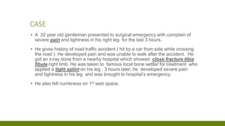
- 1. CASE ▪ A 32 year old gentleman presented to surgical emergency with complain of severe pain and tightness in his right leg for the last 3 hours. ▪ He gives history of road traffic accident ( hit by a car from side while crossing the road ). He developed pain and was unable to walk after the accident. He got an x-ray done from a nearby hospital which showed close fracture tibia fibula right limb. He was taken to famous local bone settler for treatment who applied a tight splint on his leg . 3 hours later, he developed severe pain and tightness in his leg and was brought to hospital’s emergency. ▪ He also felt numbness on 1st web space.
- 2. CONTINUED ▪ He was in painful distress. The right leg was wrapped in wooden splints spanning the entire length tightly bound with white strips of clothing. ▪ The foot was pale and cold with decreased pulses. Pulse oximeter showed 35 % saturation as opposed to contra-lateral limb which was pink , warm and showed 98 % saturation . ▪ The splint was removed. The leg was swollen, tense and tender . ▪ He felt excruciating pain on passive flexion of foot and weakness of ankle extension noted.
- 5. OBJECTIVES ▪ What is compartment syndrome? ▪ How to diagnose compartment syndrome? ▪ How to treat compartment syndrome? ▪ Acute compartment syndrome will be referred to as ACS during presentation.
- 6. INTRODUCTION ▪ Acute Compartment Syndrome: elevation of intracompartmental pressure to a level or for a duration that without decompression will cause tissue ischemia and necrosis. ▪ Exertional Compartment syndrome: elevation of intracompartmental pressure during exercise, causing pain and ischemia and resolution of pain with rest. ▪ Volkmann ischemic contracture: End stage of neglected acute compartment syndrome with irreversible muscle necrosis leading to ischemic contractures.
- 8. CONDITIONS ASSOCIATED WITH INJURY CAUSING ACS ▪ Tibial diaphyseal fracture ▪ Soft tissue injury ▪ Distal radial fracture ▪ Crush syndrome ▪ Diaphyseal fracture forearm ▪ Femoral diaphyseal fracture ▪ Tibial plateau fracture ▪ Hand fractures
- 9. CONTINUED ▪ Foot fractures ▪ Ankle fractures ▪ Elbow fracture dislocation ▪ Pelvic fracture ▪ Humeral diaphyseal fracture
- 11. PATHOGENESIS ▪ 3 theories 1. Critical closing pressure theory 2. Arteriovenous gradient theory 3. Microvascular occlusion theory ▪ Important thing is REDUCED BLOOD FLOW
- 14. EFFECTS OF RAISED TISSUE PRESSURE ON MUSCLES ▪ Reduced blood flow leads to ▪ Ischemia followed by ▪ Necrosis ▪ Depends on I. Duration II. Type of fibers III. Magnitude/ level
- 15. EFFECTS OF RAISED TISSUE PRESSURE ON NERVE ▪ Loss of neuromuscular function with raised tissue pressures ▪ Damage due to I. Ischemia II. Ischemia plus compression III. Toxic effects
- 16. EFFECTS OF RAISED TISSUE PRESSURE ON BONE ▪ Non union ▪ Because muscle ischemia reduces capacity for development of extraosseous blood supply on which long bones depend for healing.
- 17. REPERFUSION INJURY ▪ Group of complications following re-establishment of blood flow to the ischemic tissues and can occur after fasciotomy and restoration of muscle blood flow in acute compartment syndrome. ▪ Reperfusion followed by inflammatory response in ischemic tissue that cause further damage. ( toxic metabolites such as oxygen free radicals , thromboxane , interleukins , procoagulants and more).
- 18. DIAGNOSIS OF ACUTE COMPARTMENT SYNDROME ▪ Prompt diagnosis is the key to successful outcome ▪ Delay in treatment leads to I. Permanent sensory and motor deficits II. Contractures III. Infections IV. Amputations
- 19. DIAGNOSIS
- 21. PAIN ▪ is usually the early symptom in awake alert patient ▪ Severe and out of proportion to clinical situation ▪ May be absent in I. ACS associated with nerve injury II. Unconscious patient III. If regional anaesthesia used ▪ Children may not be able to express severity
- 22. PAIN WITH PASSIVE STRETCH ▪ Of the muscle involved is recognised as a symptom ▪ Increase in pain on stretching the muscle by passively moving the joints in direction opposite to that of damaged muscle's action
- 23. PARESTHESIA AND HYPOESTHESIA ▪ In territory of nerves traversing affected compartment ▪ First sign of nerve ischemia ▪ Peripheral nerves more sensitive to ischemia than muscles
- 24. PARALYSIS ▪ Late sign ▪ Can also be due to I. Inhibition by pain II. Direct injury to muscle III. Associated nerve injury
- 25. PALPABLE SWELLING ▪ Swelling is difficult to assess ▪ Always compare the uninjured contralateral limb ▪ Casts , dressings obscure compartment and prevent assessment of swelling ▪ Deep posterior compartment buried under muscle compartment, obscuring any swelling ▪ Tense palpable compartment
- 26. PULSES AND CAPILLARY REFILL ▪ Always intact unless I. Arterial injury II. Late stage of ACS ▪ DO NOT EXCLUDE DIAGNOSIS OF ACUTE COMPARTMENT SYNDROME IF DISTAL PULSES ARE PRESENT
- 27. COMPARTMENT PRESSURE MONITORING ▪ Raised tissue pressure is the primary event ▪ All compartments should be measured ▪ Measurement should be made within 5 cm of fracture ▪ Methods 1. Needle manometer method 2. Wick catheter 3. Slit catheter 4. Pressure transducer 5. Near infrared spectroscopy ( tissue oxygen saturation non invasively )
- 31. THRESHOLD FOR DECOMPRESSION ▪ Mean arterial pressure = (systolic – diastolic )/3 + diastolic ▪ Difference between MAP and tissue pressure should not be less than 30 mm Hg in normal muscle or ▪ 40 mm Hg in muscle subject to trauma. ▪ If Bp= 100/70 then MAP= (100-70)/3 + 70 = 80. ▪ Tissue pressure if = 35. ▪ Then 80 – 35 = 45 . No need of fasciotomy. ▪ Do take serial readings
- 32. THRESHOLD FOR DECOMPRESSION ▪ Delta P = difference in diastolic blood pressure and tissue pressure ▪ Delta P of 30 mm Hg is a safe threshold for decompression in ACS ▪ If Bp = 90/50 and measured tissue pressure = 25. ▪ Then delta P = 50 – 25 = 25 which is < 30 mm Hg. ▪ So decompression should be performed if the differential pressure level drops to under 30 mmHg ( Delta P )
- 34. TASK ▪ 2 minutes for completing task
- 36. TREATMENT
- 37. TREATMENT ▪ Main treatment is Fasciotomy ▪ Others 1. Remove tight casts and bandages including padding under the cast 2. Elevate limb to the level of the heart 3. Correct hypotension 4. Oxygen therapy
- 39. FASCIOTOMY ▪ Basic principles = Full and adequate decompression ▪ Skin incisions along the full length of affected compartment ▪ Assess muscle viability ▪ Debride necrotic muscle thoroughly
- 40. LEG
- 41. LEG ▪ All 4 compartments should be released ▪ Double incision four compartment fasciotomy
- 42. LEG- DOUBLE INCISION ▪ Anterior and lateral through lateral skin incision over intermuscular septum ▪ Care of not to injure superficial peroneal nerve that pierces the fascia and lies superficial to it in distal third of leg ▪ 2 posterior comparments through incision 2 cm from medial edge of tibia ▪ Superficial compartment exposed by skin retraction ▪ Deep posterior compartment exposed by posterior retraction of superficial compartment ▪ Protect saphenous vein and nerve and also posterior tibial vessels and nerves
- 43. DOUBLE INCISION FASCIOTOMY TECHNIQUE ▪ 20 cm incision in anterior compartment halfway between fibular shaft and crest of tibia ▪ Transverse incision to expose lateral intermuscular septum and identify superficial peroneal nerve just posterior to septum ▪ Release anterior compartment with metzenbaum scissors ▪ Perform fasciotomy of lateral compartment in line with fibular shaft ▪ 2nd incision 2 cm posterior to posterior margin of tibia ▪ Retract saphenous vein and nerve anteriorly ▪ Transverse incision to identify septum between deep and superficial posterior compartment. Release fascia over gastrocneumius soleus complex
- 44. DOUBLE INCISION FASCIOTOMY TECHNIQUE ▪ Make another incision over the flexor digitorum longus muscle and release entire deep posterior compartment
- 45. FASCIOTOMY OF THE LEG 2-SKIN INCISION, 4-COMPARTMENT FASCIOTOMY
- 46. FASCIOTOMY OF THE ANTERIOR AND LATERAL COMPARTMENTS OF THE LEG
- 47. FASCIOTOMY OF THE SUPERFICIAL & DEEP POSTERIOR COMPARTMENT OF THE LEG.
- 49. LEG – SINGLE INCISION ▪ Excision of fibula – a destructive process and damage to nerve can occur ▪ Single incision four compartment via lateral incision: parafibular Easy access to anterior and lateral compartment Anterior retraction of peroneal muscles allows exposure of posterior intermuscular septum Deep posterior compartment entered by incision immediately posterior to posterolateral border of fibula
- 50. SINGLE INCISION TECHNIQUE ▪ Incison in line of fibula from just distal to head of fibula to 4 cm proximal to lateral malleolus ▪ Undermine skin anteriorly ( avoid injury to peroneal nerve ▪ Fasciotomy of anterior and lateral compartments ▪ Undermine skin posteriorly and perform fasciotomy of superficial post. Compartment ▪ Identify interval between superficial and lateral compartent and develop interval ▪ Subperiosteally dissect flexor hallucis longus from fibula ▪ Retract muscle and peroneal vessels posteriorly ▪ Identify fascial attachment of posterior tibial muscle to fibula and incise fascia
- 52. THIGH
- 53. THIGH ▪ Single lateral skin incision ▪ Medial incision can be used for adductors ▪ Performed with in 5 cm of fracture site or ▪ Area of maximum swelling Decompression of thigh compartments. A, Incision from intertrochanteric line to lateral epicondyle. B, Anterior compartment is opened by incising fascia lata, and vastus lateralis is retracted medially to expose lateral intermuscular septum, which is incised to decompress posterior compartment. C, Drawing of thigh compartments and appropriate incision.
- 55. FOOT
- 56. ▪ Dorsal approach: ▪ 2 dorsal incisions centered just medial to 2nd metatarsal and lateral to 4th metatarsal ( avoid injury to sensory nerves and extensor tendons) ▪ Superficial fascia divided and interosseous are elevated off metatarsals to further decompress compartments ▪ Clamp to bluntly dissect central , medial and lateral compartments ▪ Seperate medial incision may be needed to release abductor
- 57. ▪ Medial approach: extend point below medial malleolus ( 3cm from sole ) to proximal aspect of first meta tarsel ▪ Once neurovascular bundle ( medial planter nerve an artery ) retracted out of way, fascia overlying abuctor hallucis an FB released ▪ Medial intermuscular septum opened longitudionally ▪ Remaining compartment ( central , lateral and intrinsic ) entered through blunt dissection with clamp ▪ Lateral compartment is found by retracting FDB out of way
- 58. FOOT ▪ Dorsal incisions over 2nd and 4th metatarsals for interosseous compartment and central compartment ▪ Medial and lateral compartments via deep surfaces of 1st and 5th metatarsal ▪ Hind Foot – medial incision to decompress calcaneal compartment
- 59. ARM
- 60. ARM ▪ Anterior and posterior incisions
- 61. ARM – 2 INCISION ▪ 15 cm skin incision medial to bicipital sulcus extended up anteromedial arm to acromion and fascia opened ▪ 15 cm posterolateral starting at tip of olecrenon extended up posterolaterally arm through fascia
- 62. ARM – 1 INCISION ▪ 15 cm incision over medial intermuscular septum ▪ Flaps raised anteriorly and posteriorly ▪ Fascia over anterior compartment opened midway between anterior border of biceps and medial intermuscular septum ▪ Fascia over posterior compartment opened midway between posterior border of triceps and medial intermuscular septum
- 63. FOREARM
- 64. TECHNIQUE ▪ Anterior curvilinear skin incision medial to biceps tendon crossing elbow flexion crease ▪ Then distally and radially over brachioradialis then distally and ulnarward, coursing medial to palmaris longus ▪ Then carpel tunnel release ▪ Release superficial volar compartment with scissors freeing the fascia over superificial compartment ▪ Identify flexor carpi ulnaris and retract medially along with neurovascular buncle medially ▪ Retract flexor digitorum superficialis and median nerve laterally ▪ Flexor digitorum profundus exposed. Incise fascia
- 65. ▪ Continue incision distally by incising transverse carpel ligament along ulnar border of palmaris longus tendon and median nerve ▪ For dorsal aspect, incision distal to lateral epicondyle between extensor digitorum communis and extensor carpi radialis brevis, extending 10 cm distally ▪ Apply long arm splint, with flexion not beyond 90
- 66. FOREARM ▪ Volar and dorsal fasciotomies ▪ Volar compartment first ( incision from biceps tendon to palm to allow carpel tunnel decompression ▪ Inspect muscles ▪ Usually volar incison suficient. If ICP remains elevated, then dorsal straight incision
- 67. HAND
- 68. HAND ▪ 2 dorsal incisons which allow access to interosseous compartments ▪ Additional incisions can be made on thenar and hypothenar eminences
- 69. ▪ 2 dorsal incision , 2nd and 4th metacarpels beginning at level of metacarpophalangeal joints and extending just distal to wrist ▪ Palmer radial and palmer ulnar incisions along the glabrous and non glabrous intervals ▪ Release carpel tunnel through midline palmer incision ▪ Digital fasciotomies mid lateral incisions along the radial border of ring and little finger and ulnar border of index and middle finger ▪ Prudent to relase all compartments of hand of even one compartment involved
- 70. MANAGEMENT OF FASCIOTOMY WOUNDS ▪ Never be closed primarily- left open and dressed ▪ Relook after 48 hours- for assessing viability ▪ Closure should not be attempted unless all muscles viable ▪ Techniques 1. Delayed primary closure 2. Dermatotraction 3. Split thickness graft 4. Vac
- 72. MANAGEMENT OF ASSOCIATED FRACTURES ▪ Fractures should be stabilized ▪ Fasciotomy prior to stabilization of fracture ▪ Avoid 1. Elevation of leg 2. Excessive traction ( raises pressure of 6 % / KG weight applied ) in deep posterior compartment
- 74. COMPLICATIONS OF ACS ▪ Delay to more than 6 hours can cause 1. Muscle contractures 2. Muscle weakness 3. Sensory loss 4. Infection 5. Non union 6. Amputation
- 75. LATE DIAGNOSIS ▪ If no likelihood of surviving muscles and compartment pressure low – NO NEED OF FASCIOTOMY ▪ If possibility of remaining viable muscle or if compartment pressures above critical value – FASCIOTOMY can be performed along with muscle debridement
- 76. CHRONIC COMPARTMENT SYNDROME ▪ Occurs in long distance runners ▪ On anterolateral aspect of calf brought on by muscular exertion ▪ Swelling may cause ischemia of deep peroneal nerve ▪ Diagnosis by history ▪ Confirmed by measuring compartment pressure before and after exercise ▪ Release of fascia is curative ▪ Same when occurs in forearm muscles ( arm pump in motorcyclists )
- 77. CHRONIC EXERTIONAL COMPARTMENT SYNDROME ▪ Reversible ischemia secondary to non compliant osteofascial compartment that is unresponsive to expansion of muscle volume that occurs with exercise ▪ Factors contributing are 1. Increased volume of skeletal muscles with exertion ( blood volume and edema) 2. Dynamic contraction factors due to gait ( rear – foot landing, over pronation) 3. Muscle hypertrophy 4. Anabolic steroids and creatine use increase volume ▪ Common in young adults, runners, elite athletes , military recruits
- 78. CLINICAL EVALUATION OF CECS ▪ History in evaluating lower extremity pain , exercise induced pain , feeling of tightnes after exertion and resolving after cessation of activity ▪ Paresthesia of nerves ▪ On examination, tenderness over musculature of involved compartment and muscle herniation through defect in fascia palpable
- 79. ▪ Periostosis on bone scan, with diffuse uptake often covering one third of bone ▪ Stress reactions by MRI ▪ Entrapment of peroneal nerve determined by provocative tests ( pressure over point at which superficial peroneal nerve emerge from deep fascia produces pain during active , resisted dorsiflexion and eversion of ankle ▪ Tinel sign may be postive
- 80. DIAGNOSIS ▪ Presence of one or more of criteria 1. Preexercise, resting pressure of 15 mm Hg or more 2. Pressure of 30 mm Hg or more 1 minute after exercise 3. Pressure of 20 mm Hg or more after 5 minute after exercise
- 81. TREATMENT ▪ Non operative ▪ Operative
- 82. NON OPERATIVE ▪ Relative rest ( limiting activity which avoids symptoms ▪ Anti inflammatory medications ▪ Stretching and strengthening of involved muscles
- 83. OPERATIVE ▪ Fasciotomy ( subcutaneous or endoscopic ) ▪ Double mini incision
- 86. THANK YOU