
Recommended
More Related Content
Similar to NEHA VERMA renal calculi ppt.pptx
Similar to NEHA VERMA renal calculi ppt.pptx (20)
Recently uploaded
Recently uploaded (20)
NEHA VERMA renal calculi ppt.pptx
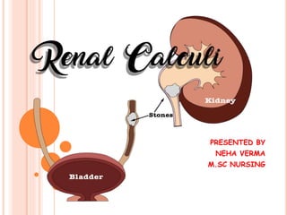
- 1. PRESENTED BY NEHA VERMA M.SC NURSING
- 2. TERMINOLOGY Urolithiasis - Stone in urinary tract Nephrolithiasis – Stone in kidney Ureterolithiasis- Stone in ureters ( most painful)
- 3. INTRODUCTION A kidney stoneor renal calculi are hard deposits of minerals or salts , that are forms in kindney and present in different sizes. It may occurs due to diet, overweight, some medical conditions, medication and often occurs when urine becomes concentrated, allowing minerals to crystallize and stick together. Stones may be formed in the kidney, urinary bladder, ureter and urethra.
- 5. DEFINITION 1. A kidney stone, also known as a renal calculus or nephrolithiasis , it is define as calculi formation in kidney and kidney part . Acc. To P.K. Panwar 2. Kidney stone / renal calculi is solid mass made up of crystals present in kidney or ureter. Acc. To M.P. Sharma
- 7. ETIOLOGY Diet pattern – 80% causes of stone. high in calcium , magnesium , phosphate Family history Dehydration UTI & prolonged catheterization Other disease of kidney – Renal failure Nephritis Dehydration Obstruction
- 8. TYPES There are mainly five types 1. Calcium oxalate stone (is the most common 80%) 2. Cystine stone 3. Struvite stone (triple stone) 4. Uric acid stone 5. Others stone
- 9. 1. CALCIUM OXALATE STONE:- It is the most common type of renal stones, about 80%. Calcium stones are the most Common stones present in urinary tract. Calcium based stone are most commonly seen in young men between the age of 20 and 30 years. It is present in foods such as spinach and vitamin C supplements and citrus fruits .
- 10. 2.CYSTINE STONE Cystine stone is rare can form in people who have cystinuria . It is cystine is a protein, about 2% of chance It can affect both women and men . (cystine source avoid meat milk, cheese, egg)
- 11. 3. STRUVITE STONE These stones are formed of struvite ( Mg amonium phasphate) This type of stone is mostly found in women who have a urinary tract infection. These stone can grow very large and can block the kidney, ureter or bladder.
- 12. 4. URIC ACID STONE It is second most common type of renal stones about10%. This type of stone is more Common in men than women. They Can occur in individual with gout or those taking chemotherapy .
- 13. OTHERS Other substances like medication like acylovir, triamterene also can cause stones.
- 14. PATHOPHYSIOLOGY due to any etiology factor urine supersaturation crystal nucleation crystal growth crystal aggregation stone formation Renal calculi
- 15. CLINICAL MANIFESTATION Usually symptoms of kidney stones may not occurs until the stones reach to the ureters. Renal colic or severe sharp pain occurs in flank area , that also radiate to lower abdomen and groin .
- 16. Pain or burning sensation while urinating. Other symptoms – Pink , red or brown urine. Cloudy or foul smelling urine. Uregency of urination ( sudden uncontrollabe, need to urinate ) Urinating small amounts of urine. Note _ kidney stones sometimes considered “the great mimicker” but common their symptoms very similar to appendicitis , gastritis, UTI, ovarian or testicular conditions.
- 17. DIAGNOSTIC TEST History collection Physical examination Blood test ( to check calcium, phosphorus, uric acid and electrolyte level) Urinalysis ( to see crystals and look for RBC in urine ) Examine of stone to determine the type of stone or blockage can be seen on : Abdominal CT scan Abdominal / kidney MRI Abdominal x-ray( less commonly ) USG Intravenous pyelogram
- 19. MANAGEMENT- MANAGEMENT OF KIDNEY STONES DEPENDING ON TYPE OF STONE AND CAUSE OF STONE . (A) Small stones with minimal symptoms _ Most small stones not require surgery, these may pass by- - drinking enough water ( 2.5- 3.5 lit/day ) Medical therapy – (<5mm treat medicine _ 10mm(50% chance ), if >10 mm required surgery) Pain relievers_ ibuprofen, naproxen Uric acid stones_ allopurinol (zyloprim) Struvite stones_ antibiotics ( amoxicilline, cotrimaxazole) Ca stones_ thiazide diuretics to prevent forming of Calcium stones. To relax smooth muscles_ Jolyn (tamsulosin +dutasteride)
- 20. (B) LARGE STONES Extracorporeal shockwave lithiotripsy- It uses sound waves to create strong vibrations (shock waves) that break stones into tiny pieces that can be passed in to urine . This procedure last about 45- 60 mins
- 21. (B) NEPHROLITHIOTOMY In this procedure a incision is made in flank area, from which a small tube with scope inserted in to the kidney and removes the stones. This procedure performed when extracorporeal shockwave lithiotripsy is unsucessful.
- 22. (C) URETEROSCOPY In this procedure, first of all surgeon placed a tube with camera into ureter by making incision into bladder. A small cage is used to snag the stone and remove it.
- 23. PREVENTION Avoid protein intake:-usually protein is restricted to 60 kg/day to decrease urinary excretion of calcium and uric acid. A sodium intake :- of 3 to 4 g/day is recommended table salt and high-sodium foods should be reduced, because sodium competes with calcium for reabsorption in the kidneys. Low calcium diets are not –generally recommended, except for true absorptive hypercalcuria evidence shows that limiting calcium, especially in women can lead to osteoporosis and does not prevent renal stone.
- 24. CONT.. Avoid intake of oxalate-containing foods (e.g. spinach, strawberries, tea, peanuts, wheat bran) During the day, drink fluids (ideally water) every day 1-2 hours Drink two glasses of water at bedtime and an additional glass at each night time awakening to prevent urine from becoming too concentrated during the night. Avoid activities leading to sudden increase in environmental temperature that may cause excessive sweating and dehydration. Contact your primary health care provide at the first sign of a urinary tract infection.
- 25. NURSING DIAGNOSIS 1-Acute pain related to obstruction in urine flow. 2-Deficient knowledge related to disease condition. 3-Impaired urinary elimination related to mechanical obstruction. 4- Risk for infection related to introduction of bacteria following manipulation of the urinary tract and obstructed urinary blood flow. 5-Altered daily living pattern related to restlessness and sudden attack of pain.
- 26. NURSING MANAGEMENT Advise the patient at least 6-8 glasses of water per day to produce a large amount of urine. This will help to pass the stone. Instrut to take a low sodium diet. Advise the patient to avoid the diet high in calcium, purine and vitamin C. Monitor urine output, frequency, consistency odour, volume and colour to evaluate the petency of urinary system. Assess the quality, intensity, severity location and frequency of pain.
- 27. Advise the patient to change life style pattern. Advise the patient to avoid prolonged sitting and standing. Administer the medications as the prescription. Check and maintain input and output chart. Teach the patient that this condition may lead to urinary tract infection so explain about sign and symptoms of UTI. Educate about the proper care of wound if surgical opening to remove stone. Use the pain control measure before it become severe.
- 28. COMPLICATION Urinary tract infection. Kidney damage or scarring (if treatment is delay for too long) Obstruction of ureter (acute unilateral obstructive uropathy).
- 29. CONCLUSION Kidney stones can develop as a result of several factors, including dehydration , the urine become concentrated and minerals can from crystals that can ultimately develop into stones.
- 30. SUMMARY- Introduction definition Etiology Risk factor Types Pathophysiology Clinical manifestation Management Prevention Complication
- 32. BIOLIOGRAPHY Brunner and Suddarth’s , “Text book of Medical Surgical Nursing” ,3rd volume , 9 th edition, Wolters Kluwer : Wolters publication, (2001), page no.1389-1402. Lippincott, “ Text book of manual of Medical Surgical Nursing,”6th volume, Edition- 10th,Wolters Kluwer:Wolters Kluwer Pvt.Ltd. ; 2009,page no.1098-1123 M.P. Sharma , “Text book of medical surgical nursing”, 1st edition, Virendra kumar: AITBS Publishers;2016,page no. 234- 238. P.K. Panwar, “ Textbook of Medical Surgical Nursing”,6th edition, Virendra Kumar Arya:AITBS Publishers; 2022,page no.345-349 https://www.ncbi.nih.gov
- 33. THANK YOU