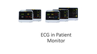
2. What is ECG ?
• An electrocardiogram — abbreviated as EKG or ECG — is a test that
measures the electrical activity of the heart. With each beat, an
electrical impulse (or “wave”) travels through the heart. This wave
causes the muscle to squeeze and pump blood from the heart.
3. What is Heart?
• The heart is the body’s engine room, about the size of a fist, located just behind
and slightly left of the breastbone, responsible for pumping life-sustaining blood
throughout the blood vessels to our body.
• Blood delivers oxygen to all the body cells to stay alive. We need healthy living
cells. Without oxygen, these cells would die. If the oxygen rich blood doesn´t
circulate as it should , we would die.
8. Electrode Position
3 lead 5 lead 10 lead
Leads available
3-lead configuration: I, II, III
5-lead configuration: I, II, III, aVR, aVL, aVF and V
10-lead configuration :I, II, III, aVR, aVL, aVF and V1 to V6
12. What Information We get from the ECG?
• Heart rate : It may be helpful if your pulse is difficult to feel or too fast or too
irregular to count accurately.
• Heart rhythm : An ECG can show heart rhythms irregularities (arrhythmias).
These conditions may occur when any part of the heart's electrical system
malfunctions.
• Heart attack : An ECG can show evidence of a previous heart attack or one that's
in progress. The patterns on the ECG may indicate which part of your heart has
been damaged, as well as the extent of the damage.
• Inadequate blood and oxygen supply to the heart : It helps to determine
whether chest pain is caused by reduced blood flow to the heart muscle, such as
with the chest pain of unstable angina.
• Structural abnormalities: An ECG can provide clues about enlargement of the
chambers or walls of the heart, heart defects and other heart problems.
13. Arrhythmia
• Heart rhythm problems (heart arrhythmias) occur when the electrical
impulses that coordinate your heartbeats don't work properly,
causing your heart to beat too fast, too slow or irregularly.
• Pulse > 100: Tachycardia
• Pulse < 60: Bradycardia
16. Why it is Best?
• GE used Marquette™ 12SL Algorithm which is 40 years legacy
• EkoPro technology for the arrhythmia detection
• Provides accurate, validated measurements of heart rate, axis,
intervals, and durations
• Offers ECG analysis including those for atrial arrhythmias, pacemaker
detection, and Offers quick quality check of ECGs
• Dedicated paediatric criteria
17.
18. What is EkPro Technology?
• The EK-Pro algorithm utilizes four simultaneous leads for analysis,
detecting and alarming for cardiac events that might otherwise go
unnoticed.
• The algorithm helps distinguish noise and artifacts from true beats,
significantly reducing false alarms, and also provides redundancy,
enabling continued function of the monitor in the event of single
electrode failure.
19. Arrhythmia detection description
ECG Signal
Detected
Begins acquiring
and analyzing
QRS complexes
Dominant QRS
complex is stored
as a reference
template
Compared with
incoming beats
to identify
possible
arrhythmias
Continuous
correlation and
contextual analysis
Make the best
possible decision
regarding the
beat’s origin
20. Multi-lead Analysis
• Assurance of Uninterrupted Monitoring Simultaneous, multi-lead analysis also provides
redundancy, so that monitoring can continue in the event of an electrode contact failure.
21. Multi-lead analysis example
LL/F lead fail , Lead II failed error shows , &
it’s changed into lead I
After solve the problem , it
automatically changed into original
setting
22. Smart lead Fail Analysis
• If 1 channel is clean and correlates
to the past rhythm, system will
not be fooled by other noisy
channels.
• Algorithm continuously evaluate
each channel for normal QRS
complexes with regular rhythm.
• Improved arrhythmia detection
performance during a noisy ECG
signal
23. Benefits
• It detect noise so True Event may not be missed out
• Assurance of Uninterrupted Monitoring
• It distinguishes noise and artifacts from true beats, and thereby
significantly reduce false alarms
• Staff may not be fooled by false alarm, So it reduced staff stress
24. Arrhythmia alarm
Arrhythmia alarm Alarm message Arrhythmia detection criteria
HR alarms Brady HR below the HR alarm limit
Tachy HR over the HR alarm limit.
Lethal alarms
Asystole HR decreased to zero, or beat
detection has not occurred in the
last 5 seconds.
V Fib / V Tach ECG waveform indicates a chaotic
ventricular rhythm.
V Tach A run of PVCs is detected with a
run length of six beats or more and
the effective HR and V Tach
duration meet the user defined
criteria.
25. Ventricular
alarms
VT > 2 A run of PVCs is detected with a run length of more than two beats but less than six
beats. In addition at least two consecutive RR intervals in the run must have an effective
HR that is equal to or exceeds the V Tach Minimum HR/min.
R on T Isolated PVC is detected within 100 ms of the peak of the T-wave of the patient’s
predominant normal beat.
V Brady Run of PVCs are detected with a run length of at least three beats.
Couplet Two consecutive PVCs are detected between normal beats, N-V-V-N
Bigeminy Every other beat is PVC (N-V-N-V-N-V )
Trigeminy Every third beat is PVC (N, N, V, N, N, V, N, N, V).
Accel. Ventric. Accelerated ventricular rhythm - Run of PVCs with a run length of at least six beats and
the requirements for user defined V Tach Criteria or V Brady are not
met.
26. Atrial
alarms
A Fib Absence of P-waves and irregular RR-interval.
Irregular Six consecutive normal RR intervals vary by 100 ms or
more.
Missing Beat Actual RR interval more than 1.8 times the average RR
interval.
Pause Coupling interval between two beats exceeds: 1 to 5
seconds (configurable)
SV Tachy A run of SVCs is detected with a run length of at least the
set SVT Length and the heart rate is at least the set HR for
SVT /min.
27. Companies with different technology &
Models
GE
Ek-Pro
arrhythmia
analysis
B1X5,BX50
series
Nihon
Kohden
eC1
BSM series
PHILIPS
Standard
MX series
MINDRAY
Glasgow
Coma
ePM 12M,
Bene View
N12
29. (2) False alarm by Type
Alarm GE Healthcare Philips Mindray Nihon Kohden
Asy 2 7 2 8
VT 5 295 35 2
VT>2 26 0 0 0
VF 0 3 84 56
VRUN 0 0 0 227
NSVT 0 0 164 0
AV 3 0 0 0
Total 36 305 285 293
NSVT - Non-Sustained Ventricular Tachycardia, a run of three to five consecutive ventricular beats at a
rate higher than or equal to VT rate
VRUN - Ventricular Beat Runs