More Related Content
Similar to Nguyen-Vu et al 2013
Similar to Nguyen-Vu et al 2013 (20)
Nguyen-Vu et al 2013
- 1. nature neuroscience advance online publication 1
b r i e f com mu n i cat i on s
in the cerebellar flocculus reflects both the vestibular stimulus and the
direction of the visual stimulus (the eye movements made to track the
visual stimulus); thus, these neurons carry the information required to
control the direction of learning6,13. We tested whether learned changes
in the VOR could be induced by pairing a vestibular stimulus with direct
activation of Purkinje cells in the absence of any visual stimulus.
When VOR learning is induced by pairing a vestibular stimulus
with a visual stimulus, the timing, or phase, of activity in Purkinje cells
relative to the vestibular stimulus encodes the required direction of
the learning6,13. We compared the effect of optogenetically stimulat-
ing floccular Purkinje cells (Supplementary Figs. 1 and 2) during the
contraversive phase of a sinusoidal vestibular stimulus with the effect
of stimulating during the ipsiversive phase (Fig. 1a).This pairing was
done in complete darkness (that is, in the absence of any visual cues
that could guide associative motor learning). As a control, the vestibular
stimulus was delivered in the absence of Purkinje cell stimulation dur-
ing the 30-min training period. This vestibular-alone training resulted
in a significant habituation of the VOR (P = 0.0006, one sample t test;
Fig. 1b), as described previously14.
Activation of Purkinje cells during the contraversive phase of the ves-
tibular stimulus to roughly mimic the response observed in the majority
of Purkinje cells during normal VOR increase training6,13 was suffi-
cient to drive learning. During training, there was a gradual increase
in the VOR response amplitude relative to the vestibular-alone control
(P = 0.0002, Tukey; Fig. 1b and Supplementary Fig. 3), with the VOR
response being larger at the end of training than the pre-training baseline
(P = 0.03, one sample t test; Fig. 1b). We assessed this VOR learning
in the absence of Purkinje cell stimulation by briefly interrupting the
optogenetic stimulation to measure the eye movement response to the
vestibular stimulus alone.
The timing of the Purkinje cell activity relative to the vestibular stimu-
lus was critical to the induction of VOR learning (P = 0.0043, contraver-
sive versus ipsiversive Purkinje cell stimulation, Tukey; Fig.1b). Purkinje
cell activation induced VOR learning when paired with the contraversive
phase of the vestibular stimulus, but, when paired with the ipsiversive
phase of the vestibular stimulus, it had no significant effect beyond that
induced by training with the vestibular stimulus alone (P = 0.42, Tukey;
Fig.1b and SupplementaryFig.3). Thus, the motor learning induced by
direct optogenetic activation of Purkinje cells was associative, as it only
occurred when Purkinje cell activation was paired with the appropriate
phase of the vestibular stimulus.
In addition to inducing learning, optogenetic activation of Purkinje
cells could elicit eye movements and have an immediate effect on eye
movement performance during training (Supplementary Figs. 4 and
5a–c). However, the learning induced by Purkinje cell activation, which
was measured after training in the absence of Purkinje cell stimulation,
was not a secondary consequence of its effects on the eye movement
Cerebellar Purkinje cell activity
drives motor learning
T D Barbara Nguyen-Vu1,2,7, Rhea R Kimpo1,7, Jacob M Rinaldi1,
Arunima Kohli1, Hongkui Zeng3, Karl Deisseroth4–6 &
Jennifer L Raymond1
The climbing fiber input to the cerebellar cortex is thought to
provide instructive signals that drive the induction of motor
skill learning. We found that optogenetic activation of Purkinje
cells, the sole output neurons of the cerebellar cortex, can
also drive motor learning in mice. This dual control over the
induction of learning by climbing fibers and Purkinje cells can
expand the learning capacity of motor circuits.
The climbing fiber input to the cerebellum from the inferior olive is
widely viewed as the source of the instructive signals controlling the
induction of cerebellum-dependent learning1. In vitro, climbing fiber
activation can induce plasticity at synapses in the cerebellar cortex, and,
in vivo, climbing fibers encode errors during a wide range of motor
learning tasks1. Notably, electrical stimulation of the inferior olive in
vivo can replace the unconditioned stimulus in a classical conditioning
procedure, providing causal evidence for a role of climbing fibers in the
induction of learned changes in behavior2. However, such causal studies
have not been replicated for motor skill learning, and recent findings
have challenged the view that instructive signals encoded by the climb-
ing fibers are the driver of cerebellum-dependent motor learning3,4.
A second, candidate neural instructive signal in the cerebellum is the
activity of Purkinje cells5. Purkinje cells encode information that could,
in principle, be used to guide the induction of plasticity6,7, and several
models have suggested a role for Purkinje cell activity in guiding the
acquisition or consolidation of motor memory5,8,9. However, causal
evidence that Purkinje cell activity can induce learning in vivo has been
lacking; previous attempts to induce learning by activating Purkinje
cells with electrical stimulation of the cerebellar cortex were unsuc-
cessful10,11. Thus, we harnessed the cell-type specificity of optogenetics
and the power of the well-characterized vestibulo-ocular reflex (VOR)
to determine whether Purkinje cell activity can drive the induction of
learned changes in behavior.
The VOR is a reflexive eye movement response to a head movement
(vestibular stimulus) that helps to stabilize images on the retina. If a ves-
tibularstimulusisrepeatedlypairedwiththemovementofavisualstimu-
lus, motor learning can adaptively modify the amplitude of the VOR to
reduce image motion on the retina. The direction of the visual stimulus
motion relative to the vestibular stimulus determines whether an adap-
tive increase or decrease in the VOR is learned12. Purkinje cell activity
1Department of Neurobiology, Stanford School of Medicine, Stanford, California, USA. 2Department of Molecular and Cellular Physiology, Stanford School of Medicine,
Stanford, California, USA. 3Allen Institute for Brain Science, Seattle, Washington, USA. 4Department of Bioengineering, Stanford University, Stanford, California, USA.
5Department of Psychiatry, Stanford School of Medicine, Stanford, California, USA. 6Howard Hughes Medical Institute, Stanford University, Stanford, California, USA.
7These authors contributed equally to this work. Correspondence should be addressed to J.L.R. (jenr@stanford.edu).
Received 8 August; accepted 16 October; published online 27 October 2013; doi:10.1038/nn.3576
npg©2013NatureAmerica,Inc.Allrightsreserved.
- 2. 2 advance online publication nature neuroscience
b r i e f com mu n i cat i on s
nuclei17–20 or both. In the Purkinje cell axons, the simple spikes elicited
optogenetically should be indistinguishable from those elicited by par-
allel fiber input; however, in the Purkinje cell dendrites, optogenetic
activation may cause more widespread calcium influx than would
normally be triggered by parallel fiber input. Moreover, in vitro, direct
depolarization of the Purkinje cells can, in some cases, substitute for
climbing fiber activity, inducing calcium influx in the Purkinje cell den-
drites and hence synaptic plasticity, particularly long-term depression at
the parallel fiber–Purkinje cell synapses15. However, the VOR learning
induced by Purkinje cell activation did not seem to result from plas-
ticity mechanisms that would normally be triggered by climbing fiber
activity, as climbing fiber activation and Purkinje cell activation did not
substitute for each other. Purkinje cell activation paired with the con-
traversive phase of the vestibular stimulus induced a learned increase in
the VOR (Fig. 1b). No such learned increase in the VOR was observed
after training, when we paired optogenetic stimulation of the climbing
fibers (Supplementary Fig. 6) with the same, contraversive phase of the
vestibular stimulus (contraversive climbing fiber versus vestibular-alone
control at 30 min, P = 0.1052, Mann-Whitney U; versus contraversive
Purkinje cell stimulation, P = 0.0278, t test; Figs. 1b and 2).
Notably, climbing fiber activation induced an associative learned
increase in the VOR when paired with the ipsiversive phase of the ves-
tibular stimulus (P = 0.0343 versus contraversive climbing fiber stimu-
lation, P = 0.0009 versus vestibular control, Mann-Whitney U; Fig. 2).
Thus, error signals carried by the climbing fibers can have a causal role in
motor skill learning, as previously demonstrated for classical condition-
ing2. However, climbing fiber stimulation was most effective at a time
relative to the vestibular stimulus when Purkinje cell stimulation had
no effect (ipsiversive stimulation; Figs. 1b and 2), just as Purkinje cell
stimulation was effective at a time when climbing fiber stimulation was
relatively ineffective (contraversive stimulation; Figs. 1b and 2). This
double dissociation suggests that Purkinje cell activation and climbing
fiber activationin vivo can inducelearningthroughdistinctmechanisms.
performance during training, because there was no correlation between
these two effects (Supplementary Fig. 5).
Our results provide causal evidence that Purkinje cell activation can
contribute to the induction of motor learning, a role that has gener-
ally been ascribed to climbing fibers. Purkinje cell activity may induce
plasticity in the cerebellar cortex15,16, downstream in the vestibular
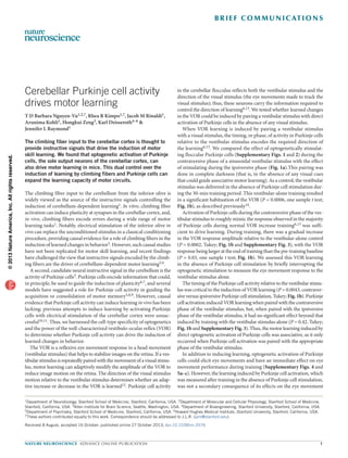
Figure 1 Purkinje cell activation induced associative motor learning. (a) Training procedures. A sinusoidal vestibular stimulus was used for training and testing
(black trace, angular velocity of the head). During training, floccular Purkinje cells in the two hemispheres were stimulated (cyan) in an alternating fashion
so that the activation of Purkinje cells in each hemisphere was paired with either the contraversive phase of the vestibular stimulus (red) or the ipsiversive
phase of the vestibular stimulus (blue). The VOR was tested pre- and post-training by measuring the eye movement response to the vestibular stimulus in the
absence of Purkinje cell stimulation. Example raw eye velocity traces illustrate results from one mouse that exhibited an increase in the VOR after training with
contraversive Purkinje cell stimulation (red) and a decrease in the VOR after ipsiversive stimulation (blue) relative to pre-training (gray). (b) Motor learning,
measured as the change in VOR amplitude post-training relative to pre-training, depended on the training condition (F2,31 = 11.59, P = 0.0002, two-way
repeated measures ANOVA). Black open squares indicate that the vestibular stimulus was presented alone during training (n = 10). Red circles and blue
squares indicate that Purkinje cell activation was paired with the contraversive or ipsiversive phase of the vestibular stimulus, respectively (n = 12). Data are
presented as mean ± s.e.m. **P = 0.0043, ***P = 0.0002, ns indicates not significant (P = 0.42).
Figure 2 Climbing fiber activation induced motor learning with different
timing than Purkinje cells. Motor learning induced by climbing fiber activation
paired with the ipsiversive (blue squares, n = 9) or contraversive (red circles,
n = 8) phase of the vestibular stimulus, compared with the control condition
in which the vestibular stimulus was presented alone during training (black
open squares, n = 7). Changes in the VOR relative to the pre-training baseline
depended on the training condition (F2,3 = 6.0, P = 0.0278, Friedman test).
Data are presented as mean ± s.e.m. *P = 0.0343, ***P = 0.0009,
ns indicates not significant (P = 0.1052).
ba
Purkinje cell
stimulation
+
Contraversive
vestibular
Purkinje cell
stimulation
+
Ipsiversive
vestibular
Training
250 ms
2˚ s–1
Left
Right
Left
Right
Learning(%VOR)
Training time (min)
15
–15
–30
***
n = 12
10 20 30
**
0
2˚ s–1
Vestibular
stimulus
Post
Test VOR
250 ms
Eye
Eye
Post
Pre
Pre
ns
Left
Right
Purkinje cell
ipsi stim
Vestibular
control
Purkinje cell
contra stim
2˚ s–1
2˚ s–1
Climbing fiber
ipsi stim
Vestibular
control
Climbing fiber
contra stim
***
10 20 30
30
15
–15
–30
0
*
ns
Training time (min)
Learning(%VOR)
npg©2013NatureAmerica,Inc.Allrightsreserved.
- 3. nature neuroscience advance online publication 3
b r i e f com mu n i cat i on s
AUTHOR CONTRIBUTIONS
T.D.B.N.-V. conducted all Purkinje cell experiments. R.R.K. conducted all climbing
fiber experiments. J.M.R. conducted pilot experiments. A.K. helped with histology.
K.D. and H.Z. provided reagents. J.L.R. supervised all aspects of the work.
COMPETING FINANCIAL INTERESTS
The authors declare no competing financial interests.
Reprints and permissions information is available online at http://www.nature.com/
reprints/index.html.
1. Ito, M. Annu. Rev. Neurosci. 12, 85–102 (1989).
2. Mauk, M.D., Steinmetz, J.E. & Thompson, R.F. Proc. Natl. Acad. Sci. USA 83, 5349–
5353 (1986).
3. Ke, M.C., Guo, C.C. & Raymond, J.L. Nat. Neurosci. 12, 1171–1179 (2009).
4. Catz, N., Dicke, P.W. & Thier, P. Curr. Biol. 15, 2179–2189 (2005).
5. Miles, F.A. & Lisberger, S.G. Annu. Rev. Neurosci. 4, 273–299 (1981).
6. Lisberger, S.G. & Fuchs, A.F. Brain Res. 69, 347–353 (1974).
7. McCormick, D.A. & Thompson, R.F. Science 223, 296–299 (1984).
8. Mauk, M.D. & Donegan, N.H. Learn. Mem. 4, 130–158 (1997).
9. Shutoh, F., Ohki, M., Kitazawa, H., Itohara, S. & Nagao, S. Neuroscience 139, 767–777
(2006).
10. Babalian, A.L. & Vidal, P.P. J. Neurophysiol. 84, 2514–2528 (2000).
11. Heuer, H.W., Tokiyama, S. & Lisberger, S.G. J. Neurophysiol. 100, 1320–1331 (2008).
12. Ito, M., Shiida, T., Yagi, N. & Yamamoto, M. Brain Res. 65, 170–174 (1974).
13. Raymond, J.L. & Lisberger, S.G. J. Neurosci. 18, 9112–9129 (1998).
14. Stahl, J.S. J. Neurophysiol. 91, 2066–2078 (2004).
15. Crepel, F. & Jaillard, D. J. Physiol. (Lond.) 432, 123–141 (1991).
16. Belmeguenai, A. et al. J. Neurosci. 30, 13630–13643 (2010).
17. Pugh, J.R. & Raman, I.M. Neuron 51, 113–123 (2006).
18. Menzies, J.R.W., Porrill, J., Dutia, M. & Dean, P. PLoS ONE 5, e13182 (2010).
19. McElvain, L.E., Bagnall, M.W., Sakatos, A. & du Lac, S. Neuron 68, 763–775 (2010).
20. Aizenman, C.D., Manis, P.B. & Linden, D.J. Neuron 21, 827–835 (1998).
We found that motor memory for an increase in the VOR could be
artificially implanted by stimulating either the climbing fibers or the
Purkinje cells. Purkinje cells and climbing fibers encode different task-
related signals during oculomotor learning6,13, yet they can achieve a
similar behavior outcome, namely, an increase in the amplitude of the
VOR. Thus, joint control of the induction of motor skill learning by
cerebellar Purkinje cells and climbing fibers may expand the capacity
of the motor circuit to learn in response to different cues.
METHODS
Methods and any associated references are available in the online
version of the paper.
Note: Any Supplementary Information and Source Data files are available in the
online version of the paper.
ACKNOWLEDGMENTS
We thank E. Knudsen, T. Moore, C. Shatz, D. Madison, G. Zhao, O. Winter,
S. Umamoto, A. Adamantidis, M. Carter, H. Nguyen and R. Hemmati for
discussions and assistance. This study was supported by grants from the
US National Institutes of Health (RO1 DC04154, RO1 NS072406 and P01
NS053862) and the James S. McDonnell Foundation to J.L.R., from the US
National Science Foundation Graduate Research Fellowship Program and
US National Institutes of Health (F31DC010547) to T.D.B.N.-V., from the
US National Institutes of Health and Defense Advanced Research Projects
Agency to K.D., from the US National Institutes of Health (K01 NS069617) to
R.R.K., and from the US National Institutes of Health (P30 DC10363 and P30
NS069375) for imaging and virus core facilities.
npg©2013NatureAmerica,Inc.Allrightsreserved.
- 4. doi:10.1038/nn.3576 nature neuroscience
coil implanted in one eye. Measurements of eye movements were taken in 40-s
blocks at a sampling rate of 500 or 1,000 Hz. Eye velocity was calculated by dif-
ferentiating eye position measurements. The gain of the VOR was measured as
the eye-movement response to a sinusoidal vestibular stimulus (1 Hz, ±10° s–1
peak velocity) in complete darkness (that is, in the absence of visual input).
The amplitude and phase of the eye movement response were extracted from
a sinusoidal fit to the eye velocity record, after excluding any segment contain-
ing a saccade or motion artifact. The VOR gain was calculated as the ratio of
the eye-to-head movement amplitudes. Learning was calculated as the percent
change in the gain of the VOR, measured in the dark, after each 5- or 10-min
block of training.
Optical stimulation was delivered to each cerebellar flocculus via a 200-µm
optical fiber, which was inserted through the implanted cannula and connected
to a blue laser (473 nm, Laserglow). Based on published results25, we estimate
that the single fiber was sufficient to illuminate most of the ~1-mm3 volume of
the cerebellar flocculus. The optical fiber was sealed along its length to prevent
light emission except for where the tip penetrated the brain. Placement of the
optical fiber was guided by the use of light pulses; when possible, the fiber was
placed at a site where the optogenetic stimulation evoked eye movements.
Separate cohorts of mice were used for the bilateral Purkinje cell stimulation
and climbing fiber stimulation experiments. Each group of mice was tested once
on each of the corresponding training conditions, with the order of experiments
randomized across animals: 1) bilateral optogenetic Purkinje cell or climbing
fiber stimulation during the contraversive phase of a vestibular stimulus; 2) bilat-
eral optogenetic Purkinje cell or climbing fiber stimulation during the ipsiversive
phase of a vestibular stimulus; 3) the vestibular stimulus alone. Contraversive
and ipsiversive refer to head motion away from or toward the side of optogenetic
stimulation, respectively. An additional cohort of mice underwent the same set
of training procedures, but with unilateral rather than bilateral stimulation of
Purkinje cells (Supplementary Fig. 3). For the Purkinje cell experiments, the
bilateral training procedures were conducted using the L7-Cre transgenic mice
crossed with the Ai32 ChR2 transgenic mice. The unilateral training procedures
were conducted using the L7-Cre transgenic mice injected with AAV virus to
express ChR2 in the Purkinje cells. Three of the mice in the unilateral stimula-
tion cohort underwent some of the training procedures more than once, and the
replications from an individual mouse were averaged and treated as a single data
point. A subset of mice that underwent unilateral stimulation also received visual-
vestibular training to increase or decrease the VOR (Supplementary Fig. 7).
The visual-vestibular training procedures were interleaved with the optogenetic
stimulation training procedures. Each mouse used in the climbing fiber stimula-
tion experiments was tested once on each training condition, with the exception
that health issues precluded the testing of one mouse on climbing fiber stimula-
tion during the contraversive phase of the vestibular stimulus, and two mice on
the vestibular alone control condition. There was a minimum of 2 d between
training sessions. A subset of mice from the climbing fiber cohort underwent
one (n = 2 mice) or two (n = 3 mice) behavioral experiments involving optoge-
netic climbing fiber stimulation before undergoing the behavioral experiments
reported in this paper.
Behavioral training consisted of pairing a 1-Hz, ±10° s–1 sinusoidal vestibular
stimulus in the dark with optogenetic stimulation during the contraversive phase
of the vestibular stimulus (head movement away from the side of stimulation) or
the ipsiversive phase of the vestibular stimulus (head movement toward the side
of stimulation). For bilateral training, the flocculi in the two hemispheres of the
cerebellum were optically stimulated in an alternating fashion so that the phase
relationship (contraversive or ipsiversive) for stimulation on each side was main-
tained relative to the vestibular stimulus. For bilateral Purkinje cell stimulation
training, the optical stimulation parameters were a 50-Hz train of 5-ms pulses for
420 ms with intensity ≤3 mW mm–2, centered on peak contraversive or ipsiversive
vestibular stimulus velocity and delivered on every cycle of the 1-Hz stimulus.
These parameters were chosen to produce an increase in Purkinje cell firing rate
roughly comparable to what occurs during visually and vestibularly driven eye
movements (mice: mean firing rate ~25–60 Hz with peak-to-peak modulation
of ~40–110 Hz26–28; other species: mean firing rate ~15–135 Hz with peak-to-
peak modulation of ~75–240 Hz13,29–31). For unilateral Purkinje cell stimulation
training, the optical stimulation parameters were similar, except we used a 66-Hz
train of 10-ms pulses (four mice) or a 50-Hz train of 10-ms pulses (nine mice).
For bilateral climbing fiber stimulation training, climbing fibers were activated
ONLINE METHODS
Mice. All experimental procedures were approved by the Administrative Panel on
Laboratory Animal Care at Stanford University. Experiments were performed on
male and female adult (≥8 weeks old) mice. All mice were housed on a reversed
12-hlight/12-hdarkcycle,andexperimentswereconductedduringthemice’sdark
cycle. For the Purkinje cell activation experiments, L7-Cre mice21 were obtained
from Jackson Laboratory. The L7/pcp2 promoter drives selective expression in
cerebellarPurkinjecells.TotargetChR2expressiontocerebellar Purkinjecells,the
L7-Cre mice were crossed with Ai32 mice22 or injected in the cerebellar flocculi
with virus (see below) carrying loxP-flanked ChR2. The Ai32 mice conditionally
express an improved channelrhodopsin-2/EYFP fusion protein (ChR2 (H134R)-
EYFP)fromtheendogenousGt(ROSA)26Sorlocus.Expressionisenhancedbythe
presence of a CAG promoter. To target ChR2 expression to the floccular climbing
fibers, an injection of viral vector (see below) carrying the ChR2 gene was targeted
to the dorsal cap of Kooy, the subnucleus of the inferior olive that projects to the
flocculi, of adult C57BL/6 male mice (≥9 weeks old, see below).
AAV virus injections. To drive ChR2 expression selectively in Purkinje cells,
adeno-associated virus (AAV) carrying loxP-flanked ChR2-EYFP under the
Ef1a promoter was injected into the cerebellum of L7-Cre mice21. The pAAV-
EF1a loxP–flanked hChR2(H134R)-EYFP-WPRE-HGHpA23 carries the
channelrhodopsin (ChR2) gene fused to enhanced yellow fluorescent protein
(ChR2-EYFP). ChR2-EYFP is in the reverse orientation between two nested
pairs of incompatible lox sites, loxP and lox2722. Following introduction into the
L7-Cre transgenics, Cre excises the lox sites and inverts the construct to allow
transcription of ChR2-EYFP via the Ef1a promoter. The Woodchuck hepatitis
virus post-transcriptional regulatory element (WPRE) enhances expression and
the human growth hormone poly A (HGHpA) tail ensures translation. The
recombinant AAV vectors were serotyped with AAV8 coat proteins and pack-
aged by the viral vector core at the University of North Carolina.
pAAV-EF1a loxP–flanked hChR2(H134R)-EYFP-WPRE-HGHpA was
injected into the cerebellar flocculi of L7-Cre mice to target ChR2 expression
specifically to Purkinje cells of the cerebellar region responsible for VOR learn-
ing. Mice were anesthetized with ketamine/dexmedetomidine followed by isoflu-
rane, and a craniotomy was made above the periotic capsule bilaterally to access
the flocculi. 1.0 µl of the AAV solution was injected into each flocculus over the
course of 10 min. Mice were allowed to recover for a minimum of 4 weeks to
allow for expression before further surgeries or experimentation.
To express ChR2 in the relevant climbing fibers, we injected AAV-CaMKIIa-
ChR2(H134R)-EYFP (Neuroscience Gene Vector and Viral Core, Stanford
University; titer ≥ 1012) into the inferior olive, stereotaxically targeting the dorsal
cap of Kooy, which projects to the cerebellar flocculi. The CaMKIIa promoter
drives expression in excitatory neurons. A 0.5–1.0-µl volume of viral particles
was pressure-injected over 15–30 min. 6–8 weeks after the injection, mice were
surgically prepared for behavioral experiments, and the climbing fibers were
activated by illuminating the cerebellar flocculi.
Implant surgery for behavioral experiments. Mice were surgically prepared
for behavioral experiments as previously described24. In brief, while under
anesthesia with ketamine/dexmedetomidine followed by isoflurane, mice were
implanted with a head post for restraining the head, an eye coil for measuring
eye movements, and craniotomies were performed to access the cerebellar floc-
culi for optogenetic stimulation and recording. A custom-built head post was
attached to the top of the skull using anchor screws and dental acrylic. A small,
copper scleral search coil (IET), 1 mm in diameter, was implanted on the tempo-
ral side of one eye beneath the conjunctiva. The search-coil leads were threaded
subcutaneously and soldered to a two-pin connector that was also cemented
with dental acrylic. Craniotomies were made on the periotic capsule bilaterally,
and cannulae (Plastic One) were implanted above the craniotomies using dental
acrylic to allow access to the cerebellar flocculi. Mice were individually housed
after surgery, and allowed to recover for 4–5 d before behavioral experiments.
VOR behavioral training and analysis. During each behavioral experiment,
the head of the mouse was immobilized by attaching the implanted head post
to a restrainer. The restrainer was attached to a turntable (Carco Electronics),
which delivered a vestibular stimulus by rotating the mouse about an earth-
vertical axis. Horizontal eye position was measured using the scleral search
npg©2013NatureAmerica,Inc.Allrightsreserved.
- 5. nature neuroscience doi:10.1038/nn.3576
For the behavioral experiments testing the effects of training with climbing
fiber stimulation, Bartlett’s test indicated that the variance of the results for the
vestibular-alone control was not equal to the variance when the climbing fibers
were stimulated, violating the equal variance assumption of the ANOVA. For
this reason, we used the nonparametric Friedman test (Prism). A significant P
value justified the use of post hoc Mann-Whitney U tests to compare training
conditions (Statview). Because ANOVA is considered to be robust to unequal
variances, we also conducted a repeated-measures two-way ANOVA on the
climbing fiber data, which, similar to the Friedman test, indicated significant
differences between training conditions (Statview).
In vivo optrode recording and analysis. Extracellular electrophysiological
recordings from Purkinje cells were made in awake mice during optogenetic
stimulation using an optrode, which consisted of a tungsten electrode clamped
to a 200-µm optical fiber identical to that used for behavioral training. Mice were
placed in the behavioral apparatus as described above. An optrode was advanced
through the implanted cannula to access the cerebellar flocculus. Purkinje cell
activity was sampled at 50 kHz using Spike 2 (CED). Purkinje cells were identi-
fied on the basis of the electrophysiological waveform and relative position in
the cerebellar laminar structure, and tested for their responsiveness to blue light
stimulation.
For mice expressing ChR2 in Purkinje cells, trains of optical stimulation were
used to test the effectiveness of the optogenetic stimulation to elevate Purkinje
cell firing rate. We compared the average, spontaneous firing rate of each
Purkinje cell during periods between trains of blue light stimulation with the
average firing rate during the trains of the blue light stimulation. The trains of
brief light pulses used in the behavioral experiments (50-Hz train of 5-ms pulses
for 420 ms with intensity ≤ 3 mW mm–2) achieved an increase in Purkinje cell
firing rate comparable in amplitude and duration to what occurs during visual
and vestibular oculomotor behaviors26–32 (Supplementary Fig. 2).
In vivo extracellular recordings of Purkinje cells in the flocculus were also
performed in mice expressing ChR2 in climbing fibers (n = 7 cells in 5 mice).
Simple and complex spikes were sorted off-line (Spike 2). In some recordings,
the optical stimulus created an electrophysiological artifact. In such cases, the
waveform of the artifact was isolated either by delivering high-frequency light
pulses (20 Hz) so that the complex spikes failed to follow every light pulse, or by
moving the electrode (~200 µm) away from the cell and delivering the standard
trains of three light pulses. The stimulus artifact was then subtracted from the
electrophysiological records to obtain complex spike waveforms.
Histology. To visualize ChR2 expression in Purkinje cells or climbing fibers,
mice were killed 4 or 6–8 weeks after virus injection, respectively. Mice were
deeply anesthetized and immediately perfused with 0.1 M phosphate-buffered
saline (PBS) followed by 4% paraformaldehye (wt/vol) in PBS. The brain was
removed and post-fixed for 2 h at 20 °C. After fixation, the brain was placed
in 30% sucrose (wt/vol) in PBS solution overnight at 4 °C. The brain was then
embedded in OTC (Sakura Fine Tek) and frozen for cryosectioning. Coronal
sections of 20 or 25 µm were made through the cerebellar flocculi, inferior
olives or both. Purkinje cells and climbing fibers expressing ChR2-EYFP were
imaged using a Nikon Eclipse E800 fluorescence microscope and identified by
anatomical location and morphology.
ChR2-EYFP expression in Purkinje cells was close to 100% for the L7-Cre
transgenic mice that were crossed to Ai32 ChR2 transgenic mice (n = 3 mice).
ChR2-EYFP–expressing Purkinje cells were counted in the flocculi recov-
ered from virus-injected L7-Cre mice that underwent behavioral experiments
(range = 10–54%, mean = 26.3 ± 3.6%, n = 11 mice). Cells were counted from
every other 20-µm section to avoid double-counting, and the percentage of cells
expressing ChR2 was calculated as the number of positive Purkinje cells in each
flocculus divided by a count of average total Purkinje cells in a flocculus from
one representative mouse. Immunohistochemistry was used to counterstain for
Purkinje cells to estimate the total number of Purkinje cells in the flocculi (esti-
mate of 1,814 Purkinje cells per flocculus). Slices were incubated with block-
ing solution containing 10% normal goat serum (vol/vol) and 1% bovine serum
albumin (wt/vol) in PBS with 0.3% Triton X-100 (PBS-T, wt/vol) for 1 h at 20 °C,
and then with primary antibodies diluted in blocking solution overnight at 4 °C
(polyclonal rabbit antibody to calbindin D-28K, AB1178, Millipore, A6455,
1:500). Slices were then washed three times with PBST and incubated with
with a 250-ms train of three pulses of light (15-ms duration, 1–2 mW mm–2)
with a 125-ms inter-pulse-interval, centered on peak ipsiversive or contraversive
vestibular stimulus velocity. At the end of each experiment with climbing fiber
stimulation training, a train of high-frequency optical stimuli (20-Hz, 2-ms pulse
duration) was delivered, to test for functional expression of ChR2 in climbing
fibers and correct placement of the optical fiber. If no eye movement was elicited
by high-frequency stimulation on at least one side, that experiment was excluded
(n = 4 experiments). This criterion was established before data collection, based
on pilot experiments not included in the present data set. Vestibular-alone control
training consisted of a 1-Hz, ±10° s–1 sinusoidal vestibular stimulus in the dark
with no optogenetic stimulation or visual stimulus. Investigators conducting the
experiments were not blind to the training conditions.
Training to induce VOR learning was conducted in 5-min blocks (Purkinje
cell stimulation) or 10-min blocks (climbing fiber stimulation). Between blocks,
the VOR was tested by delivering the vestibular stimulus in the absence of opto-
genetic stimulation for 40 s.
Visual-vestibular training to decrease the VOR gain consisted of pairing a
1-Hz, ±10° s–1 sinusoidal vestibular stimulus with 1-Hz, ±10° s–1 sinusoidal rota-
tion of an illuminated optokinetic drum (visual stimulus) in the same direction,
such that the visual stimulus was stationary relative to the mouse and required
a VOR gain of zero to stabilize the image on the retina. Training to increase the
VOR gain consisted of pairing a 1-Hz, ±10° s–1 sinusoidal vestibular stimulus
with oppositely directed 1-Hz, ±10° s–1 sinusoidal optokinetic drum rotation,
such that a VOR gain of 2 would be required to stabilize the image on the retina.
To test the immediate effects of optogenetic Purkinje cell activation on eye
movement performance during training, the eye movements made in response
to the vestibular stimulus were measured in the presence and absence of the
50-Hz blue-light stimulus trains. The effect on performance was calculated as
the percent change in eye movement amplitude observed immediately, when
Purkinje cells were stimulated during the vestibular stimulus, as compared with
the eye movement amplitude made in response to the vestibular stimulus alone
before training. The discrete, optogenetically evoked eye movements shown in
Supplementary Figure 4 were elicited using a train of 5-ms, 20-Hz light pulses
in the absence of vestibular stimulation to demonstrate the powerful effect
of Purkinje cell activation to elicit as well as modify motor performance. In
each mouse, the optogenetically evoked eye movement response was consistent
across experimental days.
Statistical analysis. For the behavioral experiments, adequate sample size was
determined from pilot experiments using unilateral stimulation of Purkinje cells
and using unilateral stimulation of climbing fibers, based on the assumption
(borne out by the results) that the effect size of bilateral stimulation would be at
least as big as the effect of unilateral stimulation.
Data are presented as mean ± s.e.m. Statistical analyses were performed using
Excel, Prism and Statview. All tests were two-sided. One-sample t tests were
used to determine if a mean was significantly different from zero. Paired t tests
were used to determine a significant change in mean across conditions or time.
Unpaired Student’s t tests were used to compare groups. Pearson’s test was used
to determine correlation.
The Lilliefors test (Matlab) was used to assess normality of the data. Based
on this test, all data were judged to have been sampled from a normal distribu-
tion. Bartlett’s test of homogeneity of variances was used to assess whether the
variance was similar in groups being compared. The results of the Lilliefors and
Bartlett’s tests were used to select appropriate statistical analyses.
For the behavioral experiments testing the effects of training with Purkinje
cell stimulation, Bartlett’s test indicated similar variance across groups, therefore,
a repeated-measures two-way ANOVA was performed, with time and training
condition (Purkinje cell stimulation during the ipsiversive phase of the vestibular
stimulus, Purkinje cell stimulation during the contraversive phase of the vestibu-
lar stimulus and vestibular-alone control) as factors. Separate ANOVAs were per-
formed on the data sets from unilateral and bilateral Purkinje cell stimulation,
which were performed in different cohorts of mice. In each case, a significant F
ratio indicated the presence of significant differences between training condi-
tions. We therefore performed post hoc tests to compare training conditions. The
bilateral data had unequal sample sizes across training conditions; we therefore
used the Tukey post hoc test (Prism). The unilateral data had equal sample sizes;
we therefore used Fisher’s LSD post hoc test (Prism).
npg©2013NatureAmerica,Inc.Allrightsreserved.
- 6. doi:10.1038/nn.3576 nature neuroscience
21. Barski, J.J., Dethleffsen, K. & Meyer, M. Genesis 28, 93–98 (2000).
22. Madisen, L. et al. Nat. Neurosci. 15, 793–802 (2012).
23. Sohal, V.S., Zhang, F., Yizhar, O. & Deisseroth, K. Nature 459, 698–702
(2009).
24. Boyden, E.S. & Raymond, J.L. Neuron 39, 1031–1042 (2003).
25. Aravanis, A.M. et al. J. Neural Eng. 4, S143–S156 (2007).
26. Barmack, N.H. & Yakhnitsa, V. J. Neurosci. 31, 9824–9835 (2011).
27. Yoshida, T., Funabiki, K. & Hirano, T. Eur. J. Neurosci. 25, 1467–1474 (2007).
28. Hoebeek, F.E. et al. Neuron 45, 953–965 (2005).
29. Lisberger, S.G. & Fuchs, A.F. J. Neurophysiol. 41, 733–763 (1978).
30. Nagao, S. Exp. Brain Res. 77, 531–540 (1989).
31. Pastor, A.M., De la Cruz, R.R. & Baker, R. Prog. Brain Res. 114, 359–381 (1997).
32. Kahlon, M. & Lisberger, S.G. J. Neurophysiol. 84, 2945–2960 (2000).
secondary antibody (Alexa Fluor 594–conjugated goat antibody to rabbit,
Invitrogen, A-11012, 1:1,000) for 1 h at 20 °C.
Brain sections from eight mice in the climbing fiber experiment were processed
forYFPimmunostainingasfollows:sectionswerewashedinPBSwith0.25%Triton
X-100 (PBS-T) and blocked with 10% normal goat serum and 5% bovine serum
albumin in PBS-T (wt/vol) overnight at 4 °C. Sections were incubated overnight at
4 °C in rabbit polyclonal antibody to GFP, which also binds to YFP (1:500 dilution,
Invitrogen). Sections were then washed with PBS-T, incubated in a goat secondary
antibody to rabbit conjugated to a fluorophore (1:250 dilution, Invitrogen) and
washed with PBS-T. Immunostained sections were mounted with Prolong Gold,
which also labeled cell nuclei with DAPI, a blue fluorescent stain (Invitrogen).
npg©2013NatureAmerica,Inc.Allrightsreserved.