SlideShare a Scribd company logo
1 of 25
Download to read offline
ARTICLES HONORING BRUCE DELMAR
Electrocardiographic Follow-Up of Biventricular
Pacemakers
S. Serge Barold, M.D.,∗ Bengt Herweg, M.D.,∗ and Michael Giudici, M.D.†
From the ∗Cardiology Division, University of South Florida College of Medicine and Tampa General Hospital,
Tampa, FL; and †Division of Cardiology, Genesis Heart Institute, Davenport, IA
Multisite pacing for the treatment of heart failure has added a new dimension to the electrocardio-
graphic evaluation of device function. During left ventricular (LV) pacing from the appropriate site
in the coronary venous system, a correctly positioned lead V1 registers a right bundle branch block
pattern with few exceptions. During biventricular stimulation associated with right ventricular (RV)
apical pacing, the QRS is often positive in lead V1. The frontal plane QRS axis is usually in the right
superior quadrant and occasionally in the left superior quadrant. Barring incorrect placement of lead
V1 (too high on the chest), lack of LV capture, LV lead displacement or marked latency (exit block
or delay from the stimulation site), ventricular fusion with the spontaneous QRS complex, a negative
QRS complex in lead V1 during biventricular pacing involving the RV apex probably reflects differ-
ent activation of an heterogeneous biventricular substrate (ischemia, scar, His-Purkinje participation
in view of the varying patterns of LV activation in spontaneous left bundle branch block) and does
not necessarily indicate a poor (electrical or mechanical) contribution from LV stimulation. In this
situation, it is imperative to rule out the presence of coronary venous pacing via the middle cardiac
vein or even unintended placement of two leads in the RV. During biventricular pacing with the RV
lead in the outflow tract, the paced QRS in lead V1 is often negative and the frontal plane paced
QRS axis is often directed to the right inferior quadrant (right axis deviation).
In patients with sinus rhythm and a relatively short PR interval, ventricular fusion with competing
native conduction during biventricular pacing may cause misinterpretation of the ECG because
narrowing of the paced QRS complex simulates appropriate biventricular capture. This represents
a common pitfall in device follow-up. Elimination of ventricular fusion by shortening the AV delay,
is often associated with clinical improvement. Anodal stimulation may complicate threshold testing
and should not be misinterpreted as pacemaker malfunction.
One must be cognizant of the various disturbances that can disrupt 1:1 atrial tracking and cause
loss of ventricular resynchronization. (1) Upper rate response. The upper rate response of biven-
tricular pacemakers differs from the traditional Wenckebach upper rate response of conventional
antibradycardia pacemakers because heart failure patients generally do not have sinus bradycardia
or AV junctional conduction delay. The programmed upper rate should be sufficiently fast to avoid
loss of resynchronization in situations associated with sinus tachycardia. (2) Below the programmed
upper rate. This may be caused by a variety of events (especially ventricular premature complexes
and favored by the presence of first-degree AV block) that alter the timing of sensed and paced events.
In such cases, atrial events become trapped into the postventricular atrial refractory period at atrial
rates below the programmed upper rate in the presence of spontaneous AV conduction. Algorithms
are available to restore resynchronization by automatic temporary abbreviation of the postventricular
atrial refractory period.
A.N.E. 2005;10(2):231–255
cardiac pacing; cardiac resynchronization; heart failure; electrocardiography; biventricular pacing;
anodal capture
The advent of multisite pacing for the treatment
of congestive heart failure (CHF) has added a
new dimension to the electrocardiographic evalu-
ation of pacemaker function.1–13
For many years,
Address for reprints: S. Serge Barold, M.D., 5806 Mariner’s Watch Drive, Tampa, FL 33615. Fax: 813 891 1908; E-mail: ssbarold@aol.com
the electrocardiogram (ECG) taken during pace-
maker follow-up has often consisted of only
single-lead recordings displayed by “high-tech”
ECG/marker systems of pacemaker programmers.
231
232 r A.N.E. r April 2005 r Vol. 10, No. 2 r Barold, et al. r Electrocardiographic Follow-Up of Biventricular Pacemakers
The “low-tech” paced 12-lead ECG was relegated
to a minor role and often neglected.14
However,
biventricular (BV) pacing has generated a well-
deserved renaissance of the 12-lead paced ECG that
has become an indispensable tool in the evaluation
of cardiac resynchronization.
NORMAL QRS PATTERNS DURING
RIGHT VENTRICULAR PACING
Pacing from the right ventricle (RV) regardless of
site almost always produces a left bundle branch
block (LBBB) pattern in the precordial leads (de-
fined as the absence of a positive complex in lead
V1 recorded in the 4th or 5th intercostal space).15,16
Pacing from the RV apex produces negative paced
QRS complexes in the inferior leads (II, III, and
aVF) simply because the activation begins in the
inferior part of the heart and travels superiorly
away from the inferior leads. The mean paced QRS
frontal plane axis is superior either in the left or
less commonly in the right superior quadrant. Dis-
placement of the electrode from the RV apex to-
ward the RV outflow tract (OT) shifts the frontal
plane paced QRS axis to the left inferior quadrant,
a site considered normal for spontaneous QRS com-
plexes. The inferior leads become positive. The axis
then shifts to the right inferior quadrant as the stim-
ulation site moves more superiorly toward the pul-
monary valve.
With the backdrop of dominant R waves in the
inferior leads, RVOT pacing may generate qR, QR,
or Qr complexes in leads I and aVL. Occasionally
with slight displacement of the pacing lead from
RV apex to the RV outflow tract, leads I and aVL
may register a qR complex in conjunction with the
typical negative complexes of RV apical stimula-
tion in the inferior leads. This qR pattern must not
be interpreted as a sign of myocardial infarction.17
RV pacing from any site never produces qR com-
plexes in V5 and V6 in the absence of myocardial
infarction or ventricular fusion with a spontaneous
QRS complex. A qR or Qr (but not QS) complex in
the precordial or inferior leads is always abnormal
in the absence of ventricular fusion. In contrast, a
q-wave is common in the lateral leads (I, aVL, V5,
and V6) during uncomplicated biventricular pacing
(using the RV apex), and should not be interpreted
as representing myocardial infarction or RVOT dis-
placement of an RV apical lead. Finally, lead I (but
not aVL) rarely displays a qR complex in uncom-
plicated RV apical pacing.
Table 1. Causes of a Dominant R wave during
Conventional Ventricular Pacing
Ventricular Fusion
• Pacing in the myocardial relative refractory period
• LV pacing from the coronary venous system
• LV endocardial or epicardial pacing
• Lead perforation of RV or ventricular septum with
left ventricular stimulation
• Uncomplicated RV pacing
DOMINANT R WAVE OF THE PACED
QRS COMPLEX DURING
CONVENTIONAL PACING
A dominant R wave in V1 during ventricular pac-
ing has been called a right bundle branch block
(RBBB) pattern of depolarization, but this terminol-
ogy is potentially misleading because this pattern
is often not related to RV activation delay14,16,18–20
(Table 1). A dominant R wave of a paced ventricular
beat in the right precordial leads occurs in approx-
imately 8–10% of patients with uncomplicated RV
apical pacing. The position of precordial leads V1
and V2 should be checked because a dominant R
wave can be sometimes recorded at the level of the
third intercostal space during uncomplicated RV
apical pacing. The pacing lead is almost certainly
in the RV (apex or distal septal site) if V1 and V2
are negative when recorded one space lower (5th
intercostal space).15,18
A dominant R wave may not
be eliminated at the level of the 5th interspace if RV
pacing originates from the midseptal region.21
Fur-
thermore, the “RBBB” pattern from pacing RV sites
results in a precordial vector change from positive
to negative by lead V3 in the precordial sequence.
Therefore, a tall R wave in V3 and V4 signifies that
a pacemaker lead is not in the RV after exclud-
ing ventricular fusion from spontaneous AV con-
duction.21
The ECG pattern with a truly posterior
RV lead has not been systematically investigated
as a potential cause of a tall R wave in V1 during
pacing.
Significance of a Small r Wave in Lead V1
During Uncomplicated RV Pacing
A small early (r) wave (sometimes wide) may oc-
casionally occur in lead V1 during uncomplicated
RV pacing. There is no evidence that this r-wave
A.N.E. r April 2005 r Vol. 10, No. 2 r Barold, et al. r Electrocardiographic Follow-Up of Biventricular Pacemakers r 233
Figure 1. Twelve-lead ECG showing LV pacing from the coronary venous system. There is typical RBBB pattern
and right axis deviation. Note the dominant R wave from V1 to V6 consistent with basal LV pacing. LV pacing
shown in all the figures was performed from the coronary venous system.
represents a conduction abnormality at the RV exit
site. Furthermore, an initial r wave during biven-
tricular pacing does not predict initial left ventric-
ular (LV) activation.1
LEFT VENTRICULAR
ENDOCARDIAL PACING
Unintended passage of a pacing lead into the LV
occurs via the subclavian artery or an atrial septal
defect (or foramen ovale). The access sites to the LV
can be easily identified by the typical widespread
RBBB pattern in the precordial leads during pac-
ing, standard chest radiographs, and echocardiog-
raphy.14,22–26
As a rule with a RBBB configuration
(tall R wave in V3 and beyond), the frontal plane
axis cannot differentiate precisely an endocardial
LV site from one in the coronary venous system.
ECG PATTERNS RECORDED
DURING LV PACING FROM THE
CORONARY VENOUS SYSTEM
An RBBB pattern in correctly positioned lead
V1 occurs with few exceptions1,2,9,11,15,26,27
(Figs. 1
and 2). With apical sites, leads V4–V6 are typi-
cally negative. With basal locations, leads V4–V6
are usually positive as with the concordant pos-
itive R waves during overt pre-excitation in left-
sided accessory pathway conduction in the Wolff–
Parkinson–White syndrome.1
Middle Cardiac Vein
Pacing usually produces an RBBB pattern but a
LBBB configuration may also occur. Rarely the pat-
tern alternates from RBBB to LBBB.15
The LBBB
configuration may represent preferential entry into
the RV from the septum perhaps on the basis of lo-
cal pathology such as a myocardial infarction scar.
The activation from the pacing site travels away
from the inferior surface of the LV and generates
negative QRS complexes in leads II, III, and aVF
with the frontal plane axis usually in the left supe-
rior quadrant.
Great Cardiac Vein
Little data are available from this site.27
The ECG
usually shows a RBBB pattern in lead V1 with axis
234 r A.N.E. r April 2005 r Vol. 10, No. 2 r Barold, et al. r Electrocardiographic Follow-Up of Biventricular Pacemakers
Figure 2. (A) Twelve-lead ECG with leads V1 and V2 recorded during LV pacing at the level of the
second intercostal space in a thin patient with an elongated chest. There is no dominant R wave in lead
V1. The ECG during biventricular pacing also failed to show a dominant R wave in V1 at the level of
the second intercostals space. (B) The dominant R-wave in V1 becomes evident only when lead V1 is
recorded in the 4th intercostal space. The R wave in V1 recorded in the 4th intercostal space during
biventricular pacing also became dominant.
deviation to the right inferior quadrant as with LV
pacing from the lateral or posterior coronary veins
especially if one of the lateral branches of the great
cardiac vein (anterior interventricular vein) is used
for pacing.
Lateral and Posterior Veins
LV pacing from the traditional site for resyn-
chronization produces a RBBB in most cases. The
frontal plane axis often points to the right inferior
A.N.E. r April 2005 r Vol. 10, No. 2 r Barold, et al. r Electrocardiographic Follow-Up of Biventricular Pacemakers r 235
quadrant (right axis deviation) and less commonly
to the right superior quadrant. In an occasional pa-
tient with uncomplicated LV pacing with a typi-
cal RBBB pattern in lead V1, the axis may point to
the left inferior or left superior quadrant. The rea-
sons for these unusual axis locations are unclear.
Lead V1 rarely shows a negative QRS complex dur-
ing uncomplicated LV pacing. This may be due to
incorrect ECG lead placement (lead V1 too high),
middle cardic vein location, or an undefined mech-
anism requiring elucidation.
ECG PATTERNS AND FOLLOW-UP
OF BIVENTRICULAR
PACEMAKERS11,28–30
A baseline 12-lead ECG should be recorded at the
time of implantation during assessment of the in-
dependent capture thresholds of the RV and LV to
identify the specific morphology of the paced QRS
complexes in a multiplicity of leads.14
This requires
having the patient connected to a multichannel 12-
lead ECG during the implantation procedure. A to-
tal of four 12-lead ECGs are required. (1) Intrinsic
rhythm and QRS complex prior to any pacing. (2)
Paced QRS associated with RV pacing. (3) Paced
QRS associated with LV pacing, and (4) Paced QRS
associated with biventricular pacing (Fig. 3A). The
four tracings should be examined to identify the
lead configuration that best demonstrates a dis-
cernible and obvious difference between the four
pacing states (inhibited, RV only, LV only, and
biventricular). This ECG lead should then be used
as the surface monitoring lead for subsequent eval-
uations. Loss of capture in one ventricle will cause
a change in the morphology of ventricular paced
beats in the 12-lead ECG similar to that of either
single-chamber RV pacing or single-chamber LV
pacing. A shift in the frontal plane axis may be use-
ful to corroborate loss of capture in one of the ven-
tricles.1,10,11
If both the native QRS and the biven-
tricular paced complex are relatively narrow, then
a widening of the paced QRS complex will iden-
tify loss of capture in one chamber with effectual
capture in the other.
First-Generation Devices with a Common
Output
In questionable cases involving first-generation
devices with a common ventricular output, loss
of capture requires monitoring the ECG with
telemetered markers and the intracardiac ventric-
ular electrogram (EGM).9–11
When there is intact
capture in both the RV and LV, the evoked re-
sponse on the ventricular electrogram will show a
monophasic complex in contrast to the two distinct
depolarizations during spontaneous AV conduction
if the native QRS is wide (LBBB or left intraventric-
ular conduction delay) (Fig. 4). With loss of capture
in one of the ventricles, ventricular pacing will per-
sist in the contralateral ventricle. The impulse will
then be conducted via the native pathways to the
other ventricle in a manner identical to traditional
single ventricular pacing systems. During threshold
testing by gradually reducing the output, LV cap-
ture is commonly lost before RV capture. In such
a case, if there is a delayed left intraventricular
conduction delay, the ventricular electrogram will
change from a monophasic complex to two discrete
complexes similar but not identical to the two com-
ponents registered in the ventricular electrogram
of spontaneous conducted (LBBB) QRS complexes
(Figs. 4 and 5). Two discrete electrographic deflec-
tions will not occur in the spontaneous ventricu-
lar electrogram of patients with underlying RBBB
or lesser degrees of LBBB. Rarely, when the pac-
ing thresholds of the RV and LV are identical, test-
ing should be started at a high output. The first
transition will be to a spontaneous QRS complex if
there is no anodal stimulation (discussed later). The
presence of biventricular pacing must then be cor-
roborated by the expected depolarization pattern in
the 12-lead ECG. Fortunately, the threshold testing
procedure has become simpler in contemporary de-
vices with programmability of separate output for
the RV and LV.
Paced QRS Duration and Status of
Mechanical Ventricular
Resynchronization
The paced QRS during biventricular pacing is of-
ten narrower than that of monochamber pacing.
Barring fusion beats, a narrower QRS implies depo-
larization from the RV and LV. Thus, measurement
of QRS duration during follow-up is helpful in the
analysis of appropriate biventricular capture and
fusion with the spontaneous QRS.1,2,11
If the biven-
tricular ECG is virtually similar to that recorded
with RV or LV pacing alone and no cause is found,
one should not automatically conclude that one of
the leads does not contribute to biventricular de-
polarization without a detailed evaluation of the
236 r A.N.E. r April 2005 r Vol. 10, No. 2 r Barold, et al. r Electrocardiographic Follow-Up of Biventricular Pacemakers
Figure 3. (A) Typical 12-lead ECG of biventricular pacing with the RV lead at the apex. Note the dominant R
wave in lead V1 and the frontal plane axis in the right superior quadrant. (B) Asynchronous biventricular pacing
(VOO) in a different patient with the RV lead at the apex. The arrows point to QRS complexes without a preceding
P wave. These complexes therefore do not represent ventricular fusion with the spontaneous conducted QRS
complex. There is a typical q wave in lead I and lead V6 shows a QR complex during pure biventricular pacing. (C)
Biventricular DDD pacing in the same patient as in panel (B). With appropriate programming of the AV delay, the
configuration of the paced QRS complexes becomes identical to that in panel (B) thereby ruling out fusion with
the spontaneous conducted QRS complex. A dominant R wave in V1 is not always the rule during biventricular
pacing with the RV lead at the apex. The frontal plane axis points to the left superior quadrant rather than the right
superior quadrant often associated with this arrangement (reproduced from Garrigue, Barold, and Clémenty11
with
permission).
A.N.E. r April 2005 r Vol. 10, No. 2 r Barold, et al. r Electrocardiographic Follow-Up of Biventricular Pacemakers r 237
Figure 3. Continued.
Figure 4. Simultaneous recording of the ECG and telemetered ventricular electrogram in a patient with left
bundle branch block and a biventricular pacemaker with a common ventricular sensing channel. The ventricu-
lar electrogram displays a monophasic pattern during biventricular capture. With loss of LV pacing, the ventricular
electrogram shows a late deflection, which represents delayed LV activation through ordinary myocardium. AS
= atrial-sensed event; VP = ventricular paced event (reproduced from Garrigue, Barold, and Clémenty11
with
permission).
238 r A.N.E. r April 2005 r Vol. 10, No. 2 r Barold, et al. r Electrocardiographic Follow-Up of Biventricular Pacemakers
Figure 5. Simultaneous recording of the ECG and telemetered ventricular electrogram during spontaneous rhythm
in a patient with left bundle branch block and a biventricular pacemaker with a common ventricular sensing channel.
The ventricular electrogram shows two discrete components corresponding to RV and delayed LV activation. The
pattern is different from the one recorded in Figure 4 during RV pacing with loss of LV capture. AS = atrial-sensed
event; VS = ventricular-sensed event (reproduced from Garrigue, Barold, and Clémenty11
with permission).
pacing system. So far, evaluation of the overall ECG
patterns of biventricular pacing has focused on si-
multaneous RV and LV stimulation. The electro-
cardiographic consequences of temporally different
RV and LV activation with programmable V–V tim-
ing in the most recent biventricular devices have
not yet been studied.31,32
Chronic studies have shown that the degree
of narrowing of the paced QRS duration is a
poor predictor of the cardiac resynchronization re-
sponse.7,11,33
In other words, the degree of QRS nar-
rowing or its absence does not correlate with the
long-term hemodynamic benefit of biventricular
pacing7,11,33
because the paced QRS does not reflect
the underlying level of mechanical dyssynchrony.
In this respect, some patients with monochamber
LV pacing exhibit an equal or superior degree of
Table 2. Change in Frontal Plane Axis of Paced QRS When Programming from Biventricular to LV and RV Pacing
Pacing Site QRS in Lead I QRS in Lead III Axis Shift
BiV → RV Greater positivity Greater negativity∗
Clockwise
BiV → LV Greater negativity Greater positivity Counterclockwise
∗QRS in lead III is more negative than in lead II.
BiV = biventricular; RV = right ventricle; LV = left ventricle.
mechanical resynchronization compared to biven-
tricular pacing despite a very wide paced QRS com-
plex.7,11,34
Usefulness of the Frontal Plane Axis of
the Paced QRS Complex
Table 2 shows the importance of the frontal plane
axis of the paced QRS complex in determining the
arrangement of pacing during testing of biventric-
ular pacemakers.9–11
Biventricular Pacing with the RV Lead
Located at the Apex
The shift in the frontal plane QRS axis during
programming the ventricular output is helpful in
A.N.E. r April 2005 r Vol. 10, No. 2 r Barold, et al. r Electrocardiographic Follow-Up of Biventricular Pacemakers r 239
Figure 6. Biventricular pacing with the RV lead in the outflow tract. There was a very prominent R wave in lead
V1 during monochamber LV pacing. Note the typical absence of a dominant R wave in lead V1 and the presence of
right axis deviation, an uncommon finding during biventricular pacing with the RV lead at the apex. The presence
of ventricular fusion with the spontaneous conducted QRS complex was ruled out. Compare this tracing with the
one in Figure 3A.
determining the site of ventricular stimulation in
patients with first-generation devices without sep-
arately programmable RV and LV outputs (Table
2). The frontal plane QRS axis usually moves supe-
riorly from the left (RV apical pacing) to the right
superior quadrant (biventricular pacing) in an anti-
clockwise fashion if the ventricular mass is predom-
inantly depolarized by the LV pacing lead1,10,11
(Fig.
3A). The frontal plane axis may occasionally reside
in the left rather than the right superior quadrant
during biventricular pacing.
The QRS is often positive in lead V1 during biven-
tricular pacing when the RV is paced from the apex.
Barring incorrect placement of lead V1 (too high
on the chest: as in Fig. 2A), lack of LV capture, LV
lead displacement, or marked latency (exit block or
delay from the stimulation site, an important but
poorly studied phenomenon with LV pacing) asso-
ciated with LV stimulation and ventricular fusion, a
negative QRS complex in lead V1 probably reflects
different activation of an heterogeneous biventric-
ular substrate (ischemia, scar, His-Purkinje partici-
pation in view of the varying patterns of LV activa-
tion in spontaneous LBBB, etc.) and does not nec-
essarily indicate a poor (electrical or mechanical)
contribution from LV stimulation. In this situation,
it is imperative to rule out the presence of coro-
nary venous pacing via the middle cardiac vein16
or even unintended placement of two leads in the
RV.35
Biventricular Pacing with the RV Lead in
the Outflow Tract
In our limited experience, we have found that
during biventricular pacing with the RV lead in the
outflow tract, the paced QRS in lead V1 is often neg-
ative and the frontal plane paced QRS axis is often
directed to the right inferior quadrant (right axis de-
viation) (Fig. 6). Further studies are required to con-
firm these preliminary findings and to determine
the significance of these ECG patterns of biventric-
ular pacing according to the RV pacing site.
Q or q and QS Configuration in Lead 1
Georger et al.29
observed a q wave in lead I in 17
of 18 patients during biventricular pacing (Fig. 3B
& C). As indicated previously, a q wave in lead I
during uncomplicated RV apical pacing is rare and
these workers observed it in only 1 patient. Loss of
the q-wave in lead I was 100% predictive of loss of
LV capture.29
It therefore appears that analysis of
the Q or q wave or a QS complex in lead I may be a
reliable way to assess LV capture during biventric-
ular pacing.
240 r A.N.E. r April 2005 r Vol. 10, No. 2 r Barold, et al. r Electrocardiographic Follow-Up of Biventricular Pacemakers
Ventricular Fusion Beats with Native
Conduction
In patients with sinus rhythm and a relatively
short PR interval, ventricular fusion with com-
peting native conduction during biventricular pac-
ing may cause misinterpretation of the ECG, and
a common pitfall in device follow-up11
(Fig. 7).
QRS shortening mandates exclusion of ventricular
fusion with the spontaneous QRS complex, espe-
cially in the setting of a relatively short PR inter-
val. The presence of ventricular fusion should be
ruled out by observing the paced QRS morphology
during progressive shortening of the AS–VP inter-
val in the VDD mode or the AP–VP interval in the
DDD mode. The AS–VP interval should be pro-
grammed (with rate-adaptive function) to ensure
pure biventricular pacing under circumstances that
might shorten the PR interval, such as increased
circulating catecholamines.
Figure 7. (A) Narrowing of the paced QRS complex (well seen in V1) due to ventricular fusion with the spontaneous
conducted QRS complex. This ECG was the initial recording taken upon arrival to the pacemaker follow-up center.
AV delay = 100 ms. The marked narrowing of the QRS complex in lead V1 suggests ventricular fusion rather than
QRS narrowing from satisfactory biventricular pacing. (B) The ECG taken 15 minutes later (same parameters and
AV delay) when the patient was more relaxed shows no evidence of ventricular fusion. (C) Immediately after the
tracing in panel (B), ventricular fusion was demonstrated only when the AV delay was lengthened to 130 ms. The
serial tracings illustrate the dynamic nature of AV conduction (emotion, catecholamines, etc.) and the importance
of appropriate programming of the AV delay to prevent ventricular fusion with the spontaneous conducted QRS
complex.
Long-Term ECG Changes
Many studies have shown that the paced QRS
duration does not vary over time as long as the LV
pacing lead does not move from its initial site.7,11,36
Yet, surface ECGs should be performed periodi-
cally because the LV lead may become displaced
into a collateral branch of the coronary sinus. Dis-
lodgement of the LV lead may result in loss of LV
capture with the ECG showing an RV pacing QRS
pattern with an increased QRS duration and su-
perior axis deviation. Ricci et al.36
suggested that
variation of the QRS duration over time may play
a determinant role if correlated with remodeling of
the ventricles by echocardiography. Finally, the un-
derlying spontaneous ECG should be exposed peri-
odically to confirm the presence of a LBBB type of
intraventricular conduction abnormality. In this re-
spect, turning off the pacemaker could potentially
improve LV function in patients who have lost their
A.N.E. r April 2005 r Vol. 10, No. 2 r Barold, et al. r Electrocardiographic Follow-Up of Biventricular Pacemakers r 241
Figure 7. Continued.
242 r A.N.E. r April 2005 r Vol. 10, No. 2 r Barold, et al. r Electrocardiographic Follow-Up of Biventricular Pacemakers
Figure 8. Anodal capture during first-generation biventricular pacing. There is anodal capture on the left (three
pacing sites). It disappears on the right (two pacing sites) with reduction of the common ventricular output revealing
pure biventricular pacing (reproduced from Garrigue, Barold, and Clémenty11
with permission).
intraventricular conduction defect through ventric-
ular remodeling.
ANODAL STIMULATION
Although anodal capture may occur with high
output traditional RV pacing, this phenomenon is
almost always not discernible electrocardiograph-
ically. Biventricular pacing systems generally uti-
lize a unipolar lead for LV pacing via a coronary
vein. The tip electrode of the LV lead is the cathode
and the proximal electrode of the bipolar RV lead
often provides the anode for LV pacing. This ar-
rangement creates a common anode for RV and LV
pacing. A high current density (from two sources)
at the common anode during biventricular pacing
may cause anodal capture manifested as a paced
QRS complex with a somewhat different configura-
tion from that derived from pure biventricular pac-
ing.37–42
Anodal capture during biventricular pac-
ing disappears by reducing the output of the pace-
maker or when the device (even at high output)
is programmed to a true unipolar system with the
common anode on the pacemaker can. Anodal cap-
ture was recognized in first-generation transvenous
biventricular pacemakers (without separately pro-
grammable RV and LV outputs) when three dis-
tinct pacing morphologies were observed exclu-
sive of fusion with the spontaneous QRS complex:
Biventricular with anodal capture (at a high out-
put), biventricular (at a lower output), and RV (with
loss of LV capture) or rarely LV (with loss of RV
capture)41,42
(Fig. 8). Anodal capture involving the
ring electrode of the bipolar RV lead can also oc-
cur in second-generation biventricular pacemakers
with separately programmable ventricular outputs.
Anodal stimulation may occur during biventricular
pacing as with first-generation devices. However,
during monochamber LV pacing at a relatively high
output, the paced QRS complex becomes identical
to that registered with biventricular pacing below
the anodal stimulation threshold37,39,41,42
(Fig. 9).
Occasionally, this type of anodal capture prevents
electrocardiographic documentation of pure LV
pacing if the LV pacing threshold is higher than
that of anodal stimulation. Anodal stimulation may
complicate threshold testing and should not be
misinterpreted as pacemaker malfunction. Further-
more, anodal stimulation during biventricular pac-
ing may interfere with a programmed interven-
tricular (V–V) delay (often programmed with the
LV preceding the RV) aimed at optimizing cardiac
resynchronization because RV and LV31,32
are acti-
vated simultaneously.37,39,41,42
If the LV threshold
A.N.E. r April 2005 r Vol. 10, No. 2 r Barold, et al. r Electrocardiographic Follow-Up of Biventricular Pacemakers r 243
Figure 9. On the left, there is anodal pacing in the DDD mode of a second-generation biventricular pace-
maker seen during monochamber LV pacing with an LV output of 3.5 V and 0.5 ms LV. The ECG pattern
was identical to that recorded during biventricular pacing. On the right, anodal stimulation alternates
with pure LV pacing when the LV output is slightly less than 3.5 V at 0.5 ms (reproduced from Herweg
and Barold38
with permission).
is not too high, appropriate programming of the
pacemaker output should eliminate anodal stimu-
lation in most cases.
UPPER RATE RESPONSE OF
BIVENTRICULAR PACEMAKERS
The upper rate response of biventricular pace-
makers differs from the traditional Wenckebach
upper rate response of conventional antibradycar-
dia pacemakers because CHF patients generally do
not have sinus bradycardia or AV junctional con-
duction delay. The upper rate response exhibits two
forms according to the location of the P wave in
the pacemaker cycle: (1) A pre-empted Wencke-
bach upper rate response with AS–VS sequences
and the P wave beyond the postventricular atrial
refractory period (PVARP).43
(2) AR–VS sequences
with the P wave sensed (but not tracked) within the
PVARP.
Pre-empted Wenckebach Upper Rate
Response
In a traditional Wenckebach upper rate response,
a dual chamber pacemaker (where upper rate inter-
val > total atrial refractory period (TARP)) deliv-
ers its ventricular stimulus only at the completion
of the (atria-driven) upper rate interval (Fig. 10).
The AV delay initiated by a sensed P wave in-
creases progressively because the ventricular chan-
nel waits to deliver its output at the end of the up-
per rate interval. Eventually, a P wave falls in the
PVARP, a pause occurs, and the ventricular paced
244 r A.N.E. r April 2005 r Vol. 10, No. 2 r Barold, et al. r Electrocardiographic Follow-Up of Biventricular Pacemakers
Figure 10. Top: Traditional pacemaker Wenckebach upper rate response. Bot-
tom: Repetitive pre-empted Wenckebach upper rate response. See text for de-
tails (reproduced from Barold, Garrigue, and Israel12
with permission).
sequence repeats itself. In patients with pacemak-
ers implanted for CHF, the Wenckeback upper rate
response (or more precisely the manifestation of up-
per rate > total atrial refractory period) assumes a
form that is not immediately recognizable because
no paced beats are evident.
In patients with normal or near normal sinus
node function and AV conduction and a relatively
short PVARP, a pacemaker Weckenbach upper rate
response takes the form of a repetitive pre-empted
process which consists of an attempted Wencke-
bach upper rate response with each cycle, asso-
ciated with continual partial or incomplete exten-
sion of the programmed AV interval12,43
(Figs. 10
and 11). The conducted spontaneous QRS complex
continually occurs before completion of the upper
rate interval. It is therefore sensed by the pace-
maker, and ventricular pacing is pre-empted. In
other words, the pacemaker cannot time out the
upper rate interval and thus cannot emit a ventric-
ular stimulus at its completion. This form of upper
rate response tends to occur in patients with rela-
tively normal AV conduction, a short programmed
AV delay, a relatively slow programmed (atrial-
driven) upper rate, and a sinus rate faster than the
programmed (atrial-driven) upper rate (Fig. 11). It
is therefore more likely to emerge on exercise or
during times of distress when adrenergic tone is
high. Consequently, the pre-empted Wenckebach
upper rate response has become important recently
A.N.E. r April 2005 r Vol. 10, No. 2 r Barold, et al. r Electrocardiographic Follow-Up of Biventricular Pacemakers r 245
Figure 11. Stored markers showing development of a pre-empted Wenckebach upper
rate response during biventricular pacing. Upper rate interval (URI) = 460 ms. (A) There
is 1:1 atrial tracking and biventricular (BV) pacing with the programmed AS–VP delay.
(B) When the spontaneous ventricular rate exceeds the programmed upper rate (VS–VS
< URI), a pre-empted Wenckebach upper rate response supervenes. AS conducts to VS
so that AS–VS becomes longer than the programmed AS–VP interval. Note that the sinus
P wave is sensed beyond the postventricular atrial refractory period. AS = atrial-sensed
event; VS = ventricular-sensed event; BV or VP = biventricular pacing event.
because biventricular pacemakers are now im-
planted in patients with CHF (or hypertrophic car-
diomyopathy) where there is commonly relatively
normal sinus node function and AV conduction.
The occurrence of a pre-empted Wenckebach re-
sponse in such patients defeats the very purpose
of this type of cardiac stimulation. Because pa-
tients with CHF are susceptible to sinus tachy-
cardia (especially during decompensation despite
beta-blocker therapy), it is particularly important to
program a relatively fast upper rate during biven-
tricular pacing to avoid a pre-empted Wenckebach
upper rate response with resultant loss of cardiac
resynchronization manifested by the emergence of
the patient’s spontaneous conducted QRS in the
electrocardiogram.
In summary, a pre-empted Wenckebach upper
rate response has no paced events and is character-
ized by three features: (1) VS–VS interval < atrial-
driven upper rate interval (VS = ventricular-sensed
event), (2) PR interval (AS–VS) > programmed AS–
VP. The spontaneous PR interval remains relatively
constant (AP = atrial paced event, AS = atrial-
sensed event), and (3) there are no unsensed (or
refractory sensed) P-waves as in a typical Wencke-
bach upper rate response in the presence of AV
block.
Upper Rate Limitation with P-Wave in
the PVARP
When the P wave falls within the PVARP, a de-
vice may not assume 1:1 atrial tracking immedi-
ately when the sinus rate drops below the pro-
grammed upper rate if the P wave remains in the
PVARP.2
The reason lies in the fact that AR–VS
(spontaneous AV conduction) > programmed AS–
VP interval. Therefore, the total atrial refractory
246 r A.N.E. r April 2005 r Vol. 10, No. 2 r Barold, et al. r Electrocardiographic Follow-Up of Biventricular Pacemakers
Time
MTR
Rate
CRT: AS - VP
AR - VS
CRT
No CRT
AS - VP
1 2
3
No CRT: AR - VS
P-P > [ (AR-VS) + PVARP]
Figure 12. Diagram showing an upper rate response
with the P wave falling within the postventricular atrial
refractory period (PVARP) in the setting of normal AV
conduction. Cardiac resynchronization (CRT) occurs with
AS–VP sequences when the sinus rate is below the max-
imum tracking rate (MTR). When the atrial rate exceeds
the MTR at point 1, the P wave falls within the PVARP
(AR marker) and CRT is lost and AR–VS sequences take
over with AR conducting to VS. When the sinus rate falls
below the MTR at point 2, no CRT occurs because the
timing cycles of the device force the continuation of AR–
VS sequences. Failure of CRT at this stage results from
the longer prevailing total atrial refractory period (TARP)
which is equal to [(AR–VS) + PVARP] longer than the pro-
grammed TARP = [(AS–VP) + PVARP]. CRT with AS–VP
sequences is restored at point 3 when the sinus inter-
val (P–P) > [(AR–VS) + PVARP], at a sinus rate substan-
tially lower than the MTR. AS = atrial-sensed event; VS =
ventricular-sensed event; VP = biventricular paced event;
AR = atrial-sensed event in the atrial refractory period of
the pacemaker where tracking cannot occur.
period during AR–VS operation, (AR–VS) interval +
PVARP must be longer than the programmed TARP
which is the sum of (AS–VP) interval + PVARP
(Fig. 12). The pacemaker will continue to oper-
ate with AR–VS cycles below the upper rate until
the sinus interval drops below the duration of the
(AR–VS) interval + PVARP interval thereby allow-
ing escape of the sinus P wave out of the PVARP.
Therefore, restoration of resynchronization will oc-
cur at a rate slower than the programmed upper
rate. This problem is worse in patients with first-
generation devices due to double counting where
1:1 atrial tracking (AS–VP pacing) will return only
when the sinus interval becomes longer than [AR–
VS] interval + PVARP + ICD (ICD = interventric-
ular conduction delay or the interval between the
RV and LV electrograms both sensed by the de-
vice).2
These considerations are important in CHF
patients who develop substantial increases in sinus
rates with exercise or states of increased circulating
catecholamines. Special algorithms based on beat to
beat PVARP shortening upon sensing a P wave in
the PVARP are now available in the latest devices
to promote 1:1 atrial tracking just below the upper
rate (discussed later).
OPTIMAL PROGRAMMING OF
BIVENTRICULAR DEVICES
Cardiac resynchronization depends on meticu-
lous programming of the device (Table 3). The most
important parameters are the AV delay, PVARP,
and the maximum tracking rate.10
Table 3. Programmability of Biventricular
Pacemakers
Parameter Management
AV delay 1. A long AV delay should not be
used.
2. Optimize the AS–VP delay and
avoid ventricular fusion with the
spontaneous conducted QRS
complex.
3. Program rate-adaptive (dynamic)
AV delay off during temporary
pacing for testing (with VDD mode
slower than sinus rate to sense
atrial activity).
4. Program rate-adaptive AV delay
for long-term pacing.
Atrial sensing
and PVARP
1. Short PVARP (aim for 250 ms).
May have to use algorithms for the
automatic termination of endless
loop tachycardia.
2. Program off the post-VPC PVARP
extension.
3. Automatic mode switching off in
devices using a relatively long
PVARP mandated by the mode
switching algorithm.
Upper rate Relatively fast upper rate so the
patient does not have
“breakthrough” ventricular sensing
within their exercise zone. Initial
upper rate of 140/min is often
appropriate in the absence of
myocardial ischemia during pacing
at this rate.
AV conduction 1. Use drugs that impair AV
conduction to avoid ventricular
fusion or double counting in
devices with a common sensing
channel.
2. Consider ablation of the AV
junction in refractory double
counting (common sensing
channel) or patients with a long PR
interval difficult to manage.
A.N.E. r April 2005 r Vol. 10, No. 2 r Barold, et al. r Electrocardiographic Follow-Up of Biventricular Pacemakers r 247
Table 4. Causes of Double Counting in Devices with
Common Sensing
1. Loss of tracking of sinus rhythm. This includes
sinus tachycardia above programmed upper
rate and sinus P waves buried in a relatively
long postventricular atrial refractory period
(especially with activation of the automatic
postventricular atrial refractory extension by a
pacemaker defined ventricular extrasystole).
Far-field sensing of left atrial activity by the LV
lead, and near-field sensing of the T-wave can
also induce double counting of the ventricular
electrogram
2. Double-counting of the QRS during
supraventricular tachyarrhythmias (with
intrinsic AV conduction) at a rate below the
cut-off point
Double-counting of QRS during ventricular
tachycardia at rates below the cut-off point
(resulting in detection of ventricular
fibrillation)
The above diagnosis of double counting of the
ventricular electrogram requires exclusion of
(a) displacement of the RV lead toward the
tricuspid valve with far-field RV sensing of
atrial activity and (b) oversensing of
diaphragmatic myopotentials by using testing
with deep respiration, coughing, laughing, and
the Valsalva maneuver
SIMULTANEOUS SENSING OF THE
VENTRICULAR ELECTROGRAM
FROM BOTH VENTRICLES
First-generation biventricular pacemakers uti-
lized a parallel dual cathodal system where pacing
and sensing occur simultaneously in the two ventri-
cles. Such systems (using a conventional DDD pace-
maker and a Y-adapter) are still being implanted in
various countries because of cost considerations.
The common sensing channel predisposes the pace-
maker to double sensing of the ventricular electro-
gram12,44
(Table 4). Double counting of the QRS
complex usually involves the conducted QRS com-
plex (as most patients have left bundle branch block
and relatively normal AV conduction).2–7
Less com-
monly, double counting of the ventricular complex
occurs when there is a loss of LV capture with
preservation of RV pacing. Both situations produce
temporal separation of the RV and LV electrograms.
The degree of separation depends on the severity
of the interventricular conduction delay and the lo-
cation of the electrodes. During AV synchrony, a
device can sense the LV electrogram only if it ex-
tends beyond the relatively short ventricular blank-
ing period initiated by the prior detection of the
RV EGM. With ventricular rhythms, the LV EGM
may precede that from the RV. The consequences
may be serious and include ventricular inhibition
(with denial of beneficial resynchronization) and in-
appropriate therapy including shocks in the case
of biventricular ICDs (excluding double counting
from lack of LV capture)44–51
(Fig. 13). The diag-
nosis is important because double counting can of-
ten be corrected by appropriate programming and
therapy.
Causes of Double Counting of the
Ventricular Electrogram
Barring isolated loss of LV pacing, Table 4 out-
lines the possible causes of double counting of
the ventricular EGM in systems with simultane-
ous sensing from the RV and LV in the presence
of an undisplaced RV lead. Double counting of the
RV electrogram is uncommon with sensing only
from the RV. In the case of biventricular pace-
makers, the ventricular blanking period (after ven-
tricular sensing) if programmable could be length-
ened to contain the entire ventricular electrogram.
This option is generally not available in biventricu-
lar ICDs where a long ventricular blanking period
might promote undersensing of ventricular tachy-
arrhythmias. A new algorithm based on program-
ming an interventricular refractory period (IRP)
(Medtronic Inc.) was designed for pacemakers (not
ICDs) to prevent double counting when sensing be-
tween the LV tip and the RV tip.12
This feature pre-
vents restarting the ventricular refractory period,
postventricular atrial blanking and refractory peri-
ods, and upper rate timers when a second sensed
depolarization is seen in the ventricular refrac-
tory period following a sensed event (as in LBBB)
(Fig. 14). When the second sensed depolarization
occurs within the interventricular refractory pe-
riod, the refractory periods and timing intervals
are not reset, thus preventing the second sensed
depolarization from limiting upper tracking rates
as seen in first-generation devices. In other words,
it rectifies the problem of inappropriate extension
of the atrial refractory period due to sensing of a
delayed LV potential during the ventricular refrac-
tory period. The interventricular refractory period
functions like a ventricular sensing blanking period
which itself is not programmable in these devices.
248 r A.N.E. r April 2005 r Vol. 10, No. 2 r Barold, et al. r Electrocardiographic Follow-Up of Biventricular Pacemakers
Figure 13. Double counting of the ventricular electrogram in a patient who had received inappropriate
shocks by an implanted Guidant Contak CD biventricular ICD that senses from both ventricles simultane-
ously. The atrial and ventricular electrograms are on top. The first 2 ventricular complexes are paced. The
atrial rate then exceeds the programmed upper rate and a repetitive pre-empted Wenckebach sequence
starts. The device then senses each conducted QRS twice. The second last cycle terminates with a paced
ventricular beat because of slight sinus slowing. AS = atrial-sensed event; VP = ventricular paced event;
VS = ventricular-sensed event; VT = ventricular tachycardia; VF = ventricular fibrillation (reproduced from
Barold, Garrigue, and Israel12
with permission).
Figure 14. Diagrammatic representation of the interventricular refractory period (IRP) of the Medtronic
InSync III biventricular pacemaker. The IRP prevents sensing of a second ventricular depolarization when
the RV and LV do not depolarize simultaneously. Thus, a sensed event in the IRP (either following a
ventricular paced event or a non-refractory sensed event) does not initiate new timing cycles. A = atrium;
S = non-refractory sensed event; P = paced event; R = refractory-sensed event. Note the short P–P
intervals representing the V–V delay or the timing difference between LV and RV stimulation (reproduced
from Barold, Garrigue, and Israel12
with permission).
A.N.E. r April 2005 r Vol. 10, No. 2 r Barold, et al. r Electrocardiographic Follow-Up of Biventricular Pacemakers r 249
Table 5. Indicators for Far-Field P wave Sensing in
Devices with Common Sensing Channels
1. Recurrence or development of symptoms of CHF
2. Inappropriately short AS–VP delay on surface
ECG
3. Unexpected inhibition of ventricular output
(DDD mode) with a PR (AS–VS) interval >
programmed AS–VP delay (at rates below the
maximum tracking rate)
4. Event markers recorded with simultaneously
telemetered ventricular electrogram and surface
ECG.
Far-Field Sensing of Atrial Depolarization
Causing Double and Triple Counting
A device with common RV and LV sensing may
sense the far-field atrial electrogram via an LV
lead located (or displaced from its original site) in
Figure 15. Far-field atrial sensing resulting in triple counting of a biventricular pacemaker. Simultaneous
recordings (from top to bottom) of the ECG, a marker channel, and telemetered ventricular electrogram
of a (dual-cathodal with a common sensing ventricular channel) Medtronic InSync DDDR biventricular
pacemaker programmed to the ODO mode (25 mm/s). There is sinus rhythm with 1:1 AV conduction and
ventricular inhibition. The LV lead detects the late portion of the P wave because of its proximity to the
coronary sinus and left atrium (VS follows each AS closely), resulting in complete inhibition of ventricular
pacing. Every P wave is conducted and produces a wide QRS sequentially sensed by the RV lead, then by
the LV lead, as a function of the distance between the leads, the long interventricular conduction time,
and the duration of the blanking period. Therefore, there are three ventricular-sensed markers associated
with each QRS complex—the first VS is the far-field atrial signal, the second VS and third VS markers
depict the two near-field components of the ventricular electrogram originating from the RV and LV leads,
respectively. In the DDDR mode, the two signals generated by ventricular depolarization were recorded
as VR events (refractory sensed) by the marker channel. VS = ventricular-sensed event; AS = atrial-sensed
event (reproduced from Lipchenka, Garrigue, Glikson, et al.52
with permission).
one of the coronary veins because of its proximity
to the AV groove and the left atrium.11,12,52–56
In
the first-generation devices with a common sens-
ing channel, far-field atrial sensing by the ventric-
ular channel inhibits biventricular pacing and in-
duces the emergence of spontaneous AV conduc-
tion (Table 5). A wide ventricular electrogram in-
ducing double counting together with the sensed
atrial signal results in triple counting at the ven-
tricular level (Fig. 15). Far-field atrial sensing can
also cause inappropriate discharge of a biventric-
ular ICD (Fig. 16). This complication is devastat-
ing in pacemaker-dependent patients who have un-
dergone ablation of the AV junction for perma-
nent atrial fibrillation prior to the implantation of a
biventricular device because of the risk of ventric-
ular asystole and inappropriate therapy including
shocks.56
250 r A.N.E. r April 2005 r Vol. 10, No. 2 r Barold, et al. r Electrocardiographic Follow-Up of Biventricular Pacemakers
Figure 16. Double jeopardy during biventricular pacing. Simultaneous recording of lead II ECG (top),
bipolar ventricular electrogram between the right and left ventricular electrodes (middle), and anno-
tated markers (bottom) showing far-field oversensing of atrial fibrillation potentials by a Guidant PRIZM
VVIR ICD modified for biventricular pacing with a Y-adaptor (25 mm/s). The recording shows a “double
whammy” with ventricular asystole and the delivery of an inappropriate shock. VT = ventricular tachy-
cardia (intervals between 300 and 500 ms); VF = ventricular fibrillation (intervals < 300 ms); VS =
ventricular-sensed event. The ventricular shock terminated atrial fibrillation and produced atrial stand-
still. Atrial fibrillation returned a few seconds later (reproduced from Garrigue, Barold, Clementy, et al.56
with permission).
Figure 17. Ventricular triggered mode of third-generation Medtronic biventricular ICD (VVIR mode). The device
triggers a biventricular output upon sensing the RV electrogram in an attempt to provide resynchronization upon
sensing. The pacemaker stimuli (RV and LV) deform the sensed ventricular premature beats. The degree of electrical
resynchronization cannot be determined from this tracing. Note that in the DDD/DDDR mode, the ventricular
triggered mode functions only with ventricular sensing in the AV delay.
A.N.E. r April 2005 r Vol. 10, No. 2 r Barold, et al. r Electrocardiographic Follow-Up of Biventricular Pacemakers r 251
Figure 18. Ventricular resynchronization upon RV sensing by the ventricular triggered mode of a
Medtronic biventricular ICD in the DDD mode. The patient has intra-atrial conduction delay so that the
atrial electrogram sensed in the atrial appendage occurs late during the isoelectric portion of the PR
interval (dotted vertical line). The AS–VS interval during AV conduction measures only 50–60 ms. The
patient did not tolerate an AS–VP interval of 40 ms to produce biventricular pacing which in all likelihood
occurred with some degree of fusion with the spontaneous conducted QRS complex. This situation calls
for one of the two options: (1) ablation of the AV junction and (2) using the triggered mode upon sens-
ing. A trial of the triggered mode produced marked clinical improvement. Consequently, the pacemaker
was programmed to the ventricular triggered mode for long-term pacing. AS is followed by VS (smaller
downward deflection) which triggers VP (larger downward deflection); AEGM = atrial electrogram; AS
= atrial-sensed event; VS = ventricular-sensed event; VP = biventricular paced event.
Devices Susceptible to Double Counting
Conventional dual chamber ICDs used in an “off-
label” fashion with a Y-connector for biventricular
pacing (simultaneous RV and LV pacing and sens-
ing) can exhibit double counting.12,50
One commer-
cially available first-generation Guidant biventric-
ular ICD (Contak CD) system used a conventional
dual cathodal system and sensed simultaneously
from the RV and LV causing double counting in
about 7% of cases.12
All contemporary devices al-
low programming of the sensing function of the
individual ventricular channels to prevent double
counting (RV and LV electrograms) or triple count-
ing (far-field P wave, RV and LV EGMs). Biventric-
ular ICDs now sense only from the RV to avoid
double sensing and inappropriate ICD therapy.
RESYNCHRONIZATION DURING
SENSING BY THE TRIGGERED
RESPONSE
The ventricular triggered mode provides car-
diac resynchronization in the presence of ventricu-
lar sensing. When a biventricular pacemaker pro-
grammed to the DDD/T mode (triggered at the ven-
tricular level) detects ventricular activity in the AV
delay, the signal triggers an output2
(Fig. 17). In
other words, a ventricular-sensed event initiates an
immediate emission of a ventricular or usually a
biventricular output (according to the programmed
settings) in conformity with the programmed up-
per rate interval.2
The stimulus will be ineffec-
tual in the chamber where sensing was initiated
because the myocardium is physiologically refrac-
tory. The triggered stimulus to the other ventricle
thus attempts to provide resynchronization activa-
tion in the setting of intraventricular dyssynchrony.
Figure 18 shows how this modality avoided AV
junctional ablation in a patient with intra-atrial con-
duction delay.
In atrial fibrillation, some devices provide some
degree of ventricular resynchronization by the trig-
gered mode and attempt regularization of the paced
beats up to the programmed maximum tacking rate.
Activation of this algorithm does not result in con-
trol of the ventricular rate, and should not be a sub-
stitute for ablation of the AV junction in patients
with drug-refractory rapid ventricular rates.
RESTORATION OF ATRIAL
TRACKING BY PVARP
ABBREVIATION
The delivery of ventricular resynchronization
and 1:1 atrial tracking can be disrupted under cer-
tain circumstances (below the upper rate) where
252 r A.N.E. r April 2005 r Vol. 10, No. 2 r Barold, et al. r Electrocardiographic Follow-Up of Biventricular Pacemakers
Figure 19. Stored markers showing the development of a locked P wave within the
postventricular atrial period (PVARP) of a Medtronic biventricular ICD. AS–VP = 130 ms,
upper rate = 130/min (upper rate interval = 460 ms). (A) Normal atrial tracking and biven-
tricular pacing at the programmed AS–VP delay. (B) The P wave is locked into the PVARP
despite a ventricular rate slower than the programmed upper rate (VS–VS = 470 ms and up-
per rate interval = 460 ms). The algorithm shown in Figure 20 would make the diagnosis of
the P wave trapped in the PVARP and would restore atrial tracking by PVARP abbreviation.
Figure 20. Medtronic’s atrial tracking recovery algorithm during biventricu-
lar pacing. The algorithm recognizes VS–AR sequences only when the VS–VS
interval is longer than the programmed upper rate interval. Abbreviation of
the postventricular atrial period promotes recovery of atrial tracking and re-
stores ventricular resynchronization. This algorithm would therefore restore
atrial tracking from point 2 to point 3. Figure 19 also shows a situation where
the algorithm would be useful.
A.N.E. r April 2005 r Vol. 10, No. 2 r Barold, et al. r Electrocardiographic Follow-Up of Biventricular Pacemakers r 253
atrial events are locked into the PVARP. The P
wave becomes trapped into the PVARP at atrial
rates below the programmed upper rate in the pres-
ence of normal AV conduction. This leads to AR–VS
cycles where AR is sensed in the PVARP (Fig. 19).
Resumption of 1:1 atrial tracking and resynchro-
nization require slowing of the sinus rate so that
the P–P or sinus interval exceeds the sum of in-
trinsic PR interval (AR–VS) + PVARP (prevailing
intrinsic TARP).2
A special algorithm to restore 1:1
atrial tracking at rates slower than the programmed
upper rate works when the devices detect AR in the
PVARP (Fig. 20). The algorithm temporarily short-
ens the PVARP and therefore the intrinsic TARP to
permit sensing of the P wave beyond the PVARP
so as to restore 1:1 atrial tracking. This algorithm
may be useful in patients with sinus tachycardia
and first-degree AV block in whom prolonged lock-
ing of the P waves inside the PVARP is an impor-
tant problem.57
Such patients are sometimes best
treated with ablation of the AV junction.
OMISSION OF LEFT VENTRICULAR
STIMULUS
Theoretically (but unlikely), lack of LV sensing
could result in competitive pacing and arrhythmia
induction. Such a situation might occur if a pre-
mature ventricular complex originates near the LV
sensing site and at specific time before the P wave:
If ventricular activation initiated by the VPC con-
ducts to the RV sensing site with a marked delay, it
will be unable to inhibit the scheduled ventricular
pacing pulse (triggered by the P wave) thereby de-
livering the ventricular stimulus beyond the abso-
lute myocardial refractory period. For this reason,
Guidant has incorporated an algorithm in their de-
vices to prevent unsafe LV pacing into the vulnera-
ble period. An LV-sensed event initiates an LV pro-
tection period during which the LV stimulus is in-
hibited. The LV protection period is programmable
between 300 and 500 ms after an LV-sensed event.
LV sensing is used only for inhibition of LV pacing.
BIVENTRICULAR PACING WITH
CONVENTIONAL PACEMAKERS
Although conventional dual chamber pacemak-
ers are not designed for biventricular pacing and
generally do not allow programming of an AV
delay of zero or near zero, they are being used
Table 6. Loss of Cardiac Resynchronization During
DDD or DDDR Pacing in the Presence of Preserved LV
Pacing
A. Intrinsic
Atrial undersensing from low amplitude atrial
potentials
T wave oversensing and other types of
ventricular oversensing such as diaphragmatic
potentials
Long PR interval
Circumstances that push the P wave into the
PVARP such as a junctional rhythm
New arrhythmia such as atrial fibrillation with a
fast ventricular rate
First-generation devices with a common sensing
channel: ventricular double counting and
sensing of far-field atrial activity
B. Extrinsic
Inappropriate programming of the AV delay or
any function that prolongs the AV delay such
as rate smoothing, AV search hysteresis, etc.
Low maximum tracking rate
Functional atrial undersensing precipitated by
an atrial premature beat or ventricular
premature beat. Long PVARP including post
VPC automatic PVARP extension
Intra-atrial conduction delay where sensing of
AS is delayed in the right atrial appendage. A
short AS–VP interval may not be able to
achieve biventricular pacing
with their shortest “AV delay” (0–30 ms) for ven-
tricular resynchronization in CHF patients with
permanent atrial fibrillation.58
Their advantages in-
clude programming flexibility, and cost consider-
ations. When a conventional dual chamber pace-
maker is used for biventricular pacing, the “atrial”
channel is generally connected to the LV and the
“ventricular” channel to the RV.58
This arrange-
ment provides (1) LV stimulation before RV activa-
tion (LV pre-excitation) and (2) protection against
ventricular asystole (but not sensing of atrial activ-
ity) related to oversensing far-field atrial activity.
The DVI(R) mode behaves like the VVI(R) mode ex-
cept that there are always two closely coupled stim-
uli (or electrocardiographically fused stimuli if the
“AV delay” is very short) thereby facilitating eval-
uation of pacemaker function. Furthermore, the
DVI(R) mode provides absolute protection against
far-field sensing of atrial activity in the case of LV
lead displacement. The short delay between LV and
RV stimulation imposed by the shortest “AV delay”
may not be a significant limitation in many patients
because it is LV pacing that generally provides the
salutary effect of biventricular pacing.
254 r A.N.E. r April 2005 r Vol. 10, No. 2 r Barold, et al. r Electrocardiographic Follow-Up of Biventricular Pacemakers
CONCLUSION
The advent of biventricular (triple-chamber) de-
vices has added new complexity to the evalua-
tion of pacemaker function and follow-up. Patients
with severe CHF benefit greatly from small im-
provements in hemodynamics. Consequently, one
must be cognizant of the various disturbances that
can disrupt ventricular resynchronization (Table 6).
The presence of interatrial conduction block or de-
lay in CHF patients complicates therapy because
it requires additional atrial resynchronization and
therefore four-chamber devices for optimal hemo-
dynamic benefit.59
The true value of ventricular
resynchronization with monochamber LV pacing
remains unclear at this time. There is more to learn
about the various patterns of the 12-lead ECG in
the assessment of lead location and the efficacy
of electrical versus mechanical resynchronization
especially with the new capability of varying the
stimulation interval (V–V) between the two ventri-
cles. The recent introduction of triple ventricular
pacing (LV and two sites in the RV) for patients re-
fractory to standard biventricular (RV + LV) resyn-
chronization promises to add more complexity to
the interpretation of pacemaker function with elec-
trocardiography.60
REFERENCES
1. Asirvatham SJ. Electrocardiogram interpretation with
biventricular pacing devices. InHayes DL, Wang PJ,
Sackner-Bernstein J, Asirvatham SJ (eds.): Resynchroniza-
tion and Defibrillation for Heart Failure. A Practical Ap-
proach.Oxford, UK, Blackwell-Futura, 2004, pp. 73–97.
2. Kay GN. Troubleshooting and programming of car-
diac resynchronization therapy. In Ellenbogen KA, Kay
GN, Wilkoff BL (eds.): Device Therapy for Congestive
Heart Failure. Philadelphia, PA, Saunders 2004, pp. 232–
293.
3. Wang P, Kramer A, Estes NA III, et al. Timing cycles for
biventricular pacing. PACE 2002;25:62–75.
4. Vardas PE. Pacing follow up techniques and trouble shoot-
ing during biventricular pacing. J Interv Card Electrophysiol
2003;9:183–187.
5. Kalinchak DM, Schoenfeld MH. Cardiac resynchroniza-
tion: A brief synopsis part II: Implant and follow-up
methodology. J Interv Card Electrophysiol 2003;9:163–
166.
6. Abraham WT, Hayes DL. Cardiac resynchronization ther-
apy for heart failure. Circulation 2003;108:2596–2603.
7. Leclercq C, Kass DA. Retiming the failing heart: Principles
and current clinical status of cardiac resynchronization. J
Am Coll Cardiol 2002;39:194–201.
8. Barold SS. What is cardiac resynchronization therapy? Am
J Med 2001;111:224–232.
9. Steinberg JS, Maniar PB, Higgins SL, et al. Noninvasive as-
sessment of the biventricular pacing system. Ann Noninva-
sive Electrocardiol 2004;9:58–70.
10. Lau CP, Barold S, Tse HF, et al. Advances in devices for car-
diac resynchronization in heart failure. J Interv Card Elec-
trophysiol 2003;9:167–181.
11. Garrigue S, Barold SS, Clémenty J. Electrocardiography
of multisite ventricular pacing. In Barold SS, Mugica J
(eds.): The Fifth Decade of Cardiac Pacing. Elmsford, NY,
Blackwell-Futura, 2004, pp. 84–100.
12. Barold SS, Garrigue S, Israel CW, et al. Arrhythmias of
biventricular pacemakers and implantable cardioverter-
defibrillators. In Barold SS, Mugica J (eds.): The Fifth
Decade of Cardiac Pacing, Elmsford, NY, Blackwell-Futura,
2004, pp. 100–117.
13. Saxon LA, Ellenbogen KA. Resynchronization therapy for
the treatment of heart failure. Circulation 2003;108:1044–
1048.
14. Barold SS, Levine PA, Ovsyshcher IE. The paced 12-lead
electrocardiogram should no longer be neglected in pace-
maker follow-up. PACE 2001;24:1455–1458.
15. Barold SS, Falkoff MD, Ong LS, et al. Electrocardiographic
analysis of normal and abnormal pacemaker function. In
Dreifus LS (ed.): Pacemaker Therapy, Cardiovascular Clin-
ics. Philadelphia, F.A. Davis, 1983:pp. 97–134.
16. Barold SS. Normal and abnormal patterns of ventricular
depolarization during cardiac pacing. In Barold SS (ed.):
Modern Cardiac Pacing, Mt Kisco, Futura, 1985, pp. 545–
569.
17. Barold SS, Falkoff MD, Ong LS, et al. Electrocardiographic
diagnosis of myocardial infarction during ventricular pac-
ing. Cardiol Clin 1987;5:403–417.
18. Klein HO, Becker B, Sareli P, et al. Unusual QRS morphol-
ogy associated with transvenous pacemakers. The pseudo
RBBB pattern. Chest 1985;87:517–521.
19. Yang YN, Yin WH, Young MS. Safe right bundle branch
block pattern during permanent right ventricular pacing. J
Electrocardiol 2003;36:67–71.
20. Klein HO, Becker B, DiSegni E, et al. The pacing electro-
gram. How important is the QRS configuration? Clin Prog
Electrophysiol 1986;4:112–136.
21. Coman JA, Trohman RG. Incidence and electrocardio-
graphic localization of safe right bundle branch block config-
urations during permanent ventricular pacing. Am J Cardiol
1995;76:781–784.
22. Sharifi M, Sorkin R, Sharifi V, et al. Inadvertent malposition
of a transvenous-inserted pacing lead in the left ventricular
chamber. Am J Cardiol 1995;76:92–95.
23. Orlov MV, Messenger JC, Tobias S, et al. Transesophageal
echocardiographic visualization of left ventricular malposi-
tioned pacemaker electrodes: Implications for lead extrac-
tion procedures. PACE 1999;22:1407–1409.
24. Blommaert D, Mucumbitsi J, De Roy L. Ventricular pacing
and right bundle branch block morphology: Diagnosis and
management. Heart 2000;83:666.
25. Van Gelder BM, Bracke FA, Oto A, et al. Diagnosis and
management of inadvertently placed pacing and ICD leads
in the left ventricle: A multicenter experience and review
of the literature. PACE 2000;23:877–883.
26. Shettigar UR, Loungani RR, Smith CA. Inadvertent perma-
nent ventricular pacing from the coronary vein: An elec-
trocardiographic, roentgenographic, and echocardiographic
assessment. Clin Cardiol 1989;12:267–274.
27. Altmiks R, Nathan AW. Left ventricular pacing via the great
cardiac vein in a patient with tricuspid and pulmonary valve
replacement. Heart 200;85:91.
28. Yong P, Duby C. A new and reliable method of individual
ventricular capture identification during biventricular pac-
ing threshold testing.PACE. 2000;23:1735–1737.
29. Georger F, Scavee C, Collet B, et al. Specific electrocardio-
graphic patterns may assess left ventricular capture during
biventricular pacing. (Abstract) PACE 2002;25:56.
A.N.E. r April 2005 r Vol. 10, No. 2 r Barold, et al. r Electrocardiographic Follow-Up of Biventricular Pacemakers r 255
30. Hart D, Luiza P, Arshad R, et al. Assessment of ven-
tricular capture in patients with cardiac resynchronization
devices: A simple surface electrocardiographic algorithm.
PACE 2003;26:1083.
31. Mortensen PT, Sogaard P, Mansour H, et al. Sequential
biventricular pacing: Evaluation of safety and efficacy.
PACE 2004;27:339–345.
32. Sogaard P, Egeblad H, Pedersen AK, et al. Sequential ver-
sus simultaneous biventricular resynchronization for severe
heart failure: Evaluation by tissue Doppler imaging. Circu-
lation 2002;106:2078–2084.
33. Kass DA. Predicting cardiac resynchronization response by
QRS duration: The long and short of it. J Am Coll Cardiol
2003;42:2125–2127.
34. Leclercq C, Faris O, Tunin R, et al. Systolic improvement
and mechanical resynchronization does not require electri-
cal synchrony in the dilated failing heart with left bundle-
branch block. Circulation 2002;106:1760–1763.
35. Kistler PM, Mond HG, Corcoran SJ. Biventricular pacing: It
isn’t always as it seems. PACE 2003;26:2185–2187.
36. Ricci R, Pignalberi C, Ansalone G, et al. Early and late QRS
morphology and width in biventricular pacing: Relationship
to lead site and electrical remodeling. J Interv Card Electro-
physiol 2002;6:279–285.
37. Herweg B, Barold SS. Anodal capture with second-
generation biventricular cardioverter-defibrillator. Acta
Cardiol 2003;58:435–436.
38. Irwin ME, Thangaroopan M, Gulamhusein SS. Electro-
cardiography of cardiac resynchronization therapy: Phe-
nomenon of left cathode and right anodal capture (Abstract).
Heart Rhythm 2004;1.
39. Steinhaus D, Suleman A, Vlach K, et al. Right ventricular
anodal capture in biventricular stimulation for heart failure:
A look at multiple lead models. (Abstract) J Am Coll Cardiol
2002;39(Suppl. A).
40. Meine M, Mueller C, Weissmueller P, et al. Anodal stim-
ulation in 3 chamber implantable cardioverter-defibrillator
(ICD) devices with unipolar left ventricular pacing leads: Is
it a problem for V-V sequential pacing? (Abstract) Europace
2004;6(Suppl. I):183.
41. Van Gelder BM, Bracke FA, Pilmeyer A, et al. Triple-site
ventricular pacing in a biventricular pacing system. PACE
2001;24:1165–1167.
42. Bulava A, Ansalone G, Ricci R, et al. Triple-site pacing with
biventricular device. Incidence of the phenomenon and car-
diac resynchronization benefit. J Interv Card Electrophysiol
2004;10:37–45.
43. Barold SS, Sayad D, Gallardo I. Upper rate response of pace-
makers implanted for nontraditional indications: The other
side of the coin. PACE 2002;25:1283–1284.
44. Barold SS, Herweg B, Gallardo I. Double counting of the
ventricular electrogram in biventricular pacemakers and
ICDs. PACE 2003;26:1645–1648.
45. Ricci R, Ansalone G, Toscano S, et al. Cardiac resynchro-
nization: Materials, technique and results. The InSync Ital-
ian Registry. Eur Heart J 2000;2(Suppl. J):J6–J15.
46. Garcia-Moran E, Mont L, Brugada J. Inappropriate tachy-
cardia detection by a biventricular implantable cardioverter
defibrillator. PACE 2002;25:123–124.
47. Betts TR, Allen S, Roberts PR, et al. Inappropriate shock
therapy in a heart failure defibrillator. PACE 2001;24:238–
240.
48. Kolb C, Zrenner B, Schreieck J, et al. Strategies to avoid in-
appropriate therapies due to ventricular double detection in
biventricular pacing implantable cardioverter/defibrillators.
Eur J Heart Fail 2003;5:523–526.
49. Schreieck J, Zrenner B, Kolb C, et al. Inappropriate shock
delivery due to ventricular double detection with a biven-
tricular pacing implantable cardioverter defibrillator. PACE
2001;24:1154–1157.
50. Kanagaratnam L, Pavia S, Schweikert R, et al. Matching
approved “nondedicated” hardware to obtain biventricular
pacing and defibrillation: Feasibility and troubleshooting.
PACE 2002;25:1066–1071.
51. Al-Ahmad A, Wang PJ, Homoud MK III, et al. Frequent ICD
shocks due to double sensing in patients with bi-ventricular
implantable cardioverter defibrillators.J Interv Card Elec-
trophysiol 2003;9:377–381.
52. Lipchenka I, Garrigue S, Glikson M, et al. Inhibition of
biventricular pacemakers by oversensing of farfield atrial
depolarization. PACE 2002;25:365–367.
53. Taieb J, Benchaa T, Foltzer E, et al. Atrioventricular cross-
talk in biventricular pacing: A potential cause of ventricular
standstill. PACE 2002;25:929–935.
54. Oguz E, Akyol A, Okmen E. Inhibition of biventricular
pacing by far-field left atrial sensing: Case report. PACE
2002;25:1517–1519.
55. Vollman D, Lüthje L, Gortler G, et al. Inhibition of
bradycardia pacing and detection of ventricular fibrilla-
tion due to far-field atrial sensing in a triple chamber
implantable cardioverter defibrillator.PACE 2002;25:1513–
1516.
56. Garrigue S, Barold SS, Clementy J. Double jeopardy in an
implantable cardioverter-defibrillator patient. J Cardiovasc
Electrophysiol 2003;14:784.
57. Barold SS. Optimal pacing in first-degree AV block. PACE
1999;22:1423–1424.
58. Barold SS, Sayad D, Gallardo I. The DVI mode of car-
diac pacing: A second coming. Am J Cardiol 2002;90:521–
523.
59. Daubert JC, Pavin D, Jauvert G, et al. Intra- and interatrial
conduction delay: Implications for cardiac pacing. PACE
2004;27:507–525.
60. Alonso C, Goscinska K, Ritter P, et al. Upgrading to triple-
ventricular pacing guided by clinical outcomes and echo as-
sessment: A pilot study. (Abstract) Europace 2004;6(3):195.

More Related Content

Similar to Pacing QRS patterns.pdf

Systematic approach to wide qrs tachycardia
Systematic approach to wide qrs tachycardiaSystematic approach to wide qrs tachycardia
Systematic approach to wide qrs tachycardiasalah_atta
 
ECG interpretation: the basics
ECG interpretation: the basicsECG interpretation: the basics
ECG interpretation: the basicsJamie Ranse
 
FASCICULAR VENTRICULAR TACHYCARDIA( VT)
FASCICULAR VENTRICULAR TACHYCARDIA( VT)FASCICULAR VENTRICULAR TACHYCARDIA( VT)
FASCICULAR VENTRICULAR TACHYCARDIA( VT)Malleswara rao Dangeti
 
Analyze an Electrocardiogram
Analyze an ElectrocardiogramAnalyze an Electrocardiogram
Analyze an ElectrocardiogramAyesha Bukhari
 
Interpretation of normal 12 leads electrocardiogram &amp; some
Interpretation of normal 12 leads electrocardiogram &amp; someInterpretation of normal 12 leads electrocardiogram &amp; some
Interpretation of normal 12 leads electrocardiogram &amp; someHarihar Adhikari
 
ECG approach to arrhythmias 2017
ECG approach to arrhythmias 2017ECG approach to arrhythmias 2017
ECG approach to arrhythmias 2017Ashutosh Pakale
 
Fascicular vt chetan new
Fascicular vt chetan newFascicular vt chetan new
Fascicular vt chetan newChetan Bhole
 
Cardiology 2.1. ECG or EKG - by Dr. Farjad Ikram
Cardiology 2.1. ECG or EKG - by Dr. Farjad IkramCardiology 2.1. ECG or EKG - by Dr. Farjad Ikram
Cardiology 2.1. ECG or EKG - by Dr. Farjad IkramFarjad Ikram
 
RIGHT BUNDLE BRANCH BLOCK.pptx
RIGHT BUNDLE BRANCH BLOCK.pptxRIGHT BUNDLE BRANCH BLOCK.pptx
RIGHT BUNDLE BRANCH BLOCK.pptxZaleDitan
 
Introduction to Electrocardiography
Introduction to ElectrocardiographyIntroduction to Electrocardiography
Introduction to ElectrocardiographyRanjeet Dalvi
 
ECG BASICS AND ARRTHYMIAS
ECG BASICS AND ARRTHYMIASECG BASICS AND ARRTHYMIAS
ECG BASICS AND ARRTHYMIASPrudhvi Krishna
 
THE WOLFF–PARKINSON–WHITE.pptx
THE WOLFF–PARKINSON–WHITE.pptxTHE WOLFF–PARKINSON–WHITE.pptx
THE WOLFF–PARKINSON–WHITE.pptxLatifaomary
 
Basics of ECG.ppt dr.k.subramanyam
Basics of ECG.ppt dr.k.subramanyamBasics of ECG.ppt dr.k.subramanyam
Basics of ECG.ppt dr.k.subramanyamAdarsh
 
7th part ECG Basics: ECG changes in IHD Dr Salah Mabrouk
7th part ECG Basics: ECG changes in IHD Dr Salah Mabrouk7th part ECG Basics: ECG changes in IHD Dr Salah Mabrouk
7th part ECG Basics: ECG changes in IHD Dr Salah MabroukDr Salah Mabrouk Khallaf
 

Similar to Pacing QRS patterns.pdf (20)

Systematic approach to wide qrs tachycardia
Systematic approach to wide qrs tachycardiaSystematic approach to wide qrs tachycardia
Systematic approach to wide qrs tachycardia
 
ECG interpretation: the basics
ECG interpretation: the basicsECG interpretation: the basics
ECG interpretation: the basics
 
FASCICULAR VENTRICULAR TACHYCARDIA( VT)
FASCICULAR VENTRICULAR TACHYCARDIA( VT)FASCICULAR VENTRICULAR TACHYCARDIA( VT)
FASCICULAR VENTRICULAR TACHYCARDIA( VT)
 
Af-training-case-svc-Boston-2009-1
Af-training-case-svc-Boston-2009-1Af-training-case-svc-Boston-2009-1
Af-training-case-svc-Boston-2009-1
 
Analyze an Electrocardiogram
Analyze an ElectrocardiogramAnalyze an Electrocardiogram
Analyze an Electrocardiogram
 
Interpretation of normal 12 leads electrocardiogram &amp; some
Interpretation of normal 12 leads electrocardiogram &amp; someInterpretation of normal 12 leads electrocardiogram &amp; some
Interpretation of normal 12 leads electrocardiogram &amp; some
 
ECG approach to arrhythmias 2017
ECG approach to arrhythmias 2017ECG approach to arrhythmias 2017
ECG approach to arrhythmias 2017
 
ECG: RBBB with LAFB
ECG: RBBB with LAFBECG: RBBB with LAFB
ECG: RBBB with LAFB
 
Fascicular vt chetan new
Fascicular vt chetan newFascicular vt chetan new
Fascicular vt chetan new
 
Ecg made easy
Ecg made easyEcg made easy
Ecg made easy
 
Cardiology 2.1. ECG or EKG - by Dr. Farjad Ikram
Cardiology 2.1. ECG or EKG - by Dr. Farjad IkramCardiology 2.1. ECG or EKG - by Dr. Farjad Ikram
Cardiology 2.1. ECG or EKG - by Dr. Farjad Ikram
 
Cardiovascular System
Cardiovascular SystemCardiovascular System
Cardiovascular System
 
RIGHT BUNDLE BRANCH BLOCK.pptx
RIGHT BUNDLE BRANCH BLOCK.pptxRIGHT BUNDLE BRANCH BLOCK.pptx
RIGHT BUNDLE BRANCH BLOCK.pptx
 
Introduction to Electrocardiography
Introduction to ElectrocardiographyIntroduction to Electrocardiography
Introduction to Electrocardiography
 
ECG BASICS AND ARRTHYMIAS
ECG BASICS AND ARRTHYMIASECG BASICS AND ARRTHYMIAS
ECG BASICS AND ARRTHYMIAS
 
Ecg in single ventricle
Ecg in single ventricleEcg in single ventricle
Ecg in single ventricle
 
Ecg fundamentals
Ecg fundamentalsEcg fundamentals
Ecg fundamentals
 
THE WOLFF–PARKINSON–WHITE.pptx
THE WOLFF–PARKINSON–WHITE.pptxTHE WOLFF–PARKINSON–WHITE.pptx
THE WOLFF–PARKINSON–WHITE.pptx
 
Basics of ECG.ppt dr.k.subramanyam
Basics of ECG.ppt dr.k.subramanyamBasics of ECG.ppt dr.k.subramanyam
Basics of ECG.ppt dr.k.subramanyam
 
7th part ECG Basics: ECG changes in IHD Dr Salah Mabrouk
7th part ECG Basics: ECG changes in IHD Dr Salah Mabrouk7th part ECG Basics: ECG changes in IHD Dr Salah Mabrouk
7th part ECG Basics: ECG changes in IHD Dr Salah Mabrouk
 

Recently uploaded

Call Girls Varanasi Just Call 9907093804 Top Class Call Girl Service Available
Call Girls Varanasi Just Call 9907093804 Top Class Call Girl Service AvailableCall Girls Varanasi Just Call 9907093804 Top Class Call Girl Service Available
Call Girls Varanasi Just Call 9907093804 Top Class Call Girl Service AvailableDipal Arora
 
Call Girls Bareilly Just Call 9907093804 Top Class Call Girl Service Available
Call Girls Bareilly Just Call 9907093804 Top Class Call Girl Service AvailableCall Girls Bareilly Just Call 9907093804 Top Class Call Girl Service Available
Call Girls Bareilly Just Call 9907093804 Top Class Call Girl Service AvailableDipal Arora
 
Call Girls Siliguri Just Call 9907093804 Top Class Call Girl Service Available
Call Girls Siliguri Just Call 9907093804 Top Class Call Girl Service AvailableCall Girls Siliguri Just Call 9907093804 Top Class Call Girl Service Available
Call Girls Siliguri Just Call 9907093804 Top Class Call Girl Service AvailableDipal Arora
 
Call Girls Bhubaneswar Just Call 9907093804 Top Class Call Girl Service Avail...
Call Girls Bhubaneswar Just Call 9907093804 Top Class Call Girl Service Avail...Call Girls Bhubaneswar Just Call 9907093804 Top Class Call Girl Service Avail...
Call Girls Bhubaneswar Just Call 9907093804 Top Class Call Girl Service Avail...Dipal Arora
 
VIP Call Girls Indore Kirti 💚😋 9256729539 🚀 Indore Escorts
VIP Call Girls Indore Kirti 💚😋  9256729539 🚀 Indore EscortsVIP Call Girls Indore Kirti 💚😋  9256729539 🚀 Indore Escorts
VIP Call Girls Indore Kirti 💚😋 9256729539 🚀 Indore Escortsaditipandeya
 
Manyata Tech Park ( Call Girls ) Bangalore ✔ 6297143586 ✔ Hot Model With Sexy...
Manyata Tech Park ( Call Girls ) Bangalore ✔ 6297143586 ✔ Hot Model With Sexy...Manyata Tech Park ( Call Girls ) Bangalore ✔ 6297143586 ✔ Hot Model With Sexy...
Manyata Tech Park ( Call Girls ) Bangalore ✔ 6297143586 ✔ Hot Model With Sexy...vidya singh
 
Lucknow Call girls - 8800925952 - 24x7 service with hotel room
Lucknow Call girls - 8800925952 - 24x7 service with hotel roomLucknow Call girls - 8800925952 - 24x7 service with hotel room
Lucknow Call girls - 8800925952 - 24x7 service with hotel roomdiscovermytutordmt
 
Top Rated Hyderabad Call Girls Erragadda ⟟ 6297143586 ⟟ Call Me For Genuine ...
Top Rated  Hyderabad Call Girls Erragadda ⟟ 6297143586 ⟟ Call Me For Genuine ...Top Rated  Hyderabad Call Girls Erragadda ⟟ 6297143586 ⟟ Call Me For Genuine ...
Top Rated Hyderabad Call Girls Erragadda ⟟ 6297143586 ⟟ Call Me For Genuine ...chandars293
 
(Rocky) Jaipur Call Girl - 09521753030 Escorts Service 50% Off with Cash ON D...
(Rocky) Jaipur Call Girl - 09521753030 Escorts Service 50% Off with Cash ON D...(Rocky) Jaipur Call Girl - 09521753030 Escorts Service 50% Off with Cash ON D...
(Rocky) Jaipur Call Girl - 09521753030 Escorts Service 50% Off with Cash ON D...indiancallgirl4rent
 
Call Girls Dehradun Just Call 9907093804 Top Class Call Girl Service Available
Call Girls Dehradun Just Call 9907093804 Top Class Call Girl Service AvailableCall Girls Dehradun Just Call 9907093804 Top Class Call Girl Service Available
Call Girls Dehradun Just Call 9907093804 Top Class Call Girl Service AvailableDipal Arora
 
Call Girls Jabalpur Just Call 9907093804 Top Class Call Girl Service Available
Call Girls Jabalpur Just Call 9907093804 Top Class Call Girl Service AvailableCall Girls Jabalpur Just Call 9907093804 Top Class Call Girl Service Available
Call Girls Jabalpur Just Call 9907093804 Top Class Call Girl Service AvailableDipal Arora
 
Call Girls Service Jaipur Grishma WhatsApp ❤8445551418 VIP Call Girls Jaipur
Call Girls Service Jaipur Grishma WhatsApp ❤8445551418 VIP Call Girls JaipurCall Girls Service Jaipur Grishma WhatsApp ❤8445551418 VIP Call Girls Jaipur
Call Girls Service Jaipur Grishma WhatsApp ❤8445551418 VIP Call Girls Jaipurparulsinha
 
Call Girls Cuttack Just Call 9907093804 Top Class Call Girl Service Available
Call Girls Cuttack Just Call 9907093804 Top Class Call Girl Service AvailableCall Girls Cuttack Just Call 9907093804 Top Class Call Girl Service Available
Call Girls Cuttack Just Call 9907093804 Top Class Call Girl Service AvailableDipal Arora
 
(👑VVIP ISHAAN ) Russian Call Girls Service Navi Mumbai🖕9920874524🖕Independent...
(👑VVIP ISHAAN ) Russian Call Girls Service Navi Mumbai🖕9920874524🖕Independent...(👑VVIP ISHAAN ) Russian Call Girls Service Navi Mumbai🖕9920874524🖕Independent...
(👑VVIP ISHAAN ) Russian Call Girls Service Navi Mumbai🖕9920874524🖕Independent...Taniya Sharma
 
Chandrapur Call girls 8617370543 Provides all area service COD available
Chandrapur Call girls 8617370543 Provides all area service COD availableChandrapur Call girls 8617370543 Provides all area service COD available
Chandrapur Call girls 8617370543 Provides all area service COD availableDipal Arora
 
Call Girls Horamavu WhatsApp Number 7001035870 Meeting With Bangalore Escorts
Call Girls Horamavu WhatsApp Number 7001035870 Meeting With Bangalore EscortsCall Girls Horamavu WhatsApp Number 7001035870 Meeting With Bangalore Escorts
Call Girls Horamavu WhatsApp Number 7001035870 Meeting With Bangalore Escortsvidya singh
 
Top Rated Bangalore Call Girls Richmond Circle ⟟ 8250192130 ⟟ Call Me For Gen...
Top Rated Bangalore Call Girls Richmond Circle ⟟ 8250192130 ⟟ Call Me For Gen...Top Rated Bangalore Call Girls Richmond Circle ⟟ 8250192130 ⟟ Call Me For Gen...
Top Rated Bangalore Call Girls Richmond Circle ⟟ 8250192130 ⟟ Call Me For Gen...narwatsonia7
 
Call Girls Mumbai Just Call 9907093804 Top Class Call Girl Service Available
Call Girls Mumbai Just Call 9907093804 Top Class Call Girl Service AvailableCall Girls Mumbai Just Call 9907093804 Top Class Call Girl Service Available
Call Girls Mumbai Just Call 9907093804 Top Class Call Girl Service AvailableDipal Arora
 
Call Girls Tirupati Just Call 9907093804 Top Class Call Girl Service Available
Call Girls Tirupati Just Call 9907093804 Top Class Call Girl Service AvailableCall Girls Tirupati Just Call 9907093804 Top Class Call Girl Service Available
Call Girls Tirupati Just Call 9907093804 Top Class Call Girl Service AvailableDipal Arora
 
Call Girls Service Surat Samaira ❤️🍑 8250192130 👄 Independent Escort Service ...
Call Girls Service Surat Samaira ❤️🍑 8250192130 👄 Independent Escort Service ...Call Girls Service Surat Samaira ❤️🍑 8250192130 👄 Independent Escort Service ...
Call Girls Service Surat Samaira ❤️🍑 8250192130 👄 Independent Escort Service ...CALL GIRLS
 

Recently uploaded (20)

Call Girls Varanasi Just Call 9907093804 Top Class Call Girl Service Available
Call Girls Varanasi Just Call 9907093804 Top Class Call Girl Service AvailableCall Girls Varanasi Just Call 9907093804 Top Class Call Girl Service Available
Call Girls Varanasi Just Call 9907093804 Top Class Call Girl Service Available
 
Call Girls Bareilly Just Call 9907093804 Top Class Call Girl Service Available
Call Girls Bareilly Just Call 9907093804 Top Class Call Girl Service AvailableCall Girls Bareilly Just Call 9907093804 Top Class Call Girl Service Available
Call Girls Bareilly Just Call 9907093804 Top Class Call Girl Service Available
 
Call Girls Siliguri Just Call 9907093804 Top Class Call Girl Service Available
Call Girls Siliguri Just Call 9907093804 Top Class Call Girl Service AvailableCall Girls Siliguri Just Call 9907093804 Top Class Call Girl Service Available
Call Girls Siliguri Just Call 9907093804 Top Class Call Girl Service Available
 
Call Girls Bhubaneswar Just Call 9907093804 Top Class Call Girl Service Avail...
Call Girls Bhubaneswar Just Call 9907093804 Top Class Call Girl Service Avail...Call Girls Bhubaneswar Just Call 9907093804 Top Class Call Girl Service Avail...
Call Girls Bhubaneswar Just Call 9907093804 Top Class Call Girl Service Avail...
 
VIP Call Girls Indore Kirti 💚😋 9256729539 🚀 Indore Escorts
VIP Call Girls Indore Kirti 💚😋  9256729539 🚀 Indore EscortsVIP Call Girls Indore Kirti 💚😋  9256729539 🚀 Indore Escorts
VIP Call Girls Indore Kirti 💚😋 9256729539 🚀 Indore Escorts
 
Manyata Tech Park ( Call Girls ) Bangalore ✔ 6297143586 ✔ Hot Model With Sexy...
Manyata Tech Park ( Call Girls ) Bangalore ✔ 6297143586 ✔ Hot Model With Sexy...Manyata Tech Park ( Call Girls ) Bangalore ✔ 6297143586 ✔ Hot Model With Sexy...
Manyata Tech Park ( Call Girls ) Bangalore ✔ 6297143586 ✔ Hot Model With Sexy...
 
Lucknow Call girls - 8800925952 - 24x7 service with hotel room
Lucknow Call girls - 8800925952 - 24x7 service with hotel roomLucknow Call girls - 8800925952 - 24x7 service with hotel room
Lucknow Call girls - 8800925952 - 24x7 service with hotel room
 
Top Rated Hyderabad Call Girls Erragadda ⟟ 6297143586 ⟟ Call Me For Genuine ...
Top Rated  Hyderabad Call Girls Erragadda ⟟ 6297143586 ⟟ Call Me For Genuine ...Top Rated  Hyderabad Call Girls Erragadda ⟟ 6297143586 ⟟ Call Me For Genuine ...
Top Rated Hyderabad Call Girls Erragadda ⟟ 6297143586 ⟟ Call Me For Genuine ...
 
(Rocky) Jaipur Call Girl - 09521753030 Escorts Service 50% Off with Cash ON D...
(Rocky) Jaipur Call Girl - 09521753030 Escorts Service 50% Off with Cash ON D...(Rocky) Jaipur Call Girl - 09521753030 Escorts Service 50% Off with Cash ON D...
(Rocky) Jaipur Call Girl - 09521753030 Escorts Service 50% Off with Cash ON D...
 
Call Girls Dehradun Just Call 9907093804 Top Class Call Girl Service Available
Call Girls Dehradun Just Call 9907093804 Top Class Call Girl Service AvailableCall Girls Dehradun Just Call 9907093804 Top Class Call Girl Service Available
Call Girls Dehradun Just Call 9907093804 Top Class Call Girl Service Available
 
Call Girls Jabalpur Just Call 9907093804 Top Class Call Girl Service Available
Call Girls Jabalpur Just Call 9907093804 Top Class Call Girl Service AvailableCall Girls Jabalpur Just Call 9907093804 Top Class Call Girl Service Available
Call Girls Jabalpur Just Call 9907093804 Top Class Call Girl Service Available
 
Call Girls Service Jaipur Grishma WhatsApp ❤8445551418 VIP Call Girls Jaipur
Call Girls Service Jaipur Grishma WhatsApp ❤8445551418 VIP Call Girls JaipurCall Girls Service Jaipur Grishma WhatsApp ❤8445551418 VIP Call Girls Jaipur
Call Girls Service Jaipur Grishma WhatsApp ❤8445551418 VIP Call Girls Jaipur
 
Call Girls Cuttack Just Call 9907093804 Top Class Call Girl Service Available
Call Girls Cuttack Just Call 9907093804 Top Class Call Girl Service AvailableCall Girls Cuttack Just Call 9907093804 Top Class Call Girl Service Available
Call Girls Cuttack Just Call 9907093804 Top Class Call Girl Service Available
 
(👑VVIP ISHAAN ) Russian Call Girls Service Navi Mumbai🖕9920874524🖕Independent...
(👑VVIP ISHAAN ) Russian Call Girls Service Navi Mumbai🖕9920874524🖕Independent...(👑VVIP ISHAAN ) Russian Call Girls Service Navi Mumbai🖕9920874524🖕Independent...
(👑VVIP ISHAAN ) Russian Call Girls Service Navi Mumbai🖕9920874524🖕Independent...
 
Chandrapur Call girls 8617370543 Provides all area service COD available
Chandrapur Call girls 8617370543 Provides all area service COD availableChandrapur Call girls 8617370543 Provides all area service COD available
Chandrapur Call girls 8617370543 Provides all area service COD available
 
Call Girls Horamavu WhatsApp Number 7001035870 Meeting With Bangalore Escorts
Call Girls Horamavu WhatsApp Number 7001035870 Meeting With Bangalore EscortsCall Girls Horamavu WhatsApp Number 7001035870 Meeting With Bangalore Escorts
Call Girls Horamavu WhatsApp Number 7001035870 Meeting With Bangalore Escorts
 
Top Rated Bangalore Call Girls Richmond Circle ⟟ 8250192130 ⟟ Call Me For Gen...
Top Rated Bangalore Call Girls Richmond Circle ⟟ 8250192130 ⟟ Call Me For Gen...Top Rated Bangalore Call Girls Richmond Circle ⟟ 8250192130 ⟟ Call Me For Gen...
Top Rated Bangalore Call Girls Richmond Circle ⟟ 8250192130 ⟟ Call Me For Gen...
 
Call Girls Mumbai Just Call 9907093804 Top Class Call Girl Service Available
Call Girls Mumbai Just Call 9907093804 Top Class Call Girl Service AvailableCall Girls Mumbai Just Call 9907093804 Top Class Call Girl Service Available
Call Girls Mumbai Just Call 9907093804 Top Class Call Girl Service Available
 
Call Girls Tirupati Just Call 9907093804 Top Class Call Girl Service Available
Call Girls Tirupati Just Call 9907093804 Top Class Call Girl Service AvailableCall Girls Tirupati Just Call 9907093804 Top Class Call Girl Service Available
Call Girls Tirupati Just Call 9907093804 Top Class Call Girl Service Available
 
Call Girls Service Surat Samaira ❤️🍑 8250192130 👄 Independent Escort Service ...
Call Girls Service Surat Samaira ❤️🍑 8250192130 👄 Independent Escort Service ...Call Girls Service Surat Samaira ❤️🍑 8250192130 👄 Independent Escort Service ...
Call Girls Service Surat Samaira ❤️🍑 8250192130 👄 Independent Escort Service ...
 

Pacing QRS patterns.pdf

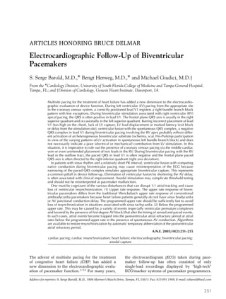
  • 1. ARTICLES HONORING BRUCE DELMAR Electrocardiographic Follow-Up of Biventricular Pacemakers S. Serge Barold, M.D.,∗ Bengt Herweg, M.D.,∗ and Michael Giudici, M.D.† From the ∗Cardiology Division, University of South Florida College of Medicine and Tampa General Hospital, Tampa, FL; and †Division of Cardiology, Genesis Heart Institute, Davenport, IA Multisite pacing for the treatment of heart failure has added a new dimension to the electrocardio- graphic evaluation of device function. During left ventricular (LV) pacing from the appropriate site in the coronary venous system, a correctly positioned lead V1 registers a right bundle branch block pattern with few exceptions. During biventricular stimulation associated with right ventricular (RV) apical pacing, the QRS is often positive in lead V1. The frontal plane QRS axis is usually in the right superior quadrant and occasionally in the left superior quadrant. Barring incorrect placement of lead V1 (too high on the chest), lack of LV capture, LV lead displacement or marked latency (exit block or delay from the stimulation site), ventricular fusion with the spontaneous QRS complex, a negative QRS complex in lead V1 during biventricular pacing involving the RV apex probably reflects differ- ent activation of an heterogeneous biventricular substrate (ischemia, scar, His-Purkinje participation in view of the varying patterns of LV activation in spontaneous left bundle branch block) and does not necessarily indicate a poor (electrical or mechanical) contribution from LV stimulation. In this situation, it is imperative to rule out the presence of coronary venous pacing via the middle cardiac vein or even unintended placement of two leads in the RV. During biventricular pacing with the RV lead in the outflow tract, the paced QRS in lead V1 is often negative and the frontal plane paced QRS axis is often directed to the right inferior quadrant (right axis deviation). In patients with sinus rhythm and a relatively short PR interval, ventricular fusion with competing native conduction during biventricular pacing may cause misinterpretation of the ECG because narrowing of the paced QRS complex simulates appropriate biventricular capture. This represents a common pitfall in device follow-up. Elimination of ventricular fusion by shortening the AV delay, is often associated with clinical improvement. Anodal stimulation may complicate threshold testing and should not be misinterpreted as pacemaker malfunction. One must be cognizant of the various disturbances that can disrupt 1:1 atrial tracking and cause loss of ventricular resynchronization. (1) Upper rate response. The upper rate response of biven- tricular pacemakers differs from the traditional Wenckebach upper rate response of conventional antibradycardia pacemakers because heart failure patients generally do not have sinus bradycardia or AV junctional conduction delay. The programmed upper rate should be sufficiently fast to avoid loss of resynchronization in situations associated with sinus tachycardia. (2) Below the programmed upper rate. This may be caused by a variety of events (especially ventricular premature complexes and favored by the presence of first-degree AV block) that alter the timing of sensed and paced events. In such cases, atrial events become trapped into the postventricular atrial refractory period at atrial rates below the programmed upper rate in the presence of spontaneous AV conduction. Algorithms are available to restore resynchronization by automatic temporary abbreviation of the postventricular atrial refractory period. A.N.E. 2005;10(2):231–255 cardiac pacing; cardiac resynchronization; heart failure; electrocardiography; biventricular pacing; anodal capture The advent of multisite pacing for the treatment of congestive heart failure (CHF) has added a new dimension to the electrocardiographic evalu- ation of pacemaker function.1–13 For many years, Address for reprints: S. Serge Barold, M.D., 5806 Mariner’s Watch Drive, Tampa, FL 33615. Fax: 813 891 1908; E-mail: ssbarold@aol.com the electrocardiogram (ECG) taken during pace- maker follow-up has often consisted of only single-lead recordings displayed by “high-tech” ECG/marker systems of pacemaker programmers. 231
  • 2. 232 r A.N.E. r April 2005 r Vol. 10, No. 2 r Barold, et al. r Electrocardiographic Follow-Up of Biventricular Pacemakers The “low-tech” paced 12-lead ECG was relegated to a minor role and often neglected.14 However, biventricular (BV) pacing has generated a well- deserved renaissance of the 12-lead paced ECG that has become an indispensable tool in the evaluation of cardiac resynchronization. NORMAL QRS PATTERNS DURING RIGHT VENTRICULAR PACING Pacing from the right ventricle (RV) regardless of site almost always produces a left bundle branch block (LBBB) pattern in the precordial leads (de- fined as the absence of a positive complex in lead V1 recorded in the 4th or 5th intercostal space).15,16 Pacing from the RV apex produces negative paced QRS complexes in the inferior leads (II, III, and aVF) simply because the activation begins in the inferior part of the heart and travels superiorly away from the inferior leads. The mean paced QRS frontal plane axis is superior either in the left or less commonly in the right superior quadrant. Dis- placement of the electrode from the RV apex to- ward the RV outflow tract (OT) shifts the frontal plane paced QRS axis to the left inferior quadrant, a site considered normal for spontaneous QRS com- plexes. The inferior leads become positive. The axis then shifts to the right inferior quadrant as the stim- ulation site moves more superiorly toward the pul- monary valve. With the backdrop of dominant R waves in the inferior leads, RVOT pacing may generate qR, QR, or Qr complexes in leads I and aVL. Occasionally with slight displacement of the pacing lead from RV apex to the RV outflow tract, leads I and aVL may register a qR complex in conjunction with the typical negative complexes of RV apical stimula- tion in the inferior leads. This qR pattern must not be interpreted as a sign of myocardial infarction.17 RV pacing from any site never produces qR com- plexes in V5 and V6 in the absence of myocardial infarction or ventricular fusion with a spontaneous QRS complex. A qR or Qr (but not QS) complex in the precordial or inferior leads is always abnormal in the absence of ventricular fusion. In contrast, a q-wave is common in the lateral leads (I, aVL, V5, and V6) during uncomplicated biventricular pacing (using the RV apex), and should not be interpreted as representing myocardial infarction or RVOT dis- placement of an RV apical lead. Finally, lead I (but not aVL) rarely displays a qR complex in uncom- plicated RV apical pacing. Table 1. Causes of a Dominant R wave during Conventional Ventricular Pacing Ventricular Fusion • Pacing in the myocardial relative refractory period • LV pacing from the coronary venous system • LV endocardial or epicardial pacing • Lead perforation of RV or ventricular septum with left ventricular stimulation • Uncomplicated RV pacing DOMINANT R WAVE OF THE PACED QRS COMPLEX DURING CONVENTIONAL PACING A dominant R wave in V1 during ventricular pac- ing has been called a right bundle branch block (RBBB) pattern of depolarization, but this terminol- ogy is potentially misleading because this pattern is often not related to RV activation delay14,16,18–20 (Table 1). A dominant R wave of a paced ventricular beat in the right precordial leads occurs in approx- imately 8–10% of patients with uncomplicated RV apical pacing. The position of precordial leads V1 and V2 should be checked because a dominant R wave can be sometimes recorded at the level of the third intercostal space during uncomplicated RV apical pacing. The pacing lead is almost certainly in the RV (apex or distal septal site) if V1 and V2 are negative when recorded one space lower (5th intercostal space).15,18 A dominant R wave may not be eliminated at the level of the 5th interspace if RV pacing originates from the midseptal region.21 Fur- thermore, the “RBBB” pattern from pacing RV sites results in a precordial vector change from positive to negative by lead V3 in the precordial sequence. Therefore, a tall R wave in V3 and V4 signifies that a pacemaker lead is not in the RV after exclud- ing ventricular fusion from spontaneous AV con- duction.21 The ECG pattern with a truly posterior RV lead has not been systematically investigated as a potential cause of a tall R wave in V1 during pacing. Significance of a Small r Wave in Lead V1 During Uncomplicated RV Pacing A small early (r) wave (sometimes wide) may oc- casionally occur in lead V1 during uncomplicated RV pacing. There is no evidence that this r-wave
  • 3. A.N.E. r April 2005 r Vol. 10, No. 2 r Barold, et al. r Electrocardiographic Follow-Up of Biventricular Pacemakers r 233 Figure 1. Twelve-lead ECG showing LV pacing from the coronary venous system. There is typical RBBB pattern and right axis deviation. Note the dominant R wave from V1 to V6 consistent with basal LV pacing. LV pacing shown in all the figures was performed from the coronary venous system. represents a conduction abnormality at the RV exit site. Furthermore, an initial r wave during biven- tricular pacing does not predict initial left ventric- ular (LV) activation.1 LEFT VENTRICULAR ENDOCARDIAL PACING Unintended passage of a pacing lead into the LV occurs via the subclavian artery or an atrial septal defect (or foramen ovale). The access sites to the LV can be easily identified by the typical widespread RBBB pattern in the precordial leads during pac- ing, standard chest radiographs, and echocardiog- raphy.14,22–26 As a rule with a RBBB configuration (tall R wave in V3 and beyond), the frontal plane axis cannot differentiate precisely an endocardial LV site from one in the coronary venous system. ECG PATTERNS RECORDED DURING LV PACING FROM THE CORONARY VENOUS SYSTEM An RBBB pattern in correctly positioned lead V1 occurs with few exceptions1,2,9,11,15,26,27 (Figs. 1 and 2). With apical sites, leads V4–V6 are typi- cally negative. With basal locations, leads V4–V6 are usually positive as with the concordant pos- itive R waves during overt pre-excitation in left- sided accessory pathway conduction in the Wolff– Parkinson–White syndrome.1 Middle Cardiac Vein Pacing usually produces an RBBB pattern but a LBBB configuration may also occur. Rarely the pat- tern alternates from RBBB to LBBB.15 The LBBB configuration may represent preferential entry into the RV from the septum perhaps on the basis of lo- cal pathology such as a myocardial infarction scar. The activation from the pacing site travels away from the inferior surface of the LV and generates negative QRS complexes in leads II, III, and aVF with the frontal plane axis usually in the left supe- rior quadrant. Great Cardiac Vein Little data are available from this site.27 The ECG usually shows a RBBB pattern in lead V1 with axis
  • 4. 234 r A.N.E. r April 2005 r Vol. 10, No. 2 r Barold, et al. r Electrocardiographic Follow-Up of Biventricular Pacemakers Figure 2. (A) Twelve-lead ECG with leads V1 and V2 recorded during LV pacing at the level of the second intercostal space in a thin patient with an elongated chest. There is no dominant R wave in lead V1. The ECG during biventricular pacing also failed to show a dominant R wave in V1 at the level of the second intercostals space. (B) The dominant R-wave in V1 becomes evident only when lead V1 is recorded in the 4th intercostal space. The R wave in V1 recorded in the 4th intercostal space during biventricular pacing also became dominant. deviation to the right inferior quadrant as with LV pacing from the lateral or posterior coronary veins especially if one of the lateral branches of the great cardiac vein (anterior interventricular vein) is used for pacing. Lateral and Posterior Veins LV pacing from the traditional site for resyn- chronization produces a RBBB in most cases. The frontal plane axis often points to the right inferior
  • 5. A.N.E. r April 2005 r Vol. 10, No. 2 r Barold, et al. r Electrocardiographic Follow-Up of Biventricular Pacemakers r 235 quadrant (right axis deviation) and less commonly to the right superior quadrant. In an occasional pa- tient with uncomplicated LV pacing with a typi- cal RBBB pattern in lead V1, the axis may point to the left inferior or left superior quadrant. The rea- sons for these unusual axis locations are unclear. Lead V1 rarely shows a negative QRS complex dur- ing uncomplicated LV pacing. This may be due to incorrect ECG lead placement (lead V1 too high), middle cardic vein location, or an undefined mech- anism requiring elucidation. ECG PATTERNS AND FOLLOW-UP OF BIVENTRICULAR PACEMAKERS11,28–30 A baseline 12-lead ECG should be recorded at the time of implantation during assessment of the in- dependent capture thresholds of the RV and LV to identify the specific morphology of the paced QRS complexes in a multiplicity of leads.14 This requires having the patient connected to a multichannel 12- lead ECG during the implantation procedure. A to- tal of four 12-lead ECGs are required. (1) Intrinsic rhythm and QRS complex prior to any pacing. (2) Paced QRS associated with RV pacing. (3) Paced QRS associated with LV pacing, and (4) Paced QRS associated with biventricular pacing (Fig. 3A). The four tracings should be examined to identify the lead configuration that best demonstrates a dis- cernible and obvious difference between the four pacing states (inhibited, RV only, LV only, and biventricular). This ECG lead should then be used as the surface monitoring lead for subsequent eval- uations. Loss of capture in one ventricle will cause a change in the morphology of ventricular paced beats in the 12-lead ECG similar to that of either single-chamber RV pacing or single-chamber LV pacing. A shift in the frontal plane axis may be use- ful to corroborate loss of capture in one of the ven- tricles.1,10,11 If both the native QRS and the biven- tricular paced complex are relatively narrow, then a widening of the paced QRS complex will iden- tify loss of capture in one chamber with effectual capture in the other. First-Generation Devices with a Common Output In questionable cases involving first-generation devices with a common ventricular output, loss of capture requires monitoring the ECG with telemetered markers and the intracardiac ventric- ular electrogram (EGM).9–11 When there is intact capture in both the RV and LV, the evoked re- sponse on the ventricular electrogram will show a monophasic complex in contrast to the two distinct depolarizations during spontaneous AV conduction if the native QRS is wide (LBBB or left intraventric- ular conduction delay) (Fig. 4). With loss of capture in one of the ventricles, ventricular pacing will per- sist in the contralateral ventricle. The impulse will then be conducted via the native pathways to the other ventricle in a manner identical to traditional single ventricular pacing systems. During threshold testing by gradually reducing the output, LV cap- ture is commonly lost before RV capture. In such a case, if there is a delayed left intraventricular conduction delay, the ventricular electrogram will change from a monophasic complex to two discrete complexes similar but not identical to the two com- ponents registered in the ventricular electrogram of spontaneous conducted (LBBB) QRS complexes (Figs. 4 and 5). Two discrete electrographic deflec- tions will not occur in the spontaneous ventricu- lar electrogram of patients with underlying RBBB or lesser degrees of LBBB. Rarely, when the pac- ing thresholds of the RV and LV are identical, test- ing should be started at a high output. The first transition will be to a spontaneous QRS complex if there is no anodal stimulation (discussed later). The presence of biventricular pacing must then be cor- roborated by the expected depolarization pattern in the 12-lead ECG. Fortunately, the threshold testing procedure has become simpler in contemporary de- vices with programmability of separate output for the RV and LV. Paced QRS Duration and Status of Mechanical Ventricular Resynchronization The paced QRS during biventricular pacing is of- ten narrower than that of monochamber pacing. Barring fusion beats, a narrower QRS implies depo- larization from the RV and LV. Thus, measurement of QRS duration during follow-up is helpful in the analysis of appropriate biventricular capture and fusion with the spontaneous QRS.1,2,11 If the biven- tricular ECG is virtually similar to that recorded with RV or LV pacing alone and no cause is found, one should not automatically conclude that one of the leads does not contribute to biventricular de- polarization without a detailed evaluation of the
  • 6. 236 r A.N.E. r April 2005 r Vol. 10, No. 2 r Barold, et al. r Electrocardiographic Follow-Up of Biventricular Pacemakers Figure 3. (A) Typical 12-lead ECG of biventricular pacing with the RV lead at the apex. Note the dominant R wave in lead V1 and the frontal plane axis in the right superior quadrant. (B) Asynchronous biventricular pacing (VOO) in a different patient with the RV lead at the apex. The arrows point to QRS complexes without a preceding P wave. These complexes therefore do not represent ventricular fusion with the spontaneous conducted QRS complex. There is a typical q wave in lead I and lead V6 shows a QR complex during pure biventricular pacing. (C) Biventricular DDD pacing in the same patient as in panel (B). With appropriate programming of the AV delay, the configuration of the paced QRS complexes becomes identical to that in panel (B) thereby ruling out fusion with the spontaneous conducted QRS complex. A dominant R wave in V1 is not always the rule during biventricular pacing with the RV lead at the apex. The frontal plane axis points to the left superior quadrant rather than the right superior quadrant often associated with this arrangement (reproduced from Garrigue, Barold, and Clémenty11 with permission).
  • 7. A.N.E. r April 2005 r Vol. 10, No. 2 r Barold, et al. r Electrocardiographic Follow-Up of Biventricular Pacemakers r 237 Figure 3. Continued. Figure 4. Simultaneous recording of the ECG and telemetered ventricular electrogram in a patient with left bundle branch block and a biventricular pacemaker with a common ventricular sensing channel. The ventricu- lar electrogram displays a monophasic pattern during biventricular capture. With loss of LV pacing, the ventricular electrogram shows a late deflection, which represents delayed LV activation through ordinary myocardium. AS = atrial-sensed event; VP = ventricular paced event (reproduced from Garrigue, Barold, and Clémenty11 with permission).
  • 8. 238 r A.N.E. r April 2005 r Vol. 10, No. 2 r Barold, et al. r Electrocardiographic Follow-Up of Biventricular Pacemakers Figure 5. Simultaneous recording of the ECG and telemetered ventricular electrogram during spontaneous rhythm in a patient with left bundle branch block and a biventricular pacemaker with a common ventricular sensing channel. The ventricular electrogram shows two discrete components corresponding to RV and delayed LV activation. The pattern is different from the one recorded in Figure 4 during RV pacing with loss of LV capture. AS = atrial-sensed event; VS = ventricular-sensed event (reproduced from Garrigue, Barold, and Clémenty11 with permission). pacing system. So far, evaluation of the overall ECG patterns of biventricular pacing has focused on si- multaneous RV and LV stimulation. The electro- cardiographic consequences of temporally different RV and LV activation with programmable V–V tim- ing in the most recent biventricular devices have not yet been studied.31,32 Chronic studies have shown that the degree of narrowing of the paced QRS duration is a poor predictor of the cardiac resynchronization re- sponse.7,11,33 In other words, the degree of QRS nar- rowing or its absence does not correlate with the long-term hemodynamic benefit of biventricular pacing7,11,33 because the paced QRS does not reflect the underlying level of mechanical dyssynchrony. In this respect, some patients with monochamber LV pacing exhibit an equal or superior degree of Table 2. Change in Frontal Plane Axis of Paced QRS When Programming from Biventricular to LV and RV Pacing Pacing Site QRS in Lead I QRS in Lead III Axis Shift BiV → RV Greater positivity Greater negativity∗ Clockwise BiV → LV Greater negativity Greater positivity Counterclockwise ∗QRS in lead III is more negative than in lead II. BiV = biventricular; RV = right ventricle; LV = left ventricle. mechanical resynchronization compared to biven- tricular pacing despite a very wide paced QRS com- plex.7,11,34 Usefulness of the Frontal Plane Axis of the Paced QRS Complex Table 2 shows the importance of the frontal plane axis of the paced QRS complex in determining the arrangement of pacing during testing of biventric- ular pacemakers.9–11 Biventricular Pacing with the RV Lead Located at the Apex The shift in the frontal plane QRS axis during programming the ventricular output is helpful in
  • 9. A.N.E. r April 2005 r Vol. 10, No. 2 r Barold, et al. r Electrocardiographic Follow-Up of Biventricular Pacemakers r 239 Figure 6. Biventricular pacing with the RV lead in the outflow tract. There was a very prominent R wave in lead V1 during monochamber LV pacing. Note the typical absence of a dominant R wave in lead V1 and the presence of right axis deviation, an uncommon finding during biventricular pacing with the RV lead at the apex. The presence of ventricular fusion with the spontaneous conducted QRS complex was ruled out. Compare this tracing with the one in Figure 3A. determining the site of ventricular stimulation in patients with first-generation devices without sep- arately programmable RV and LV outputs (Table 2). The frontal plane QRS axis usually moves supe- riorly from the left (RV apical pacing) to the right superior quadrant (biventricular pacing) in an anti- clockwise fashion if the ventricular mass is predom- inantly depolarized by the LV pacing lead1,10,11 (Fig. 3A). The frontal plane axis may occasionally reside in the left rather than the right superior quadrant during biventricular pacing. The QRS is often positive in lead V1 during biven- tricular pacing when the RV is paced from the apex. Barring incorrect placement of lead V1 (too high on the chest: as in Fig. 2A), lack of LV capture, LV lead displacement, or marked latency (exit block or delay from the stimulation site, an important but poorly studied phenomenon with LV pacing) asso- ciated with LV stimulation and ventricular fusion, a negative QRS complex in lead V1 probably reflects different activation of an heterogeneous biventric- ular substrate (ischemia, scar, His-Purkinje partici- pation in view of the varying patterns of LV activa- tion in spontaneous LBBB, etc.) and does not nec- essarily indicate a poor (electrical or mechanical) contribution from LV stimulation. In this situation, it is imperative to rule out the presence of coro- nary venous pacing via the middle cardiac vein16 or even unintended placement of two leads in the RV.35 Biventricular Pacing with the RV Lead in the Outflow Tract In our limited experience, we have found that during biventricular pacing with the RV lead in the outflow tract, the paced QRS in lead V1 is often neg- ative and the frontal plane paced QRS axis is often directed to the right inferior quadrant (right axis de- viation) (Fig. 6). Further studies are required to con- firm these preliminary findings and to determine the significance of these ECG patterns of biventric- ular pacing according to the RV pacing site. Q or q and QS Configuration in Lead 1 Georger et al.29 observed a q wave in lead I in 17 of 18 patients during biventricular pacing (Fig. 3B & C). As indicated previously, a q wave in lead I during uncomplicated RV apical pacing is rare and these workers observed it in only 1 patient. Loss of the q-wave in lead I was 100% predictive of loss of LV capture.29 It therefore appears that analysis of the Q or q wave or a QS complex in lead I may be a reliable way to assess LV capture during biventric- ular pacing.
  • 10. 240 r A.N.E. r April 2005 r Vol. 10, No. 2 r Barold, et al. r Electrocardiographic Follow-Up of Biventricular Pacemakers Ventricular Fusion Beats with Native Conduction In patients with sinus rhythm and a relatively short PR interval, ventricular fusion with com- peting native conduction during biventricular pac- ing may cause misinterpretation of the ECG, and a common pitfall in device follow-up11 (Fig. 7). QRS shortening mandates exclusion of ventricular fusion with the spontaneous QRS complex, espe- cially in the setting of a relatively short PR inter- val. The presence of ventricular fusion should be ruled out by observing the paced QRS morphology during progressive shortening of the AS–VP inter- val in the VDD mode or the AP–VP interval in the DDD mode. The AS–VP interval should be pro- grammed (with rate-adaptive function) to ensure pure biventricular pacing under circumstances that might shorten the PR interval, such as increased circulating catecholamines. Figure 7. (A) Narrowing of the paced QRS complex (well seen in V1) due to ventricular fusion with the spontaneous conducted QRS complex. This ECG was the initial recording taken upon arrival to the pacemaker follow-up center. AV delay = 100 ms. The marked narrowing of the QRS complex in lead V1 suggests ventricular fusion rather than QRS narrowing from satisfactory biventricular pacing. (B) The ECG taken 15 minutes later (same parameters and AV delay) when the patient was more relaxed shows no evidence of ventricular fusion. (C) Immediately after the tracing in panel (B), ventricular fusion was demonstrated only when the AV delay was lengthened to 130 ms. The serial tracings illustrate the dynamic nature of AV conduction (emotion, catecholamines, etc.) and the importance of appropriate programming of the AV delay to prevent ventricular fusion with the spontaneous conducted QRS complex. Long-Term ECG Changes Many studies have shown that the paced QRS duration does not vary over time as long as the LV pacing lead does not move from its initial site.7,11,36 Yet, surface ECGs should be performed periodi- cally because the LV lead may become displaced into a collateral branch of the coronary sinus. Dis- lodgement of the LV lead may result in loss of LV capture with the ECG showing an RV pacing QRS pattern with an increased QRS duration and su- perior axis deviation. Ricci et al.36 suggested that variation of the QRS duration over time may play a determinant role if correlated with remodeling of the ventricles by echocardiography. Finally, the un- derlying spontaneous ECG should be exposed peri- odically to confirm the presence of a LBBB type of intraventricular conduction abnormality. In this re- spect, turning off the pacemaker could potentially improve LV function in patients who have lost their
  • 11. A.N.E. r April 2005 r Vol. 10, No. 2 r Barold, et al. r Electrocardiographic Follow-Up of Biventricular Pacemakers r 241 Figure 7. Continued.
  • 12. 242 r A.N.E. r April 2005 r Vol. 10, No. 2 r Barold, et al. r Electrocardiographic Follow-Up of Biventricular Pacemakers Figure 8. Anodal capture during first-generation biventricular pacing. There is anodal capture on the left (three pacing sites). It disappears on the right (two pacing sites) with reduction of the common ventricular output revealing pure biventricular pacing (reproduced from Garrigue, Barold, and Clémenty11 with permission). intraventricular conduction defect through ventric- ular remodeling. ANODAL STIMULATION Although anodal capture may occur with high output traditional RV pacing, this phenomenon is almost always not discernible electrocardiograph- ically. Biventricular pacing systems generally uti- lize a unipolar lead for LV pacing via a coronary vein. The tip electrode of the LV lead is the cathode and the proximal electrode of the bipolar RV lead often provides the anode for LV pacing. This ar- rangement creates a common anode for RV and LV pacing. A high current density (from two sources) at the common anode during biventricular pacing may cause anodal capture manifested as a paced QRS complex with a somewhat different configura- tion from that derived from pure biventricular pac- ing.37–42 Anodal capture during biventricular pac- ing disappears by reducing the output of the pace- maker or when the device (even at high output) is programmed to a true unipolar system with the common anode on the pacemaker can. Anodal cap- ture was recognized in first-generation transvenous biventricular pacemakers (without separately pro- grammable RV and LV outputs) when three dis- tinct pacing morphologies were observed exclu- sive of fusion with the spontaneous QRS complex: Biventricular with anodal capture (at a high out- put), biventricular (at a lower output), and RV (with loss of LV capture) or rarely LV (with loss of RV capture)41,42 (Fig. 8). Anodal capture involving the ring electrode of the bipolar RV lead can also oc- cur in second-generation biventricular pacemakers with separately programmable ventricular outputs. Anodal stimulation may occur during biventricular pacing as with first-generation devices. However, during monochamber LV pacing at a relatively high output, the paced QRS complex becomes identical to that registered with biventricular pacing below the anodal stimulation threshold37,39,41,42 (Fig. 9). Occasionally, this type of anodal capture prevents electrocardiographic documentation of pure LV pacing if the LV pacing threshold is higher than that of anodal stimulation. Anodal stimulation may complicate threshold testing and should not be misinterpreted as pacemaker malfunction. Further- more, anodal stimulation during biventricular pac- ing may interfere with a programmed interven- tricular (V–V) delay (often programmed with the LV preceding the RV) aimed at optimizing cardiac resynchronization because RV and LV31,32 are acti- vated simultaneously.37,39,41,42 If the LV threshold
  • 13. A.N.E. r April 2005 r Vol. 10, No. 2 r Barold, et al. r Electrocardiographic Follow-Up of Biventricular Pacemakers r 243 Figure 9. On the left, there is anodal pacing in the DDD mode of a second-generation biventricular pace- maker seen during monochamber LV pacing with an LV output of 3.5 V and 0.5 ms LV. The ECG pattern was identical to that recorded during biventricular pacing. On the right, anodal stimulation alternates with pure LV pacing when the LV output is slightly less than 3.5 V at 0.5 ms (reproduced from Herweg and Barold38 with permission). is not too high, appropriate programming of the pacemaker output should eliminate anodal stimu- lation in most cases. UPPER RATE RESPONSE OF BIVENTRICULAR PACEMAKERS The upper rate response of biventricular pace- makers differs from the traditional Wenckebach upper rate response of conventional antibradycar- dia pacemakers because CHF patients generally do not have sinus bradycardia or AV junctional con- duction delay. The upper rate response exhibits two forms according to the location of the P wave in the pacemaker cycle: (1) A pre-empted Wencke- bach upper rate response with AS–VS sequences and the P wave beyond the postventricular atrial refractory period (PVARP).43 (2) AR–VS sequences with the P wave sensed (but not tracked) within the PVARP. Pre-empted Wenckebach Upper Rate Response In a traditional Wenckebach upper rate response, a dual chamber pacemaker (where upper rate inter- val > total atrial refractory period (TARP)) deliv- ers its ventricular stimulus only at the completion of the (atria-driven) upper rate interval (Fig. 10). The AV delay initiated by a sensed P wave in- creases progressively because the ventricular chan- nel waits to deliver its output at the end of the up- per rate interval. Eventually, a P wave falls in the PVARP, a pause occurs, and the ventricular paced
  • 14. 244 r A.N.E. r April 2005 r Vol. 10, No. 2 r Barold, et al. r Electrocardiographic Follow-Up of Biventricular Pacemakers Figure 10. Top: Traditional pacemaker Wenckebach upper rate response. Bot- tom: Repetitive pre-empted Wenckebach upper rate response. See text for de- tails (reproduced from Barold, Garrigue, and Israel12 with permission). sequence repeats itself. In patients with pacemak- ers implanted for CHF, the Wenckeback upper rate response (or more precisely the manifestation of up- per rate > total atrial refractory period) assumes a form that is not immediately recognizable because no paced beats are evident. In patients with normal or near normal sinus node function and AV conduction and a relatively short PVARP, a pacemaker Weckenbach upper rate response takes the form of a repetitive pre-empted process which consists of an attempted Wencke- bach upper rate response with each cycle, asso- ciated with continual partial or incomplete exten- sion of the programmed AV interval12,43 (Figs. 10 and 11). The conducted spontaneous QRS complex continually occurs before completion of the upper rate interval. It is therefore sensed by the pace- maker, and ventricular pacing is pre-empted. In other words, the pacemaker cannot time out the upper rate interval and thus cannot emit a ventric- ular stimulus at its completion. This form of upper rate response tends to occur in patients with rela- tively normal AV conduction, a short programmed AV delay, a relatively slow programmed (atrial- driven) upper rate, and a sinus rate faster than the programmed (atrial-driven) upper rate (Fig. 11). It is therefore more likely to emerge on exercise or during times of distress when adrenergic tone is high. Consequently, the pre-empted Wenckebach upper rate response has become important recently
  • 15. A.N.E. r April 2005 r Vol. 10, No. 2 r Barold, et al. r Electrocardiographic Follow-Up of Biventricular Pacemakers r 245 Figure 11. Stored markers showing development of a pre-empted Wenckebach upper rate response during biventricular pacing. Upper rate interval (URI) = 460 ms. (A) There is 1:1 atrial tracking and biventricular (BV) pacing with the programmed AS–VP delay. (B) When the spontaneous ventricular rate exceeds the programmed upper rate (VS–VS < URI), a pre-empted Wenckebach upper rate response supervenes. AS conducts to VS so that AS–VS becomes longer than the programmed AS–VP interval. Note that the sinus P wave is sensed beyond the postventricular atrial refractory period. AS = atrial-sensed event; VS = ventricular-sensed event; BV or VP = biventricular pacing event. because biventricular pacemakers are now im- planted in patients with CHF (or hypertrophic car- diomyopathy) where there is commonly relatively normal sinus node function and AV conduction. The occurrence of a pre-empted Wenckebach re- sponse in such patients defeats the very purpose of this type of cardiac stimulation. Because pa- tients with CHF are susceptible to sinus tachy- cardia (especially during decompensation despite beta-blocker therapy), it is particularly important to program a relatively fast upper rate during biven- tricular pacing to avoid a pre-empted Wenckebach upper rate response with resultant loss of cardiac resynchronization manifested by the emergence of the patient’s spontaneous conducted QRS in the electrocardiogram. In summary, a pre-empted Wenckebach upper rate response has no paced events and is character- ized by three features: (1) VS–VS interval < atrial- driven upper rate interval (VS = ventricular-sensed event), (2) PR interval (AS–VS) > programmed AS– VP. The spontaneous PR interval remains relatively constant (AP = atrial paced event, AS = atrial- sensed event), and (3) there are no unsensed (or refractory sensed) P-waves as in a typical Wencke- bach upper rate response in the presence of AV block. Upper Rate Limitation with P-Wave in the PVARP When the P wave falls within the PVARP, a de- vice may not assume 1:1 atrial tracking immedi- ately when the sinus rate drops below the pro- grammed upper rate if the P wave remains in the PVARP.2 The reason lies in the fact that AR–VS (spontaneous AV conduction) > programmed AS– VP interval. Therefore, the total atrial refractory
  • 16. 246 r A.N.E. r April 2005 r Vol. 10, No. 2 r Barold, et al. r Electrocardiographic Follow-Up of Biventricular Pacemakers Time MTR Rate CRT: AS - VP AR - VS CRT No CRT AS - VP 1 2 3 No CRT: AR - VS P-P > [ (AR-VS) + PVARP] Figure 12. Diagram showing an upper rate response with the P wave falling within the postventricular atrial refractory period (PVARP) in the setting of normal AV conduction. Cardiac resynchronization (CRT) occurs with AS–VP sequences when the sinus rate is below the max- imum tracking rate (MTR). When the atrial rate exceeds the MTR at point 1, the P wave falls within the PVARP (AR marker) and CRT is lost and AR–VS sequences take over with AR conducting to VS. When the sinus rate falls below the MTR at point 2, no CRT occurs because the timing cycles of the device force the continuation of AR– VS sequences. Failure of CRT at this stage results from the longer prevailing total atrial refractory period (TARP) which is equal to [(AR–VS) + PVARP] longer than the pro- grammed TARP = [(AS–VP) + PVARP]. CRT with AS–VP sequences is restored at point 3 when the sinus inter- val (P–P) > [(AR–VS) + PVARP], at a sinus rate substan- tially lower than the MTR. AS = atrial-sensed event; VS = ventricular-sensed event; VP = biventricular paced event; AR = atrial-sensed event in the atrial refractory period of the pacemaker where tracking cannot occur. period during AR–VS operation, (AR–VS) interval + PVARP must be longer than the programmed TARP which is the sum of (AS–VP) interval + PVARP (Fig. 12). The pacemaker will continue to oper- ate with AR–VS cycles below the upper rate until the sinus interval drops below the duration of the (AR–VS) interval + PVARP interval thereby allow- ing escape of the sinus P wave out of the PVARP. Therefore, restoration of resynchronization will oc- cur at a rate slower than the programmed upper rate. This problem is worse in patients with first- generation devices due to double counting where 1:1 atrial tracking (AS–VP pacing) will return only when the sinus interval becomes longer than [AR– VS] interval + PVARP + ICD (ICD = interventric- ular conduction delay or the interval between the RV and LV electrograms both sensed by the de- vice).2 These considerations are important in CHF patients who develop substantial increases in sinus rates with exercise or states of increased circulating catecholamines. Special algorithms based on beat to beat PVARP shortening upon sensing a P wave in the PVARP are now available in the latest devices to promote 1:1 atrial tracking just below the upper rate (discussed later). OPTIMAL PROGRAMMING OF BIVENTRICULAR DEVICES Cardiac resynchronization depends on meticu- lous programming of the device (Table 3). The most important parameters are the AV delay, PVARP, and the maximum tracking rate.10 Table 3. Programmability of Biventricular Pacemakers Parameter Management AV delay 1. A long AV delay should not be used. 2. Optimize the AS–VP delay and avoid ventricular fusion with the spontaneous conducted QRS complex. 3. Program rate-adaptive (dynamic) AV delay off during temporary pacing for testing (with VDD mode slower than sinus rate to sense atrial activity). 4. Program rate-adaptive AV delay for long-term pacing. Atrial sensing and PVARP 1. Short PVARP (aim for 250 ms). May have to use algorithms for the automatic termination of endless loop tachycardia. 2. Program off the post-VPC PVARP extension. 3. Automatic mode switching off in devices using a relatively long PVARP mandated by the mode switching algorithm. Upper rate Relatively fast upper rate so the patient does not have “breakthrough” ventricular sensing within their exercise zone. Initial upper rate of 140/min is often appropriate in the absence of myocardial ischemia during pacing at this rate. AV conduction 1. Use drugs that impair AV conduction to avoid ventricular fusion or double counting in devices with a common sensing channel. 2. Consider ablation of the AV junction in refractory double counting (common sensing channel) or patients with a long PR interval difficult to manage.
  • 17. A.N.E. r April 2005 r Vol. 10, No. 2 r Barold, et al. r Electrocardiographic Follow-Up of Biventricular Pacemakers r 247 Table 4. Causes of Double Counting in Devices with Common Sensing 1. Loss of tracking of sinus rhythm. This includes sinus tachycardia above programmed upper rate and sinus P waves buried in a relatively long postventricular atrial refractory period (especially with activation of the automatic postventricular atrial refractory extension by a pacemaker defined ventricular extrasystole). Far-field sensing of left atrial activity by the LV lead, and near-field sensing of the T-wave can also induce double counting of the ventricular electrogram 2. Double-counting of the QRS during supraventricular tachyarrhythmias (with intrinsic AV conduction) at a rate below the cut-off point Double-counting of QRS during ventricular tachycardia at rates below the cut-off point (resulting in detection of ventricular fibrillation) The above diagnosis of double counting of the ventricular electrogram requires exclusion of (a) displacement of the RV lead toward the tricuspid valve with far-field RV sensing of atrial activity and (b) oversensing of diaphragmatic myopotentials by using testing with deep respiration, coughing, laughing, and the Valsalva maneuver SIMULTANEOUS SENSING OF THE VENTRICULAR ELECTROGRAM FROM BOTH VENTRICLES First-generation biventricular pacemakers uti- lized a parallel dual cathodal system where pacing and sensing occur simultaneously in the two ventri- cles. Such systems (using a conventional DDD pace- maker and a Y-adapter) are still being implanted in various countries because of cost considerations. The common sensing channel predisposes the pace- maker to double sensing of the ventricular electro- gram12,44 (Table 4). Double counting of the QRS complex usually involves the conducted QRS com- plex (as most patients have left bundle branch block and relatively normal AV conduction).2–7 Less com- monly, double counting of the ventricular complex occurs when there is a loss of LV capture with preservation of RV pacing. Both situations produce temporal separation of the RV and LV electrograms. The degree of separation depends on the severity of the interventricular conduction delay and the lo- cation of the electrodes. During AV synchrony, a device can sense the LV electrogram only if it ex- tends beyond the relatively short ventricular blank- ing period initiated by the prior detection of the RV EGM. With ventricular rhythms, the LV EGM may precede that from the RV. The consequences may be serious and include ventricular inhibition (with denial of beneficial resynchronization) and in- appropriate therapy including shocks in the case of biventricular ICDs (excluding double counting from lack of LV capture)44–51 (Fig. 13). The diag- nosis is important because double counting can of- ten be corrected by appropriate programming and therapy. Causes of Double Counting of the Ventricular Electrogram Barring isolated loss of LV pacing, Table 4 out- lines the possible causes of double counting of the ventricular EGM in systems with simultane- ous sensing from the RV and LV in the presence of an undisplaced RV lead. Double counting of the RV electrogram is uncommon with sensing only from the RV. In the case of biventricular pace- makers, the ventricular blanking period (after ven- tricular sensing) if programmable could be length- ened to contain the entire ventricular electrogram. This option is generally not available in biventricu- lar ICDs where a long ventricular blanking period might promote undersensing of ventricular tachy- arrhythmias. A new algorithm based on program- ming an interventricular refractory period (IRP) (Medtronic Inc.) was designed for pacemakers (not ICDs) to prevent double counting when sensing be- tween the LV tip and the RV tip.12 This feature pre- vents restarting the ventricular refractory period, postventricular atrial blanking and refractory peri- ods, and upper rate timers when a second sensed depolarization is seen in the ventricular refrac- tory period following a sensed event (as in LBBB) (Fig. 14). When the second sensed depolarization occurs within the interventricular refractory pe- riod, the refractory periods and timing intervals are not reset, thus preventing the second sensed depolarization from limiting upper tracking rates as seen in first-generation devices. In other words, it rectifies the problem of inappropriate extension of the atrial refractory period due to sensing of a delayed LV potential during the ventricular refrac- tory period. The interventricular refractory period functions like a ventricular sensing blanking period which itself is not programmable in these devices.
  • 18. 248 r A.N.E. r April 2005 r Vol. 10, No. 2 r Barold, et al. r Electrocardiographic Follow-Up of Biventricular Pacemakers Figure 13. Double counting of the ventricular electrogram in a patient who had received inappropriate shocks by an implanted Guidant Contak CD biventricular ICD that senses from both ventricles simultane- ously. The atrial and ventricular electrograms are on top. The first 2 ventricular complexes are paced. The atrial rate then exceeds the programmed upper rate and a repetitive pre-empted Wenckebach sequence starts. The device then senses each conducted QRS twice. The second last cycle terminates with a paced ventricular beat because of slight sinus slowing. AS = atrial-sensed event; VP = ventricular paced event; VS = ventricular-sensed event; VT = ventricular tachycardia; VF = ventricular fibrillation (reproduced from Barold, Garrigue, and Israel12 with permission). Figure 14. Diagrammatic representation of the interventricular refractory period (IRP) of the Medtronic InSync III biventricular pacemaker. The IRP prevents sensing of a second ventricular depolarization when the RV and LV do not depolarize simultaneously. Thus, a sensed event in the IRP (either following a ventricular paced event or a non-refractory sensed event) does not initiate new timing cycles. A = atrium; S = non-refractory sensed event; P = paced event; R = refractory-sensed event. Note the short P–P intervals representing the V–V delay or the timing difference between LV and RV stimulation (reproduced from Barold, Garrigue, and Israel12 with permission).
  • 19. A.N.E. r April 2005 r Vol. 10, No. 2 r Barold, et al. r Electrocardiographic Follow-Up of Biventricular Pacemakers r 249 Table 5. Indicators for Far-Field P wave Sensing in Devices with Common Sensing Channels 1. Recurrence or development of symptoms of CHF 2. Inappropriately short AS–VP delay on surface ECG 3. Unexpected inhibition of ventricular output (DDD mode) with a PR (AS–VS) interval > programmed AS–VP delay (at rates below the maximum tracking rate) 4. Event markers recorded with simultaneously telemetered ventricular electrogram and surface ECG. Far-Field Sensing of Atrial Depolarization Causing Double and Triple Counting A device with common RV and LV sensing may sense the far-field atrial electrogram via an LV lead located (or displaced from its original site) in Figure 15. Far-field atrial sensing resulting in triple counting of a biventricular pacemaker. Simultaneous recordings (from top to bottom) of the ECG, a marker channel, and telemetered ventricular electrogram of a (dual-cathodal with a common sensing ventricular channel) Medtronic InSync DDDR biventricular pacemaker programmed to the ODO mode (25 mm/s). There is sinus rhythm with 1:1 AV conduction and ventricular inhibition. The LV lead detects the late portion of the P wave because of its proximity to the coronary sinus and left atrium (VS follows each AS closely), resulting in complete inhibition of ventricular pacing. Every P wave is conducted and produces a wide QRS sequentially sensed by the RV lead, then by the LV lead, as a function of the distance between the leads, the long interventricular conduction time, and the duration of the blanking period. Therefore, there are three ventricular-sensed markers associated with each QRS complex—the first VS is the far-field atrial signal, the second VS and third VS markers depict the two near-field components of the ventricular electrogram originating from the RV and LV leads, respectively. In the DDDR mode, the two signals generated by ventricular depolarization were recorded as VR events (refractory sensed) by the marker channel. VS = ventricular-sensed event; AS = atrial-sensed event (reproduced from Lipchenka, Garrigue, Glikson, et al.52 with permission). one of the coronary veins because of its proximity to the AV groove and the left atrium.11,12,52–56 In the first-generation devices with a common sens- ing channel, far-field atrial sensing by the ventric- ular channel inhibits biventricular pacing and in- duces the emergence of spontaneous AV conduc- tion (Table 5). A wide ventricular electrogram in- ducing double counting together with the sensed atrial signal results in triple counting at the ven- tricular level (Fig. 15). Far-field atrial sensing can also cause inappropriate discharge of a biventric- ular ICD (Fig. 16). This complication is devastat- ing in pacemaker-dependent patients who have un- dergone ablation of the AV junction for perma- nent atrial fibrillation prior to the implantation of a biventricular device because of the risk of ventric- ular asystole and inappropriate therapy including shocks.56
  • 20. 250 r A.N.E. r April 2005 r Vol. 10, No. 2 r Barold, et al. r Electrocardiographic Follow-Up of Biventricular Pacemakers Figure 16. Double jeopardy during biventricular pacing. Simultaneous recording of lead II ECG (top), bipolar ventricular electrogram between the right and left ventricular electrodes (middle), and anno- tated markers (bottom) showing far-field oversensing of atrial fibrillation potentials by a Guidant PRIZM VVIR ICD modified for biventricular pacing with a Y-adaptor (25 mm/s). The recording shows a “double whammy” with ventricular asystole and the delivery of an inappropriate shock. VT = ventricular tachy- cardia (intervals between 300 and 500 ms); VF = ventricular fibrillation (intervals < 300 ms); VS = ventricular-sensed event. The ventricular shock terminated atrial fibrillation and produced atrial stand- still. Atrial fibrillation returned a few seconds later (reproduced from Garrigue, Barold, Clementy, et al.56 with permission). Figure 17. Ventricular triggered mode of third-generation Medtronic biventricular ICD (VVIR mode). The device triggers a biventricular output upon sensing the RV electrogram in an attempt to provide resynchronization upon sensing. The pacemaker stimuli (RV and LV) deform the sensed ventricular premature beats. The degree of electrical resynchronization cannot be determined from this tracing. Note that in the DDD/DDDR mode, the ventricular triggered mode functions only with ventricular sensing in the AV delay.
  • 21. A.N.E. r April 2005 r Vol. 10, No. 2 r Barold, et al. r Electrocardiographic Follow-Up of Biventricular Pacemakers r 251 Figure 18. Ventricular resynchronization upon RV sensing by the ventricular triggered mode of a Medtronic biventricular ICD in the DDD mode. The patient has intra-atrial conduction delay so that the atrial electrogram sensed in the atrial appendage occurs late during the isoelectric portion of the PR interval (dotted vertical line). The AS–VS interval during AV conduction measures only 50–60 ms. The patient did not tolerate an AS–VP interval of 40 ms to produce biventricular pacing which in all likelihood occurred with some degree of fusion with the spontaneous conducted QRS complex. This situation calls for one of the two options: (1) ablation of the AV junction and (2) using the triggered mode upon sens- ing. A trial of the triggered mode produced marked clinical improvement. Consequently, the pacemaker was programmed to the ventricular triggered mode for long-term pacing. AS is followed by VS (smaller downward deflection) which triggers VP (larger downward deflection); AEGM = atrial electrogram; AS = atrial-sensed event; VS = ventricular-sensed event; VP = biventricular paced event. Devices Susceptible to Double Counting Conventional dual chamber ICDs used in an “off- label” fashion with a Y-connector for biventricular pacing (simultaneous RV and LV pacing and sens- ing) can exhibit double counting.12,50 One commer- cially available first-generation Guidant biventric- ular ICD (Contak CD) system used a conventional dual cathodal system and sensed simultaneously from the RV and LV causing double counting in about 7% of cases.12 All contemporary devices al- low programming of the sensing function of the individual ventricular channels to prevent double counting (RV and LV electrograms) or triple count- ing (far-field P wave, RV and LV EGMs). Biventric- ular ICDs now sense only from the RV to avoid double sensing and inappropriate ICD therapy. RESYNCHRONIZATION DURING SENSING BY THE TRIGGERED RESPONSE The ventricular triggered mode provides car- diac resynchronization in the presence of ventricu- lar sensing. When a biventricular pacemaker pro- grammed to the DDD/T mode (triggered at the ven- tricular level) detects ventricular activity in the AV delay, the signal triggers an output2 (Fig. 17). In other words, a ventricular-sensed event initiates an immediate emission of a ventricular or usually a biventricular output (according to the programmed settings) in conformity with the programmed up- per rate interval.2 The stimulus will be ineffec- tual in the chamber where sensing was initiated because the myocardium is physiologically refrac- tory. The triggered stimulus to the other ventricle thus attempts to provide resynchronization activa- tion in the setting of intraventricular dyssynchrony. Figure 18 shows how this modality avoided AV junctional ablation in a patient with intra-atrial con- duction delay. In atrial fibrillation, some devices provide some degree of ventricular resynchronization by the trig- gered mode and attempt regularization of the paced beats up to the programmed maximum tacking rate. Activation of this algorithm does not result in con- trol of the ventricular rate, and should not be a sub- stitute for ablation of the AV junction in patients with drug-refractory rapid ventricular rates. RESTORATION OF ATRIAL TRACKING BY PVARP ABBREVIATION The delivery of ventricular resynchronization and 1:1 atrial tracking can be disrupted under cer- tain circumstances (below the upper rate) where
  • 22. 252 r A.N.E. r April 2005 r Vol. 10, No. 2 r Barold, et al. r Electrocardiographic Follow-Up of Biventricular Pacemakers Figure 19. Stored markers showing the development of a locked P wave within the postventricular atrial period (PVARP) of a Medtronic biventricular ICD. AS–VP = 130 ms, upper rate = 130/min (upper rate interval = 460 ms). (A) Normal atrial tracking and biven- tricular pacing at the programmed AS–VP delay. (B) The P wave is locked into the PVARP despite a ventricular rate slower than the programmed upper rate (VS–VS = 470 ms and up- per rate interval = 460 ms). The algorithm shown in Figure 20 would make the diagnosis of the P wave trapped in the PVARP and would restore atrial tracking by PVARP abbreviation. Figure 20. Medtronic’s atrial tracking recovery algorithm during biventricu- lar pacing. The algorithm recognizes VS–AR sequences only when the VS–VS interval is longer than the programmed upper rate interval. Abbreviation of the postventricular atrial period promotes recovery of atrial tracking and re- stores ventricular resynchronization. This algorithm would therefore restore atrial tracking from point 2 to point 3. Figure 19 also shows a situation where the algorithm would be useful.
  • 23. A.N.E. r April 2005 r Vol. 10, No. 2 r Barold, et al. r Electrocardiographic Follow-Up of Biventricular Pacemakers r 253 atrial events are locked into the PVARP. The P wave becomes trapped into the PVARP at atrial rates below the programmed upper rate in the pres- ence of normal AV conduction. This leads to AR–VS cycles where AR is sensed in the PVARP (Fig. 19). Resumption of 1:1 atrial tracking and resynchro- nization require slowing of the sinus rate so that the P–P or sinus interval exceeds the sum of in- trinsic PR interval (AR–VS) + PVARP (prevailing intrinsic TARP).2 A special algorithm to restore 1:1 atrial tracking at rates slower than the programmed upper rate works when the devices detect AR in the PVARP (Fig. 20). The algorithm temporarily short- ens the PVARP and therefore the intrinsic TARP to permit sensing of the P wave beyond the PVARP so as to restore 1:1 atrial tracking. This algorithm may be useful in patients with sinus tachycardia and first-degree AV block in whom prolonged lock- ing of the P waves inside the PVARP is an impor- tant problem.57 Such patients are sometimes best treated with ablation of the AV junction. OMISSION OF LEFT VENTRICULAR STIMULUS Theoretically (but unlikely), lack of LV sensing could result in competitive pacing and arrhythmia induction. Such a situation might occur if a pre- mature ventricular complex originates near the LV sensing site and at specific time before the P wave: If ventricular activation initiated by the VPC con- ducts to the RV sensing site with a marked delay, it will be unable to inhibit the scheduled ventricular pacing pulse (triggered by the P wave) thereby de- livering the ventricular stimulus beyond the abso- lute myocardial refractory period. For this reason, Guidant has incorporated an algorithm in their de- vices to prevent unsafe LV pacing into the vulnera- ble period. An LV-sensed event initiates an LV pro- tection period during which the LV stimulus is in- hibited. The LV protection period is programmable between 300 and 500 ms after an LV-sensed event. LV sensing is used only for inhibition of LV pacing. BIVENTRICULAR PACING WITH CONVENTIONAL PACEMAKERS Although conventional dual chamber pacemak- ers are not designed for biventricular pacing and generally do not allow programming of an AV delay of zero or near zero, they are being used Table 6. Loss of Cardiac Resynchronization During DDD or DDDR Pacing in the Presence of Preserved LV Pacing A. Intrinsic Atrial undersensing from low amplitude atrial potentials T wave oversensing and other types of ventricular oversensing such as diaphragmatic potentials Long PR interval Circumstances that push the P wave into the PVARP such as a junctional rhythm New arrhythmia such as atrial fibrillation with a fast ventricular rate First-generation devices with a common sensing channel: ventricular double counting and sensing of far-field atrial activity B. Extrinsic Inappropriate programming of the AV delay or any function that prolongs the AV delay such as rate smoothing, AV search hysteresis, etc. Low maximum tracking rate Functional atrial undersensing precipitated by an atrial premature beat or ventricular premature beat. Long PVARP including post VPC automatic PVARP extension Intra-atrial conduction delay where sensing of AS is delayed in the right atrial appendage. A short AS–VP interval may not be able to achieve biventricular pacing with their shortest “AV delay” (0–30 ms) for ven- tricular resynchronization in CHF patients with permanent atrial fibrillation.58 Their advantages in- clude programming flexibility, and cost consider- ations. When a conventional dual chamber pace- maker is used for biventricular pacing, the “atrial” channel is generally connected to the LV and the “ventricular” channel to the RV.58 This arrange- ment provides (1) LV stimulation before RV activa- tion (LV pre-excitation) and (2) protection against ventricular asystole (but not sensing of atrial activ- ity) related to oversensing far-field atrial activity. The DVI(R) mode behaves like the VVI(R) mode ex- cept that there are always two closely coupled stim- uli (or electrocardiographically fused stimuli if the “AV delay” is very short) thereby facilitating eval- uation of pacemaker function. Furthermore, the DVI(R) mode provides absolute protection against far-field sensing of atrial activity in the case of LV lead displacement. The short delay between LV and RV stimulation imposed by the shortest “AV delay” may not be a significant limitation in many patients because it is LV pacing that generally provides the salutary effect of biventricular pacing.
  • 24. 254 r A.N.E. r April 2005 r Vol. 10, No. 2 r Barold, et al. r Electrocardiographic Follow-Up of Biventricular Pacemakers CONCLUSION The advent of biventricular (triple-chamber) de- vices has added new complexity to the evalua- tion of pacemaker function and follow-up. Patients with severe CHF benefit greatly from small im- provements in hemodynamics. Consequently, one must be cognizant of the various disturbances that can disrupt ventricular resynchronization (Table 6). The presence of interatrial conduction block or de- lay in CHF patients complicates therapy because it requires additional atrial resynchronization and therefore four-chamber devices for optimal hemo- dynamic benefit.59 The true value of ventricular resynchronization with monochamber LV pacing remains unclear at this time. There is more to learn about the various patterns of the 12-lead ECG in the assessment of lead location and the efficacy of electrical versus mechanical resynchronization especially with the new capability of varying the stimulation interval (V–V) between the two ventri- cles. The recent introduction of triple ventricular pacing (LV and two sites in the RV) for patients re- fractory to standard biventricular (RV + LV) resyn- chronization promises to add more complexity to the interpretation of pacemaker function with elec- trocardiography.60 REFERENCES 1. Asirvatham SJ. Electrocardiogram interpretation with biventricular pacing devices. InHayes DL, Wang PJ, Sackner-Bernstein J, Asirvatham SJ (eds.): Resynchroniza- tion and Defibrillation for Heart Failure. A Practical Ap- proach.Oxford, UK, Blackwell-Futura, 2004, pp. 73–97. 2. Kay GN. Troubleshooting and programming of car- diac resynchronization therapy. In Ellenbogen KA, Kay GN, Wilkoff BL (eds.): Device Therapy for Congestive Heart Failure. Philadelphia, PA, Saunders 2004, pp. 232– 293. 3. Wang P, Kramer A, Estes NA III, et al. Timing cycles for biventricular pacing. PACE 2002;25:62–75. 4. Vardas PE. Pacing follow up techniques and trouble shoot- ing during biventricular pacing. J Interv Card Electrophysiol 2003;9:183–187. 5. Kalinchak DM, Schoenfeld MH. Cardiac resynchroniza- tion: A brief synopsis part II: Implant and follow-up methodology. J Interv Card Electrophysiol 2003;9:163– 166. 6. Abraham WT, Hayes DL. Cardiac resynchronization ther- apy for heart failure. Circulation 2003;108:2596–2603. 7. Leclercq C, Kass DA. Retiming the failing heart: Principles and current clinical status of cardiac resynchronization. J Am Coll Cardiol 2002;39:194–201. 8. Barold SS. What is cardiac resynchronization therapy? Am J Med 2001;111:224–232. 9. Steinberg JS, Maniar PB, Higgins SL, et al. Noninvasive as- sessment of the biventricular pacing system. Ann Noninva- sive Electrocardiol 2004;9:58–70. 10. Lau CP, Barold S, Tse HF, et al. Advances in devices for car- diac resynchronization in heart failure. J Interv Card Elec- trophysiol 2003;9:167–181. 11. Garrigue S, Barold SS, Clémenty J. Electrocardiography of multisite ventricular pacing. In Barold SS, Mugica J (eds.): The Fifth Decade of Cardiac Pacing. Elmsford, NY, Blackwell-Futura, 2004, pp. 84–100. 12. Barold SS, Garrigue S, Israel CW, et al. Arrhythmias of biventricular pacemakers and implantable cardioverter- defibrillators. In Barold SS, Mugica J (eds.): The Fifth Decade of Cardiac Pacing, Elmsford, NY, Blackwell-Futura, 2004, pp. 100–117. 13. Saxon LA, Ellenbogen KA. Resynchronization therapy for the treatment of heart failure. Circulation 2003;108:1044– 1048. 14. Barold SS, Levine PA, Ovsyshcher IE. The paced 12-lead electrocardiogram should no longer be neglected in pace- maker follow-up. PACE 2001;24:1455–1458. 15. Barold SS, Falkoff MD, Ong LS, et al. Electrocardiographic analysis of normal and abnormal pacemaker function. In Dreifus LS (ed.): Pacemaker Therapy, Cardiovascular Clin- ics. Philadelphia, F.A. Davis, 1983:pp. 97–134. 16. Barold SS. Normal and abnormal patterns of ventricular depolarization during cardiac pacing. In Barold SS (ed.): Modern Cardiac Pacing, Mt Kisco, Futura, 1985, pp. 545– 569. 17. Barold SS, Falkoff MD, Ong LS, et al. Electrocardiographic diagnosis of myocardial infarction during ventricular pac- ing. Cardiol Clin 1987;5:403–417. 18. Klein HO, Becker B, Sareli P, et al. Unusual QRS morphol- ogy associated with transvenous pacemakers. The pseudo RBBB pattern. Chest 1985;87:517–521. 19. Yang YN, Yin WH, Young MS. Safe right bundle branch block pattern during permanent right ventricular pacing. J Electrocardiol 2003;36:67–71. 20. Klein HO, Becker B, DiSegni E, et al. The pacing electro- gram. How important is the QRS configuration? Clin Prog Electrophysiol 1986;4:112–136. 21. Coman JA, Trohman RG. Incidence and electrocardio- graphic localization of safe right bundle branch block config- urations during permanent ventricular pacing. Am J Cardiol 1995;76:781–784. 22. Sharifi M, Sorkin R, Sharifi V, et al. Inadvertent malposition of a transvenous-inserted pacing lead in the left ventricular chamber. Am J Cardiol 1995;76:92–95. 23. Orlov MV, Messenger JC, Tobias S, et al. Transesophageal echocardiographic visualization of left ventricular malposi- tioned pacemaker electrodes: Implications for lead extrac- tion procedures. PACE 1999;22:1407–1409. 24. Blommaert D, Mucumbitsi J, De Roy L. Ventricular pacing and right bundle branch block morphology: Diagnosis and management. Heart 2000;83:666. 25. Van Gelder BM, Bracke FA, Oto A, et al. Diagnosis and management of inadvertently placed pacing and ICD leads in the left ventricle: A multicenter experience and review of the literature. PACE 2000;23:877–883. 26. Shettigar UR, Loungani RR, Smith CA. Inadvertent perma- nent ventricular pacing from the coronary vein: An elec- trocardiographic, roentgenographic, and echocardiographic assessment. Clin Cardiol 1989;12:267–274. 27. Altmiks R, Nathan AW. Left ventricular pacing via the great cardiac vein in a patient with tricuspid and pulmonary valve replacement. Heart 200;85:91. 28. Yong P, Duby C. A new and reliable method of individual ventricular capture identification during biventricular pac- ing threshold testing.PACE. 2000;23:1735–1737. 29. Georger F, Scavee C, Collet B, et al. Specific electrocardio- graphic patterns may assess left ventricular capture during biventricular pacing. (Abstract) PACE 2002;25:56.
  • 25. A.N.E. r April 2005 r Vol. 10, No. 2 r Barold, et al. r Electrocardiographic Follow-Up of Biventricular Pacemakers r 255 30. Hart D, Luiza P, Arshad R, et al. Assessment of ven- tricular capture in patients with cardiac resynchronization devices: A simple surface electrocardiographic algorithm. PACE 2003;26:1083. 31. Mortensen PT, Sogaard P, Mansour H, et al. Sequential biventricular pacing: Evaluation of safety and efficacy. PACE 2004;27:339–345. 32. Sogaard P, Egeblad H, Pedersen AK, et al. Sequential ver- sus simultaneous biventricular resynchronization for severe heart failure: Evaluation by tissue Doppler imaging. Circu- lation 2002;106:2078–2084. 33. Kass DA. Predicting cardiac resynchronization response by QRS duration: The long and short of it. J Am Coll Cardiol 2003;42:2125–2127. 34. Leclercq C, Faris O, Tunin R, et al. Systolic improvement and mechanical resynchronization does not require electri- cal synchrony in the dilated failing heart with left bundle- branch block. Circulation 2002;106:1760–1763. 35. Kistler PM, Mond HG, Corcoran SJ. Biventricular pacing: It isn’t always as it seems. PACE 2003;26:2185–2187. 36. Ricci R, Pignalberi C, Ansalone G, et al. Early and late QRS morphology and width in biventricular pacing: Relationship to lead site and electrical remodeling. J Interv Card Electro- physiol 2002;6:279–285. 37. Herweg B, Barold SS. Anodal capture with second- generation biventricular cardioverter-defibrillator. Acta Cardiol 2003;58:435–436. 38. Irwin ME, Thangaroopan M, Gulamhusein SS. Electro- cardiography of cardiac resynchronization therapy: Phe- nomenon of left cathode and right anodal capture (Abstract). Heart Rhythm 2004;1. 39. Steinhaus D, Suleman A, Vlach K, et al. Right ventricular anodal capture in biventricular stimulation for heart failure: A look at multiple lead models. (Abstract) J Am Coll Cardiol 2002;39(Suppl. A). 40. Meine M, Mueller C, Weissmueller P, et al. Anodal stim- ulation in 3 chamber implantable cardioverter-defibrillator (ICD) devices with unipolar left ventricular pacing leads: Is it a problem for V-V sequential pacing? (Abstract) Europace 2004;6(Suppl. I):183. 41. Van Gelder BM, Bracke FA, Pilmeyer A, et al. Triple-site ventricular pacing in a biventricular pacing system. PACE 2001;24:1165–1167. 42. Bulava A, Ansalone G, Ricci R, et al. Triple-site pacing with biventricular device. Incidence of the phenomenon and car- diac resynchronization benefit. J Interv Card Electrophysiol 2004;10:37–45. 43. Barold SS, Sayad D, Gallardo I. Upper rate response of pace- makers implanted for nontraditional indications: The other side of the coin. PACE 2002;25:1283–1284. 44. Barold SS, Herweg B, Gallardo I. Double counting of the ventricular electrogram in biventricular pacemakers and ICDs. PACE 2003;26:1645–1648. 45. Ricci R, Ansalone G, Toscano S, et al. Cardiac resynchro- nization: Materials, technique and results. The InSync Ital- ian Registry. Eur Heart J 2000;2(Suppl. J):J6–J15. 46. Garcia-Moran E, Mont L, Brugada J. Inappropriate tachy- cardia detection by a biventricular implantable cardioverter defibrillator. PACE 2002;25:123–124. 47. Betts TR, Allen S, Roberts PR, et al. Inappropriate shock therapy in a heart failure defibrillator. PACE 2001;24:238– 240. 48. Kolb C, Zrenner B, Schreieck J, et al. Strategies to avoid in- appropriate therapies due to ventricular double detection in biventricular pacing implantable cardioverter/defibrillators. Eur J Heart Fail 2003;5:523–526. 49. Schreieck J, Zrenner B, Kolb C, et al. Inappropriate shock delivery due to ventricular double detection with a biven- tricular pacing implantable cardioverter defibrillator. PACE 2001;24:1154–1157. 50. Kanagaratnam L, Pavia S, Schweikert R, et al. Matching approved “nondedicated” hardware to obtain biventricular pacing and defibrillation: Feasibility and troubleshooting. PACE 2002;25:1066–1071. 51. Al-Ahmad A, Wang PJ, Homoud MK III, et al. Frequent ICD shocks due to double sensing in patients with bi-ventricular implantable cardioverter defibrillators.J Interv Card Elec- trophysiol 2003;9:377–381. 52. Lipchenka I, Garrigue S, Glikson M, et al. Inhibition of biventricular pacemakers by oversensing of farfield atrial depolarization. PACE 2002;25:365–367. 53. Taieb J, Benchaa T, Foltzer E, et al. Atrioventricular cross- talk in biventricular pacing: A potential cause of ventricular standstill. PACE 2002;25:929–935. 54. Oguz E, Akyol A, Okmen E. Inhibition of biventricular pacing by far-field left atrial sensing: Case report. PACE 2002;25:1517–1519. 55. Vollman D, Lüthje L, Gortler G, et al. Inhibition of bradycardia pacing and detection of ventricular fibrilla- tion due to far-field atrial sensing in a triple chamber implantable cardioverter defibrillator.PACE 2002;25:1513– 1516. 56. Garrigue S, Barold SS, Clementy J. Double jeopardy in an implantable cardioverter-defibrillator patient. J Cardiovasc Electrophysiol 2003;14:784. 57. Barold SS. Optimal pacing in first-degree AV block. PACE 1999;22:1423–1424. 58. Barold SS, Sayad D, Gallardo I. The DVI mode of car- diac pacing: A second coming. Am J Cardiol 2002;90:521– 523. 59. Daubert JC, Pavin D, Jauvert G, et al. Intra- and interatrial conduction delay: Implications for cardiac pacing. PACE 2004;27:507–525. 60. Alonso C, Goscinska K, Ritter P, et al. Upgrading to triple- ventricular pacing guided by clinical outcomes and echo as- sessment: A pilot study. (Abstract) Europace 2004;6(3):195.