
Recommended
More Related Content
What's hot
What's hot (20)
Similar to Surgical Anatomy of Mandible
Similar to Surgical Anatomy of Mandible (20)
More from Priyanka Pai
More from Priyanka Pai (13)
Recently uploaded
Recently uploaded (20)
Surgical Anatomy of Mandible
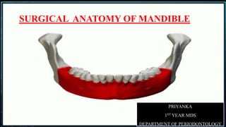
- 1. SURGICALANATOMY OF MANDIBLE SURGICAL ANATOMY OF MANDIBLE
- 2. CONTENTS : Introduction Anatomy of mandible -parts of mandible -body of mandible FEATURES OF EXTERNAL SURFACE OF BODY FEATURES OF INTERNAL SURFACE OF BODY Growth & development of mandible OSSIFICATION Sex & age determination Attachments & relations Muscles controlling mandible Nerve supply of mandible Blood supply of mandible Lymphatic’s Anatomical spaces Clinical and antomical consideration in mandible for surgical procedure. Surgical consideration in implant. Conclusion. Reference.
- 3. INTRODUCTION : Mandible is the bone of lower jaw. The word mandible derived from Latin word mandibula-"jawbone" or inferior maxillary bone. Only movable bone in the skull. It provides structural and protective support for the oral cavity. The mandible is articulated in ball and socket fashion at the condylar process. Strength resides in its dense cortical plates
- 4. SURGICAL ANATOMY knowledge of anatomical facts which have local significance in relation to surgical therapy Primary emphasis is placed on an understanding and awareness of important structures that may be encountered during surgery or which place limits on the nature of the planned surgery, rather than on a detailed and precise knowledge of systematic anatomy.
- 5. ANATOMY MANDIBLE : PARTS OF MANDIBLE BODY OF THE MANDIBLE RAMI BORDERS EXETERN AL INTERNAL SURFACES UPPER LOWER
- 6. Symphysis Menti: faint median ridge on external surface of body. Mental Protuberance-symphysis menti expands below into a triangular elevation Mental Tubercles- the base limit of point of chin on each side Mental foramen: below premolar teeth; provides passage to mental nerve and vessels External Oblique line: continuation of anterior border of ramus, runs downwards and forwards towards mental tubercle. Incisive fossa: shallow depression just below incisor teeth. FEATURES OF EXTERNAL SURFACE OF BODY
- 8. Genial tubercles-inner aspect of symphysis menti possesses four tubercles Mylohyoid line-prominent oblique ridge that runs obliquely downwards and forwards from behind the 3rd molar tooth to the symphysis menti below the genial tubercles Submandibular fossa-slightly hollowed out area below the posterior part of the mylohyoid line and lodges submandibular gland Sublingual fossa-shallow area above the anterior part of the mylohyoid line and lodges sublingual gland. Mylohyoid groove-below the posterior end of the mylohyoid line Digastric fossa- Inferior border or base of the mandible presents a small depression
- 10. Body ;Superior & Inferior borders • The upper border, the alveolar part which lodges the teeth. The lower border ,the base extends posteriolaterally from the symphysis into that of ramus behind the third molar.
- 11. RAMUS OF MANDIBLE Is a quadrilateral vertical plate of bone that projects upwards from posterior part of body. FEATURES: 2 surfaces: lateral and medial 4 borders: Superior, inferior, anterior and posterior 2 processes: Coronoid and condylar processes
- 12. RAMUS :Lateral surface/external surface: Flat and bears a number of oblique ridges
- 13. RAMUS : Medial surface/Internal surface Mandibular foramen Lingula Mylohyoid groove
- 14. CORONOID PROCESS
- 17. • 4th week of intra-uterine life,a prominent bulge appears on the ventral aspect of the embryo • Below the bulge a shallow depression-----the primitive mouth --------- stomodeum. • The floor of the stomodeum is formed by the buccopharyngeal membrane that separates the stomodeum from the foregut PRENATAL
- 26. INTRAMEMBRANOUS BONE FORMATION The first structure to develop in the primodium of the lower jaw is the mandibular division of trigeminal nerve that precedes the mesenchymal condensation forming the first [mandibular] arch. At around 36 – -38 days of intrauterine life there is ectomesenchymal condensation Some mesenchymal cells enlarges , acquire a basophilic cytoplasm and form osteoblasts These osteoblasts secrete a gelatinous matrix called osteoid and result in ossification of an osteogenic membrane.
- 27. The resulting intramembranous bone lies lateral to meckel’s cartilage of first [mandibular ] arch. In the 6th week of the intrauterine life a single ossification centre for each half of the mandible arises in the bifurcation of inferior alveolar nerve into mental and incisive branches
- 28. During 7th week of intrauterine life bone begin to develop lateral to meckel’s cartilage & continues until the posterior aspect is covered with bone. Between 8th & 12th week of intrauterine life mandibular growth accelerate , as a result mandibular length increases Ossification stops at a piont , which later become mandibular lingula, the remaining part of meckels cartilage continues to form sphenomandibular ligament & spinous process of sphenoid. Secondary accessory cartilage appear between 10th & 14th week of intrauterine life to form head of condyle , part of coronoid process & mental protuberance.
- 30. ENDROCHONDRAL BONE FORMATION Endrocondral bone formation is seen in 3 areas of mandible 1)The condylar process 2) The coronoid process 3) The mental process THE CONDYLAR PROCESS At 5th week of intrauterine life , an area of mesenchymal condensation is seen above the ventral part of developing mandible At about 10th week it develops in cone shaped cartilage. It migrate inferior & fuses with mandi bular ramus at about 4 month
- 31. This cone shaped cartilage is replaced by bone but its upper end persists acting as growth cartilage & articular cartilage
- 32. THE CORONOID PROCESS Secondary accessory cartilage appear in region of coronoid process at about 10- 14 week of intrauterine life. This cartilage become incorporated into expanding intramembranous bone of ramus & dissapear before birth
- 33. THE MENTAL REGION In mental region , on either side of symphysis , one or two small cartilage appear and ossify in seventh week of intrauterine life to become mental ossicles. These ossicles become incorporated into intramembranous bone when symphysis ossify completely
- 34. NEONATAL MANDIBLE Rami - low and wide Coronoid - large and projects above the condyle. Body - open shell containing tooth buds. Mandibular - low in body
- 35. GROWTH OF MANDIBLE In adults developmentally and functionally divided into – • Body • Condyle • Coronoid • Ramus • Angle • Lingual tuberosity • Chin Condylar cartilage Posterior border of rami Alveolar ridge Symphysis – limited. MAJOR GROWTH SITES
- 36. RAMUS The ramus moves progressively posterior by a combination Of deposition and resorption Resorption occur on the anterior part deposition occur on the posterior region POSTERIOR DIRECTION
- 37. CORPUS /BODY OF MANDIBLE Body of mandible lengthens as the ramus exhibits bone deposition on the posterior aspect and resorption on the anterior aspect.
- 38. ANGLE OF MANDIBLE On the lingual side : Resorption takes place on the posterio-inferior aspect. • Deposition occurs on the anterio- superior aspect. On the buccal side: Resorption occurs on the anterio-superior part. Deposition occurs on the posterio-inferior part. Flaring of the angle of the mandible as age advances
- 39. THE LINGUAL TUBEROSITY Grows posterior and medial by deposition Resorptive field below –lingual fossa
- 40. THE ALVEOLAR PROCESS As the teeth erupt the alveolar process develops and increase in height by bone deposition at the margins. the height and thickness of the body of the mandible. absence of teeth -- fails to develop and it resorb
- 41. THE CONDYLE The role of condyle in the growth of mandible has remained controversy. growth of soft tissue including the muscles and connective tissue carries the mandible forward away from the cranial base (cary awray phenomenon)
- 42. THE THE CORONOID PROCESS ONOID PROCESS It follows Enlow”s “V” principle In longitudinal section – from posterior aspect, deposition occurs on the lingual surface of the left and right coronoid process
- 43. THE THE CHIN 1-2 YR – Chin prominence is seen The mental protuberance forms by bone deposition. The change in the contour occur by following two mechanism: 1.The area just above the chin and the base of the alveolar process, is a resorptive. 2. There is forward translation of chin as mandible grows forward
- 46. LIGAMENTS ATTACHED TO MANDIBLE 1. Stylomandibular ligament- attached to angle of mandible 2. Temporomandibular- attached to lateral aspect of neck of mandible 3. Sphenomandibular ligament- attached to lingula 4. Pterygomandibular raphae/ligament- attached behind the last molar tooth to upper end of mylohyoid line.
- 48. MUSCLES CONTROLLING MANDIBULAR MOVEMENTS VERTICAL MOVEMENTS Elevate mandible: • Masseter • Temporalis • Medial pterygoid Depress mandible: • Lateral pteyigoid • Geniohyoid • Ant. Diagastric • Mylohyoid ACCESSORY MUSCLES -GENIOHYOID ANT DIAGASTRIC MYLOHYOID STYLOHYOID MUSCLES OF MASTICATION • MASSETER • TEMPORALIS • MEDIAL PTERYGOID • LATERAL PTERYGOID
- 49. SIDE TO SIDE MOVEMENT Lateral pterygoid Medial pterygoid SAGITTAL MOVEMENTS Protrusion Lat. & medial pterygoid together Retrusion Post. Fibres of temporalis
- 52. SUPRAHYOID MUSCLES : •Importance: facial deformities such as mandibular retrognathia and open bite ,the actions of suprahyoid muscle maybe determinantal to the stability of the surgical repairs, and therefore they may have to be partially released along their inferior mandibular insertions • ( Steinhauser,1973)
- 53. FORAMINAAND RELATION 1) Mental foramina - mental nerve and vessels 2) Mandibular notch - massetric nerve and vessels 3) Medial side of neck - auriculo temporal nerve 4) Mylohyoid groove - mylohyoid nerve and vessels 5) Mylohyoid groove in front of ramus - lingual nerve 6) Mandibular canal and foramina - inferior alveolar nerve and vessels
- 54. TEMPORAMANDIBULAR JOINT This is a synovial joint of condylar variety. ARTICULAR SURFACES: The upper articular surface is formed by the following part of temporal bone: A:articular tubercle B: anterior part of mandibular fossa The inferior articular surface is formed by the head of the mandible. The articular surface is covered with fibro cartilage. The joint cavity is divided into upper and lower parts by an intra – articular disc.
- 56. NERVE SUPPY OF MANDIBLE 1. Lingual nerve- runs on the inner surface of body close to medial side of root of 3rd molar. 2. Inferior alveolar nerve- enters mandibular foramen and pass through mandibular canal. 3. Mylohyoid nerve- runs in the mylohyoid groove. 4. Mental nerve- comes out of mental foramen. 5. Nerve to masseter- runs through mandibular notch. 6. Auriculo temporal- runs to medial side of neck. 7. Marginal mandibular nerve- across the lower border of mandible.
- 58. BLOOD SUPPLY OF THE MANDIBLE Arterial supply • Mainly by Maxillary artery, Branch of external carotid artery ,by its branches, mainly through inferior alveolar artery
- 60. As the external carotid artery ascends the face, it will branch into six arteries: the superior thyroid artery, lingual artery, ascending pharyngeal artery, facial artery, occipital artery, and posterior auricular artery. The external carotid artery will terminate and become the superficial temporal artery and the maxillary artery. The maxillary artery is what branches into the inferior alveolar artery The inferior alveolar artery is a small muscular artery that branches from the first portion of the maxillary artery. The course of the inferior alveolar artery is similar to the inferior alveolar nerve
- 61. LINGUAL ARTERY The lingual artery arises from the anterior surface of the external carotid artery at the level of the hyoid bone between the superior thyroid and facial arteries It first runs upward and medialward to the greater cornua of hyoid bone. It then passes deep to the hyoglossal muscle extending downward and forward to form a characteristic loop Finallly ascending almost perpendicularly to the tongue turns forward on its lower surface as far as the tip under the deep lingual artery .
- 62. • The preparation of a grafting osteotomy in the midline can potentially resect these blood vessels if they fall in the path of the vertical preparation. • If this occurs, the sectioned extension of the lingual artery can prolapse back into the floor of the mouth. • The severed vessel may release arterial blood flow in the sublingual space, potentially raising the tongue to a point that compromises the airway. • Immediate emergency intervention to maintain the airway is critical, and in some cases this requires use of a tracheostomy until the blood flow has been controlled.
- 63. FACIAL ARTERY The facial artery arises from the anterior surface of the external carotid artery slightly above the origin of the lingual artery and has a tortuous route along the nasolabial fold towards the medial canthus of the eye. It moves beneath the digastric and stylohyoid muscles and it will pass through the submandibular gland. The artery will then curve over the body of the mandible (deep to platysma), as the anteroinferior angle of the masseter, will ascend forwards and upwards across the cheek, to the angle of the mouth and along the side of the nose. It terminates near the medial aspect of the eye. In the region of the head, the facial artery runs roughly parallel to the facial vein, although not adjacent to it.
- 64. Venous supply of mandible Drains into •Internal jugular vein • external jugular vein through maxillary vein •Facial vein and pterygoid plexus
- 65. LYMPHATICS Most of the mandible & lower teeth drain into the submandibular group of lymph nodes . Except a small wedge in the symphysis region & the lower incisors which drain into the submental group of lymph nodes. From the submental group the lymph drains to the submandibular group of nodes. Most of the submandibular nodes ultimately drain to the jugulo- omohyoid group of deep cervical lymph nodes. Few extremely posterior submandibular nodes drain to jugulo- digastric group of deep cervical lymph nodes.
- 67. ANATOMICAL SPACE
- 70. SUBMENTAL SPACE. Boundaries Deep or lateral : Anterior belly of digastric Superficial or medial : Investing layer of deep cervical fascia Superior : Mylohyoid muscle Inferior: Investing layer of deep cervical fascia Anterior : Inferior border of mandible Posterior : Hyoid bone
- 71. CLINICAL FEATURES Extraoral findigs Distinct,firm swelling in midline,beneath the chin. Skin overlying the swellig is board like and taut. Fluctuation may be present. Intraoral findings The anterior teet,are eiether nonvital,fractured or carious. The offending tooth may exhibit tenderness to percussionn and may show mobility . The patient may experience considerable discomfort on swallowing.
- 73. SUBLINGUAL SPACE sub mucosal connective tissue of the floor of the mouth. Contains : sublingual gland Wharton's Duct The commonest cause : • dental caries • sialoadenitis • infection tracking via the submandibular duct from the submandibular gland The posterior border of the sublingual space is open and communicate with submandibular space
- 74. Inferiorly: Mylohyoid muscle Laterally : Medial surface of mandible Medially : Hyoglossus,genioglossus & geniohyoid Posteriorly :Submandibular space Laterally and inferiorly : Mylohyoid muscle & lingual side of mandible
- 77. SUBMANDIBULAR SPACE If the apex of the tooth is inferior to the muscle(third molar), the submandibular space is involved
- 78. Anteromedially: mylohyoid muscle Posteromedially : hyoglossus muscle Superolaterally : medial surface of mandible Anterolaterally: anterior belly of digastric Posteromedially : posterior elly of digastric,stylohyoid & stylopharyngeus muscle Superficial : platysma and skin Deep : Mylohyoid,hyoglossus & superior constrictor
- 79. Causes: Infection from mandibular molar Infection from sublingual space Infection from middle third of tongue Posterior part of floor of the mouth From submental space/submental lymph nodes Infection from the submandibular gland. CONTENTS: Superficial portion of submandibular gland. Submental and submandibular lymph nodes. Facial artery and vein Fat and belly of diagastric.
- 81. PTERYGO -MANDIBULAR SPACE Superiorly : lower head of lateral pterygoid muscle Laterally : medial surface of ramus. Medially: medial pterygoid muscle Posteriorly :deep part of parotid Anteriorly :pterygomandibular raphe
- 82. Infection primarily from the third molar or from infected needle track. CONTENTS: Inferior alveolar neurovascular bundle. Lingual Auriculotemporal nerve Mylohyoid nerve and vessels
- 83. PARAPHARYNGEAL SPACE The parapharyngeal space lies at the base of the skull medial to the medial pterygoid muscle and is delineated by fascial membranes. It communicates anteriorly - buccal, sublingual, and pterygomandibular spaces. Medially with the retropharyngeal space. Inferiorly with the spaces of the neck.
- 84. CLINICALAND ANTOMICAL CONSIDERATION IN MANDIBLE FOR SURGICAL PROCEDURE. INFERIOR ALVEOLAR NERVE BLOCK LANDMARKS Coronoid notch Pterygomandibular raphe Occlusal plane of mandibular posteriors
- 85. LANDMARKS Coronoid notch Pterygomandibular raphe Occlusal plane of mandibular posteriors TECHNIQUE: 3 Parameter to consider (1) the height of the injection (2) the anteroposterior placement of the needle (3) the depth of penetration Depoite : IAN :1.5 Lingual nerve:.2
- 86. COMPLICATIONS Hematoma Trismus Facial paralysis Due to L.A penetration to parotid gland capsule Rx:Transient ,self correcting with in 3 hr or less CAUSES: Trauma to muscle Or blood vessels in infratemporal fossa. Contaminated LA large volume of
- 87. BUCCAL NERVE BLOCK Anesthetized: Soft tissue and periosteum buccal to the mandibular molar teeth Insertion: Distal, Buccal of last molar Land mark: Mucobuccal fold Deposit : 0.3mL
- 88. MENTAL NERVE BLOCK Anesthetized :Provides sensory input for the lower lip skin, mucous membrane, pulpal/alveolar tissue for the premolars, canine, and incisors on side blocked AREA OF INJECTION: Mucobuccal fold at or anterior to the mental foramen. This lies between the mandibular premolars Deposite: 0.5-1.ml of L.A
- 89. SURGICAL ANATOMY Location of muscle attachments Thinness /absence of radicular bone Mentalis muscle → Prevent surgeon from ↑ zone of attached gingiva / deepening the vestibule ANTERIOR FACIAL REGION
- 90. The plate of bone overlying the facial and lingual root surfaces of the anterior teeth is usually quite thin. When surgical therapy is required in this area, a technique may be chosen which leaves the bone covered with periosteum and connective tissue to prevent possible postoperative osseous and gingival recession over these roots. A prominent mental tuberosity on occasion may also limit the depth of the vestibule by forming a flat projection in the midline of the mandible. Deepening of the vestibular fornix may not be possible in such a
- 91. As mucogingival problems are very common in the anterior region. The coronal level of the muscle attachment is often approached in attempts to apically position the mucogingival junction. Since it is a thick attachment, its more coronal fibers may be removed to gain the necessary depth for adequate surgical results.
- 92. Large or high Genial tubercle upon which several muscles attach Tubercle could approximate deep osseous defects → Prevent lingual osseous Recontouring during periodontal surgery ANTERIOR LINGUAL REGION
- 93. POSTERIOR FACIAL REGION Periodontal surgery in the mandibular posterior facial region is most often complicated by the presence of a prominent external oblique ridge If the periodontal osseous defects extend below the level of the ridge, osseous recontouring in an attempt to eliminate these defects would require extensive and unwanted removal of large amounts of bone.
- 94. Loose areolar mucosa found attached by a narrow ring of gingiva – distal surface of last Molar
- 95. Surgical correction of distal defects in these areas which attempt to widen the band of attached tissue are hampered by the vertical bony prominence of the ramus.
- 96. Buccinator muscle forms medial wall of buccal space If perforated while elevating a Buccal flap → Buccal space entered → Infection Buccal space ↔ Parapharyngeal space → Spread into other spaces ( Head & Neck ) Thin attachment BM Limit extension of vestibule
- 97. An additional operative hazard exists in the molar region where the facial artery passes under the inferior border of the mandible. The vessel normally continues its course deep within the cheek and is not disturbed while elevating mucosal flaps from the mandible.
- 98. . MENTAL FORAMEN
- 99. POSTERIOR LINGUAL REGION An unusually wide mylohyoid ridge or a lingual mandibular torus offer the same complications in osseous surgery as previously mentioned concerning the oblique ridge on the facial surface.
- 100. The while performing surgery on the superficial structures which lie just under the thin mucosa which forms the floor of the mouth. The lingual nerve is most easily damaged as it lies very close to the mucosal surface in the region of the second and third molars. Major precaution --lingual aspect of the mandible is to avoid incising
- 101. Whenever the attached gingiva is elevated from the lingual aspect of a mandible, or when the mucosal lining of the floor of the mouth is perforated, the sublingual space . Submandibular gland and duct are less likely to be violated because of their deeper position. mylohyoid muscle attachment would result in opening into the submandibular
- 102. With wide zones of keratinized gingiva present or a short mandible, a sulcular incision may be used. A periosteal elevator is used to reflect the mucoperiosteal flap toward the base of the mandible to the level of the “pogonion” (most anterior point of mandible), thus ensuring that the inferior border of the mandible remains intact. This will leave the most facial aspect of the periosteal attachment intact and prevent “ptosis” of the chin by avoiding “degloving” of the mandible.
- 103. MANDIBULAR FRACTURE INCIDENCE OF MANDIBULAR FRACTURES •Body fractures 33.6% •Subcondylar fracture 33.4% •Fractures at the angle 17.4% •Alveolar fractures 6.7% •Ramus fractures 5.4% •Midline fractures 2.9% •Fracture of coronoid process 1.3% Oikarinen & Malmstrom 1969
- 104. TYPES OF FRACTURE Simple Greenstick fracture (rare, exclusively in children) Fracture with no displacement (Linear) Fracture with minimal displacement Displaced fracture Comminuted fracture Extensive breakage with possible bone and soft tissue loss Compound fracture Severe and tooth bearing area fractures Pathological fracture (osteomyelities, neoplasm and generalized skeletal disease)
- 106. Ludwig’s angina: Perimandibular spaces are bilaterally involved in an infection. Rapidly spreading cellulitis that can obstruct the airway and it spreads posteriorly to the deep fascial spaces of the neck. Patients usually has sever indurated swelling with :Tongue elevation Trismus Drooling Difficulty in swallowing and breathing Rx: I&D Antibiotics
- 113. SIGNIFICANCE OF LINGUAL NERVE DURING PERIODONTAL/IMPLANT SURGERY • The lingual nerve is a branch of the mandibular nerve. This nerve provides sensory innervation to the mucous membranes of the anterior two thirds of the tongue and lingual tissues. • The proximity of this nerve to the mandibular third molar region is a concern when performing flap surgery in this area. • On average, it is located 3 mm apical to the osseous crest and 2 mm horizontally from the lingual cortical plate in the third molar area. BEHNIA H et al 2005
- 114. • To reduce the chance of injuring this nerve, some procedures have been advocated.Incisions distal to the third molar should be made on the buccal aspect of the ridge and always on the bone. The elevator should be used to protect the nerve in the flap, and the tissue should be managed gently.
- 115. AGE DETERMINATION OF MANDIBLE Mostly determined by 1) Eruption of temporary & permanent teeth 2) Condition of socket of teeth
- 116. 1) Mental foramena Near to lower border midway Near upper border 2)Angle of mandible Obtuse ( near 180 ) about Right angle(110 -120) Obtuse (140) 3) Coronoid & condyloid process Coronoid is larger & above condyloid Condyloid is above coronoid Condyloid is above coronoid 4)Mandibular canal Above mylohyoid line Parallel to mylohyoid line Run close to upper border 5)Symphysis menti Present & 2 halves are united by fibrous band As faint ridge only on upper part Not recognizable or absent
- 122. SURGICAL CONSIDERATION FOR IMPLANT Implants can strengthen the jawline and create a more balanced facial structure by augmenting the mandibular body, angle, and ramus
- 124. MANDIBULAR ANTERIOR REGION •Minimum of 7 mm from inferior border of mandible to the crestal ridge is needed •In resorbed ridge mental foramina located on top of the ridge;care is necessary to prevent damage to it and possible paresthesia. •This region between mental foramina has adequate bone for 4-6 implants. MYLOHYOID RIDGE Careful palpation – a concavity below the mylohyoid ridge. Implant placed in the posterior mandible are at risk of entering this region,which is highly vascularized ,with resultant risk of haemorrhage.
- 125. APPLIED ANATOMY -implant installation planning should be done on three-dimensional edentulous jaw segment (EJS) pattern -EJS consists of alveolar and basal bone -EJS describes planned implant bed relation to present anatomical borders such as mandibular vital structures • The vertical dimension of the planned implant site in mandible is determined by the distance between crestal ridge of the alveolar process and mandibular vital structures (EJS height) -mental foramen (MF) , -mandibular canal (MC), -mandibular incisive canal (MIC), -anterior loop of mental nerve (AL) • horizontal dimensions are determined by the
- 126. Mental Foramen(MF) -round/oval -2.5mm-5.5mm diameter -location I LPM –caucasians II LPM- Mongoloids - Vertcal positio irt premolars I PM- 38.6% coronal 15.4% apex 46% below apex II PM – 24.5% coronal 13.9% apex 61.6% below apex
- 127. AGE CHANGES IN LOCATION OF MANDIBULAR FORAMEN • For greater accuracy in anesthetic procedures, dentists should relate the locational changes in the mandibular foramen with age when performing block anesthesia for the inferior alveolar nerve • 3 yrs …….. 4.12 mm below (mandibular occlusal plane) • 9 yrs …….. At level • Adult ……. 4.16 mm above • Old age ….. Further ABOVE
- 128. Mandibular zone of safety( Misch 1980) - An area with in the bone that can safely support implants without fear of impingement on the mandibular neuro vascular bundle
- 129. ANOMALIES OF DEVELOPMENT CONGENITAL • Agnathia • micrognathia • Macrognathia • facial hemihypertrophy • Facial hemiatropy DEVELOPMENTAL • Infantile cortical hyperostosis • Achondroplasia • Torus mandibularis • Stafne’s cyst • Odontogenic cyst
- 132. Some of the syndromes associated with mandibular abnorma • Down’s syndrome •Marfan syndrome •turner syndrome •Klinfelter syndrome •Pierre-robin syndrome •Treacher- collins syndrome
- 134. PATHOLOGY OF JAW ODONTOGENIC CYSTS Inflammatory. Radicular cysts Developmental: Dentigerous cysts Lateral periodontal cysts Odontogenic keratocyst Radicular cysts OKC
- 135. OSTEORADIONECROSIS (ORN) – INJURY TO BONE Osteoradionecrosis (ORN) is a condition of nonvital bone in a site of radiation injury. The absence of reserve reparative capacity is a result of the prior radiation injury. trauma such as denture- related injury, ulcers, or tooth extraction GRADE I:Bone exposure respond to HBO GRADE II: Bone exposure not respond to HBO,need sequestromy/ saucerisation. GRADE III: pathological fracture
- 136. CONCLUSION The mandibular movement is considered as the chewing apparatus of masticatory system. All the events taking place during development of mandible play an important role in determining the final structure of mandible, any deviation of which can give rise to various abnormalties in oro facial region. Finally, three things we should keep in mind: (a) constantly revise your knowledge of anatomy; (b) constantly think of the local anatomy in relation to injury and disease; and (c) never take a knife in your hand without picturing in your mind's eye the structures in and adjacent to your operative field, however small that field may be .
- 137. REFERENCES • -Gray’s anatomy ; williams ;37th edition • Human anatomy ; Chaurasia B.D. 3RD Edition • Human embryology; Inderbir singh ;7th edition • Killey’s fracture of mandible ;Peter bank: 4th edition • Anatomical considerations in periodontal surgery by micheal a et al vol 42 jop 1991 • SURGICAL ANATOMY OF THE JAWS • Lecture delivered at the Royal College of Surgeons of England on 28th May 1963 by Ian H. Heslop, M.B., B.S., B.D.S., F.D.S.R.C.S.
- 138. • Vishram Singh Textbook of Anatomy -Head, Neck, and Brain- 2nd edition • -Juodzbalys G, Wang HL. Guidelines for the Identification of the Mandibular Vital Structures: Practical Clinical Applications of Anatomy and Radiological Examination Methods. J Oral Maxillofac Res 2010 (Apr-Jun);1(2):e1 doi:10.5037/jomr.2010.1201 • --Greenstein G, Cavallaro J, Tarnow D. Practical application of anatomy for the dental implant surgeon. J Periodontol. 2008;79(10):1833-1846. doi:10.1902/jop.2008.080086 • John Nguyen; Hieu Duong. Anatomy, Head and Neck, Inferior Alveolar Arteries
- 139. THANK YOU