
Recommended
More Related Content
Similar to 5.TISSUES.pdf
Similar to 5.TISSUES.pdf (20)
Recently uploaded
Recently uploaded (20)
5.TISSUES.pdf
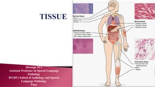
- 1. Shrunga M S Assistant Professor In Speech Language Pathology BV(DU) School of Audiology and Speech Language Pathology Pune
- 2. A group of cells having similar origin, structure and function is called as tissue
- 4. It is the layer of cells which covers the external surface (skin) or the lines the internal surface of gastrointestinal, respiratory and urogenital tract
- 5. Functions 1. Protection 2. Transport 3. Secretion 4. Excretion 5. Absorption 6. Lubrication 7. Sensory
- 7. SIMPLE SQUAMOUS SIMPLE CUBOIDAL SIMPLE COLUMNAR
- 8. Irregular flat cells with the height less than the width Distribution: 1. Alveoli of the lungs 2. Bowman’s capsule and loop of Henle of kidney 3. Mesothelium lining the peritoneum, pleura and pericardial cavities 4. Endothelial cells lining blood vessels
- 9. Height and width are nearly equal and nuclei are central in position Distribution: 1. Thyroid follicle 2. Ducts of many glands 3. Surface of ovary
- 10. Height of the cells is greater than the width. Nuclei are elongated and placed towards the base Distribution: 1. Small bronchi and bronchioles, uterine tube 2. Ependyma lining the cavities of the brain. Efferent ductules of the testes
- 12. Some cells are shorter and do not reach the lumen, while tall cells reach the lumen The nuclei of the cells therefore lie at different levels This gives the impression of stratification(false stratification)
- 14. Distribution: 1. Male urethra(membranous and penile part) 2. Auditory Tube 3. Vas deferens Pseudostratified Ciliated • Distribution: Upper part of the respiratory tract(trachea and larger bronchi)
- 16. Epithelium is made up of many layers of cells Basal cells resting on the basement membrane are columnar or low cuboidal Superficial cells are squamous and flat, hence called stratified squamous epithelium Distribution: Epithelium lining mouth, pharynx and esophagus, anal canal etc
- 17. The superficial layer consists of non- living cells with keratin in their cytoplasm They are rough and water resistant Distribution : Epidermis of the skin
- 18. Stratified Cuboidal Epithelium It consists of few layers of cuboidal cells Distribution: Ducts of sweat glands Stratified Columnar Epithelium It consists of two or more layers of cells Basal cells are polyhedral Superficial cells are columnar Distribution Male urethra
- 19. This is stratified epithelium with three to four layers of cells The deepest cells are columnar or cuboidal The middle layer is made up if polyhedral or pear shaped cells The cells of the surface are large and are shaped like an umbrella Distribution: Renal pelvis Ureter Urinary bladder Urethra(proximal part)
- 20. The epithelial cells are specialised to perform secretary function Such epithelial cells constitutes glands
- 23. Connective tissues has formed elements (fibres and cells) and amorphous substances This tissue bind various other tissues of the body CELLS FIBRES GROUND SUBSTANCE
- 25. CELLS: 1. Fibroblast: they help in healing of wounds 2. Macrophages: they are phagocytic in function 3. Plasma cells: they produce antibodies 4. Mast cells: they secrets anticoagulants 5. Pigment cells: they are present in iris of the eye 6. Reticular cells: they are phagocytic in nature 7. Fat cells(Adipocytes): they are contain lots of fat
- 26. Fibres: Collagen: They appear white and called as white fibres It contain a protein called as collagen Elastic They appear yellow It can be stretched and are made up of protein called elastin Reticular They are similar to collagen fibers They form the skeletal framework of the lymphatic organs
- 28. CONNECTIVE TISSUES AREOLAR TISSUE ADIPOSE TISSUE MYXOMATOUS TISSUE CARTILAGE BONE LOOSE DENSE
- 29. AEROLAR TISSUE It is the most common connective tissue where collagen and elastic fibres are loosely arranged The ground substance is semifluid in nature They have plenty of fibroblasts and macrophages They are traversed by nerves and vessels
- 30. Distribution: Subcutaneous tissue Submucous coat in gastro-intestinal tract Between muscles, vessels and nerves Spaces between the organs Inside the organ between lobes and lobules
- 31. ADIPOSE TISSUE: This is an aggregation of fat cells It serves as insulating material to conserve body heat, as storage depot for food, as a protective pad around organs It is abundant in females(under the hormone influence) Distribution: Subcutaneous tissue Bone marrow and bone orbit
- 32. Myxomatous tissue The matrix consists of mucoid substances with few collagen fibres It also shows star shaped fibroblasts Present in the umbilical cord and vitreous body of the eye
- 33. Cartilage is a specialised dense connective tissue General Features: They are rigid, provide protection and support the organs They are present in the body where elasticity and rigidity is required They are avascular structures
- 37. Bones are highly vascular living connective tissue in which matrix is calcified by deposition of calcium phosphate Functions of bone: Supporting framework and shape for the body It protects vital organ Transmission of the body weight Attaches to the muscle and act as levers of the joints helping in locomotion It is the store house of calcium salts
- 38. POSITION SHAPE GROSS STRUCTURE DEVELOPMEN T • AXIAL • APPENDICULAR • LONG BONES • SHORT BONES • FLAT BONES • IRREGULAR BONES • COMPACT • SPONGY • DIPLOIC • MEMBRANEOUS • CARTILAGINOU S