Parsing the Practicalities of Pathologic Response Assessment After Neoadjuvant Immunotherapy to Facilitate Progress in Early-Stage Cancers
•
0 likes•76 views

Chair & Moderator, Prof. Solange Peters, MD, PhD, Mark M. Awad, MD, PhD, and Jonathan D. Spicer, MD, PhD, FRCSC, prepared useful Practice Aids pertaining to Cancer Immunotherapy for this CME/MOC/CC activity titled “Parsing the Practicalities of Pathologic Response Assessment After Neoadjuvant Immunotherapy to Facilitate Progress in Early-Stage Cancers.” For the full presentation, downloadable Practice Aids, and complete CME/MOC/CC information, and to apply for credit, please visit us at https://bit.ly/3uRHyjk. CME/MOC/CC credit will be available until May 9, 2023.

Recommended
Recommended
More Related Content
Similar to Parsing the Practicalities of Pathologic Response Assessment After Neoadjuvant Immunotherapy to Facilitate Progress in Early-Stage Cancers
Similar to Parsing the Practicalities of Pathologic Response Assessment After Neoadjuvant Immunotherapy to Facilitate Progress in Early-Stage Cancers (20)
More from PVI, PeerView Institute for Medical Education
More from PVI, PeerView Institute for Medical Education (20)
Recently uploaded
Recently uploaded (20)
Parsing the Practicalities of Pathologic Response Assessment After Neoadjuvant Immunotherapy to Facilitate Progress in Early-Stage Cancers
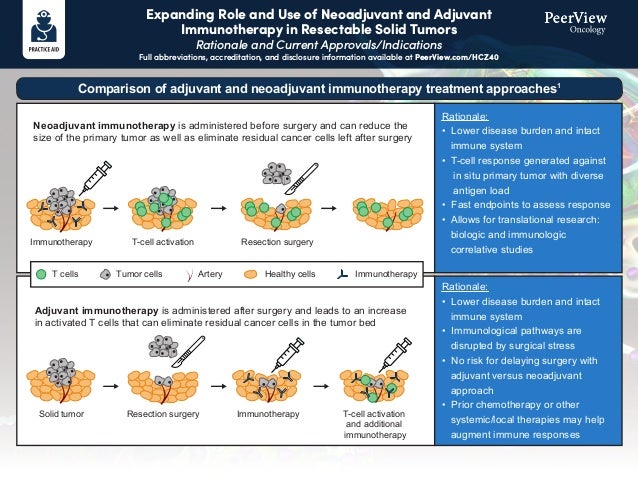
- 1. Expanding Role and Use of Neoadjuvant and Adjuvant Immunotherapy in Resectable Solid Tumors Rationale and Current Approvals/Indications Full abbreviations, accreditation, and disclosure information available at PeerView.com/HCZ40 Neoadjuvant immunotherapy is administered before surgery and can reduce the size of the primary tumor as well as eliminate residual cancer cells left after surgery Rationale: • Lower disease burden and intact immune system • T-cell response generated against in situ primary tumor with diverse antigen load • Fast endpoints to assess response • Allows for translational research: biologic and immunologic correlative studies Rationale: • Lower disease burden and intact immune system • Immunological pathways are disrupted by surgical stress • No risk for delaying surgery with adjuvant versus neoadjuvant approach • Prior chemotherapy or other systemic/local therapies may help augment immune responses Adjuvant immunotherapy is administered after surgery and leads to an increase in activated T cells that can eliminate residual cancer cells in the tumor bed Comparison of adjuvant and neoadjuvant immunotherapy treatment approaches1 Immunotherapy T cells T-cell activation Resection surgery Solid tumor Resection surgery Immunotherapy T-cell activation and additional immunotherapy Tumor cells Artery Healthy cells Immunotherapy
- 2. Expanding Role and Use of Neoadjuvant and Adjuvant Immunotherapy in Resectable Solid Tumors Rationale and Current Approvals/Indications Full abbreviations, accreditation, and disclosure information available at PeerView.com/HCZ40 Overall survival (OS) is the gold-standard outcome measure for phase 3 trials, but the protracted length of these clinical trials in resectable cancers makes this research daunting and expensive One strategy to expedite clinical trials, including those assessing immunotherapies in early-stage cancer settings, is the use of newer, innovative surrogate measurements for endpoints Because pathologic response reflects a therapy’s ability to eradicate tumor cells more directly than radiographic evaluation using RECIST criteria, it may better correlate with clinically meaningful outcomes Several trials in different tumor types have correlated the novel endpoints with more traditional endpoints such as OS, DFS, and RFS, but additional confirmatory studies are needed Many adjuvant approvals have been based on disease-free survival (DFS) Trials in neoadjuvant settings provide an opportunity to assess pathologic response as an early surrogate marker for survival outcomes, and pathologic response criteria such as major pathological response (MPR) and pathologic complete response (pCR) have been assessed in neoadjuvant immunotherapy trials; there are various definitions, but generally: MPR: ≤10% of viable tumor in the treated tumor bed pCR: complete absence of viable tumor in the treated tumor bed Recently, immune-related pathologic response criteria (irPRC) have also been developed with the aim of assessing the full spectrum of response to immunotherapy in resection specimens Different scoring systems exist or are in development for evaluating pathologic response in various tumor types (eg, melanoma, lung cancer, bladder cancer) Relevant endpoints for neoadjuvant and adjuvant immunotherapy clinical trials2-8
- 3. Expanding Role and Use of Neoadjuvant and Adjuvant Immunotherapy in Resectable Solid Tumors Rationale and Current Approvals/Indications Full abbreviations, accreditation, and disclosure information available at PeerView.com/HCZ40 Adjuvant treatment of patients with urothelial carcinoma who are at high risk of recurrence after undergoing radical resection Adjuvant treatment of patients with completely resected esophageal or gastroesophageal junction cancer with residual pathologic disease who have received neoadjuvant chemoradiotherapy Adjuvant treatment of patients with melanoma with lymph node involvement or metastatic disease who have undergone complete resection Current perioperative approvals and indications of immunotherapies in solid tumors9 Nivolumab Adjuvant treatment of patients with cutaneous melanoma with pathologic involvement of regional lymph nodes of more than 1 mm who have undergone complete resection, including total lymphadenectomy Ipilimumab Newest Represents the first FDA approval for neoadjuvant therapy for early-stage NSCLC Neoadjuvant treatment of patients with resectable non–small cell lung cancer with platinum-doublet chemotherapy
- 4. Expanding Role and Use of Neoadjuvant and Adjuvant Immunotherapy in Resectable Solid Tumors Rationale and Current Approvals/Indications Full abbreviations, accreditation, and disclosure information available at PeerView.com/HCZ40 Neoadjuvant treatment of high-risk, early-stage triple-negative breast cancer with chemotherapy adjuvant treatment after surgery as a single agent Adjuvant treatment of patients with renal cell carcinoma at intermediate-to-high or high risk of recurrence following nephrectomy, or following nephrectomy and resection of metastatic lesions Adjuvant treatment of patients with stage IIB, IIC, or III melanoma following complete resection Patients with BCG-unresponsive, high-risk, non–muscle invasive bladder cancer with carcinoma in situ with or without papillary tumors who are ineligible for or have elected not to undergo cystectomy Current perioperative approvals and indications of immunotherapies in solid tumors9 Pembrolizumab Adjuvant treatment following resection and platinum-based chemotherapy in patients with stage II to IIIA non–small cell lung cancer whose tumors have PD-L1 expression on ≥1% of tumor cells Atezolizumab 1. Krishnamoorthy M et al. J Natl Cancer Inst. 2021;113:823-832. 2. Topalian SL et al. Science 2020;367:eaax0182. 3. Benitez JC et al. Clin Cancer Res. 2020;26:5068-5077. 4. Bilusic M, Gulley JL. J Natl Cancer Inst. 2021;113:799-800. 5. Krishnamoorthy M et al. J Natl Cancer Inst. 2021;113:823- 832. 6. O’Donnell JS et al. Clin Cancer Res. 2019;25:5743-5751. 7. Hellmann MD et al. Lancet Oncol. 2014;15:e42-50. 8. Cottrell TR et al. Ann Oncol. 2018;29:1853-1860. 9. https://www.fda.gov/drugs/resources-information-approved-drugs/oncology-cancer-hematologic-malignancies- approval-notifications.
- 5. IASLC Multidisciplinary Recommendations for Pathologic Assessment of Lung Cancer Specimens Following Neoadjuvant Therapy1 Full abbreviations, accreditation, and disclosure information available at PeerView.com/HCZ40 Recommendation 1 The term “tumor bed” is the area where the original pretreatment tumor was considered to be located. It can be challenging to determine whether necrosis and stromal inflammation and/or fibrosis are due to regression secondary to neoadjuvant therapy, native tumor characteristics, or a combination. For this reason we favor the term tumor bed. It is suggested to simply describe the major components of the tumor bed as (1) viable tumor, (2) necrosis, or (3) stroma (which can include inflammation or fibrosis). See page 3 for an example. Recommendation 2 It is essential that information be provided from the surgical team to the pathology laboratory on whether the patient received neoadjuvant therapy in order for this specimen processing protocol to be followed. If there is more than one tumor in the specimen, it is of critical importance to also provide this information. It is good clinical practice to correctly label the specimen with the lobe(s) resected and to clarify any issues that may be needed for pathologic staging such as the pericardium, diaphragm, or chest wall. Recommendation 3 Lung cancer resection specimens following neoadjuvant therapy should be sampled to optimize comprehensive gross and histologic assessment of the lung tumor bed for pathologic response. The tumor should be cut in its greatest dimension to maximize the tumor bed cross section. In cases where identification and/or orientation of the tumor are difficult, review of the preoperative CT scan can be helpful. Tumors 3 cm or less in size should be completely sampled. For larger tumors greater than 3 cm, the tumor should be cut across in serial sections 0.5 cm thick, and after gross inspection, the most representative cross section showing viable tumor should be sampled. At least one cross section of the entire tumor (0.5 cm thick) with a gross photograph and histologic mapping should be made. Histologic sections at the tumor periphery should include 1 cm of adjacent lung parenchyma. Pathologic response cannot be assessed in small biopsies; a resection specimen is required. Recommendation 4 To determine the border of the tumor bed, the edge of the tumor needs to be distinguished from the surrounding non-neoplastic lung parenchyma. This can be facilitated by review of the gross specimen and the histologic slides from the periphery of the tumor bed. Recommendation 5 Determination of the pathologic response to therapy should be made after review of all H&E slides of tumor by estimating the percentages of (1) viable tumor, (2) necrosis, and (3) stroma, which includes both fibrosis and inflammation, so each of these three components add up to 100%. Each component should be assessed in 10% increments unless the amount is below 5% when an estimate of single percentages should be recorded. While this is primarily done by review of histologic sections of the tumor bed, correlation with the gross findings, in some cases facilitated by a gross photograph, may be important in markedly necrotic and/or cavitated tumors where it is not possible to reflect this change in histologic sections. Note: Although it may be useful to record the amount of each of these components on each individual histologic slide, it needs to be kept in mind that the amount of tumor bed varies on each slide, so these percentages cannot be summed and averaged as if they were in equal amounts. This is a semiquantitative process. There is no validated quantitative method that is available that can be implemented in a timely fashion for clinical decisions. A proposed synoptic template for reporting pathologic findings for resected lung cancers following neoadjuvant therapy is summarized on page 2.
- 6. IASLC Multidisciplinary Recommendations for Pathologic Assessment of Lung Cancer Specimens Following Neoadjuvant Therapy1 Full abbreviations, accreditation, and disclosure information available at PeerView.com/HCZ40 Recommended Synoptic Template for Recording Lung Cancers Following Neoadjuvant Therapy Primary tumor Type of neoadjuvant therapy • No known presurgical therapy: _____ • Type of neoadjuvant therapy – Chemotherapy: ____________________ – Radiotherapy: ____________________ – Immunotherapy (please specify): ____________________ – TKI (please specify): ____________________ – Other (please specify): ____________________ Treatment effect in primary tumor • Percentage of viable tumor (record in 10% increments except below 10%; then record single digits between 1%-5%): _____ • No residual viable tumor identified: _____ • Percentage of necrosis: _____ • Percentage of stroma (includes fibrosis and inflammation): _____ Grade of inflammation (choose the appropriate grade) _____ Mild _____ Moderate _____ Marked Method (choose what was used for evaluation) _____ Correlation was made with a gross photograph of tumor cut surface: Yes _____ No _____ _____ Evaluation was aided by use of tumor mapping to match a gross photograph to histologic sections: Yes _____ No _____ _____ Evaluation was aided by radiologic pathologic correlation: Yes _____ No _____ Treatment effect in lymph node metastases • Total number of lymph node stations examined _____ • Total number of lymph nodes examined: _____ • No carcinoma present: _____ • Total number of lymph nodes with metastatic carcinoma: _____ • Lymph node stations involved by tumor with treatment-related changes: _____ • Lymph node stations with treatment-related changes without viable tumor: _____ • Largest tumor focus: mm at station number: _____ • Extracapsular extension present: _____ • No extracapsular extension: ____________________ Comments: ___________________________________________________________________________________________ _____________________________________________________________________________________________________
- 7. IASLC Multidisciplinary Recommendations for Pathologic Assessment of Lung Cancer Specimens Following Neoadjuvant Therapy1 Full abbreviations, accreditation, and disclosure information available at PeerView.com/HCZ40 Histologic Components of the Tumor Bed (A) Schematic image showing how percentage compositions are assigned. The tumor bed is divided into viable tumor area, necrosis, and stroma. Stroma includes inflammation and fibrosis. (B) A representative hematoxylin-and-eosin stained slide image (left) and a corresponding color illustration of the distribution of the components (right). The blue, red, and black areas represent viable tumor, necrosis, and stroma, respectively. Tumor bed = X + Y + Z = 100% Viable tumor (X%) Necrosis (Y%) Stroma (Z%) Viable tumor = 50% Necrosis = 40% Stroma = 10% A B C Tumor bed
- 8. IASLC Multidisciplinary Recommendations for Pathologic Assessment of Lung Cancer Specimens Following Neoadjuvant Therapy1 Full abbreviations, accreditation, and disclosure information available at PeerView.com/HCZ40 Recommendation 6 Definition of major pathologic response (MPR) MPR is defined as the reduction of viable tumor to the amount beneath an established clinically significant cutoff based on prior evidence according to the individual histologic type of lung cancer and a specific therapy. The historical definition of MPR for all histologic types of lung cancer is ≤ 10% of viable tumor with no viable tumor required for pathologic complete response (pCR). MPR is calculated as the estimated size of viable tumor divided by the size of the tumor bed. For the moment, this is the cutoff being used in multiple active clinical trials. However, recent data suggests the MPR in the conventional chemotherapy setting may differ according to histologic type: ie, adenocarcinoma versus squamous cell carcinoma. If after review of histologic sections, the percentage of viable tumor is near the cutoff for major pathologic response, additional histologic sections should be submitted. The pathology report should record the total number of blocks of tumor bed that were examined even if the blocks did not consist entirely of tumor bed but also included some uninvolved lung. For colloid adenocarcinomas, where tumor cells are only focal, the mucin pools should be included in the percentage of viable tumor. However, if there are only areas of extracellular mucin without any apparent viable tumor cells within the mucin, we suggest regarding this as stroma. Further study is needed to address this point. Major pathologic response can also be classified for the lung primary in the setting where the lung primary shows little or no viable tumor, but lymph nodes show viable metastatic carcinoma (ypT0, N1, 2 or 3). However, the prognostic and therapeutic implications of this clinical setting are not known. Recommendation 7 Definition of pathologic complete response (pCR) pCR is defined as lack of any viable tumor cells on review of H&E slides after complete evaluation of a resected lung cancer specimen including all sampled regional lymph nodes. Such tumors would be staged as ypT0N0 according to the 8th edition AJCC and UICC staging systems. Note: If no tumor is seen in the initial sections and tissue from the tumor bed remains, additional histologic sections should be made. The number of additional sections should be whatever seems reasonable in the individual setting depending on the size of the tumor bed and the capacity of the individual pathology laboratory. If the histologic changes in the initial sections obtained do not show findings that fit for the effects of therapy, the possibility that the wrong area was sampled should be considered. In such cases the gross specimen may need to be re-evaluated using radiologic pathologic correlation and if additional lesions are identified, these should be sampled. The pathology report should record the total number of blocks of tumor bed that were examined even if the entire block did not consist of tumor bed. The identification of incidental lesions of squamous cell carcinoma in situ, atypical adenomatous hyperplasia, adenocarcinoma in situ, or minimally invasive adenocarcinoma in the surrounding lung parenchyma that are clearly separate from the main tumor for which neoadjuvant therapy was administered does not disqualify a case for classification as MPR or CPR. This proposal is based on clinical judgement, as currently no clinical data exist to make a specific recommendation. In the setting of multiple tumors where a second invasive predominant lung carcinoma is present that was regarded preoperatively to be an intrapulmonary metastasis but is determined to be a second synchronous primary after clinical, radiologic, pathologic, and/or molecular assessment, it is unclear whether the terms MPR or CPR should be used if the main tumor otherwise meets the above criteria. No data exists currently to address this question.
- 9. IASLC Multidisciplinary Recommendations for Pathologic Assessment of Lung Cancer Specimens Following Neoadjuvant Therapy1 Full abbreviations, accreditation, and disclosure information available at PeerView.com/HCZ40 Recommendation 8 In the absence of more systemic data regarding evaluation of tumors following immunotherapy and molecular targeted therapy, the same approach to pathologic assessment of resected lung cancers in the neoadjuvant setting in evaluating the percentage of viable tumor, necrosis, and stroma should be used regardless of the type of neoadjuvant therapy administered whether it was radiation, chemotherapy, targeted therapy, immunotherapy, chemoradiation, chemoimmunotherapy, or chemotherapy-targeted therapy. There also may be different features that can be addressed depending on the type of therapy such as immune cell infiltrates in patients who received immunotherapy. Recommendation 9 In most cases, the lymph nodes are small enough to completely sample, but if there is a very large metastasis or tumor bed (>2 cm), the lymph node can be bisected and the central slice through the tumor can be submitted in designated cassettes. This should also be done during grossing of intraoperative frozen sections of lymph nodes. Depending on individual laboratory resources, more extensive or even complete sampling can also be done. Then the same approach can be used for histologic evaluation that is used for the resected lung cancer reporting percent viable tumor, necrosis, and stroma. Complete pathologic response in a lymph node can be recognized if there is a well-defined scar and/or area of tumor necrosis in the absence of identifiable viable tumor cells. Recommendation 10 The following recommendations are made for T factor staging of neoadjuvant lung cancer resection specimens. Tumor size: If the viable tumor forms a discrete mass where the size can be measured with a ruler either grossly or microscopically (where it can be measured on a single H&E slide), this is the preferred approach (Figure 2A-D). However, if the viable tumor cannot be measured with a ruler either due to grossly indistinct borders, multiple foci interspersed among necrosis and/or stroma, or if it is present on multiple slides, the viable invasive tumor size should be estimated using the following formula. Viable invasive tumor size (cm) = tumor bed size X percentage viable invasive tumor Estimating invasive size by adjusting for lepidic component In tumors that have a component of lepidic growth, tumor size estimation should use the principles introduced in the 8th Edition TNM classification that record both total size and invasive size, but only use invasive size for T-factor determination. Thus, in the neoadjuvant setting, viable tumor size estimation for such cases may need two adjustments: one for invasive size excluding the lepidic component and a second for the percent viable tumor as outlined above. However, the clinical implications of this adjustment for the lepidic component are not known in the neoadjuvant setting. T3: multiple tumors considered to represent intrapulmonary metastases If there is more than one tumor within a lobe, the pathological response or percent viable tumor should be reported for each tumor unless the number of intrapulmonary metastases are too numerous to count. Recommendation 11 Ongoing neoadjuvant studies with targeted therapies and immunotherapies in resectable NSCLC represent a unique source of information, and the International Association for the Study of Lung Cancer strongly recommends and will promote the design and implementation of an international database to collect uniformly clinical and pathologic information with the ultimate goal of fostering collaboration and to facilitate the identification of surrogate end points of long-term survival. 1. Travis WD et al. J Thorac Oncol. 2020;15:709-740.