2. P.J. Kaboli et al. / Pharmacological Research 97 (2015) 104–121 105
3. miRNA-based therapeutic strategies for breast cancer . . . . . . . . . . . . . . . . . . . . . . . . . . . . . . . . . . . . . . . . . . . . . . . . . . . . . . . . . . . . . . . . . . . . . . . . . . . . . . . . . . . . . . . . . . . . . 111
3.1. Nucleic acid-based strategies. . . . . . . . . . . . . . . . . . . . . . . . . . . . . . . . . . . . . . . . . . . . . . . . . . . . . . . . . . . . . . . . . . . . . . . . . . . . . . . . . . . . . . . . . . . . . . . . . . . . . . . . . . . . . . . . 111
3.1.1. miRNA replacement therapy . . . . . . . . . . . . . . . . . . . . . . . . . . . . . . . . . . . . . . . . . . . . . . . . . . . . . . . . . . . . . . . . . . . . . . . . . . . . . . . . . . . . . . . . . . . . . . . . . . . . . . 111
3.1.2. Anti-miRNA therapy . . . . . . . . . . . . . . . . . . . . . . . . . . . . . . . . . . . . . . . . . . . . . . . . . . . . . . . . . . . . . . . . . . . . . . . . . . . . . . . . . . . . . . . . . . . . . . . . . . . . . . . . . . . . . . . 111
3.2. Drug-based strategies . . . . . . . . . . . . . . . . . . . . . . . . . . . . . . . . . . . . . . . . . . . . . . . . . . . . . . . . . . . . . . . . . . . . . . . . . . . . . . . . . . . . . . . . . . . . . . . . . . . . . . . . . . . . . . . . . . . . . . . 112
3.2.1. Stilbenes . . . . . . . . . . . . . . . . . . . . . . . . . . . . . . . . . . . . . . . . . . . . . . . . . . . . . . . . . . . . . . . . . . . . . . . . . . . . . . . . . . . . . . . . . . . . . . . . . . . . . . . . . . . . . . . . . . . . . . . . . . . 112
3.2.2. Curcumin. . . . . . . . . . . . . . . . . . . . . . . . . . . . . . . . . . . . . . . . . . . . . . . . . . . . . . . . . . . . . . . . . . . . . . . . . . . . . . . . . . . . . . . . . . . . . . . . . . . . . . . . . . . . . . . . . . . . . . . . . . . 113
3.2.3. Flavonoids. . . . . . . . . . . . . . . . . . . . . . . . . . . . . . . . . . . . . . . . . . . . . . . . . . . . . . . . . . . . . . . . . . . . . . . . . . . . . . . . . . . . . . . . . . . . . . . . . . . . . . . . . . . . . . . . . . . . . . . . . . 113
3.2.4. HDAC inhibitors. . . . . . . . . . . . . . . . . . . . . . . . . . . . . . . . . . . . . . . . . . . . . . . . . . . . . . . . . . . . . . . . . . . . . . . . . . . . . . . . . . . . . . . . . . . . . . . . . . . . . . . . . . . . . . . . . . . . 113
3.3. The effects of miRNAs on response to conventional therapy . . . . . . . . . . . . . . . . . . . . . . . . . . . . . . . . . . . . . . . . . . . . . . . . . . . . . . . . . . . . . . . . . . . . . . . . . . . . . . . 113
3.3.1. Increased levels of ABC transporters . . . . . . . . . . . . . . . . . . . . . . . . . . . . . . . . . . . . . . . . . . . . . . . . . . . . . . . . . . . . . . . . . . . . . . . . . . . . . . . . . . . . . . . . . . . . . . 113
3.3.2. Alteration of drug targets . . . . . . . . . . . . . . . . . . . . . . . . . . . . . . . . . . . . . . . . . . . . . . . . . . . . . . . . . . . . . . . . . . . . . . . . . . . . . . . . . . . . . . . . . . . . . . . . . . . . . . . . . . 113
3.3.3. Alterations in DNA repair pathways. . . . . . . . . . . . . . . . . . . . . . . . . . . . . . . . . . . . . . . . . . . . . . . . . . . . . . . . . . . . . . . . . . . . . . . . . . . . . . . . . . . . . . . . . . . . . . . 114
3.3.4. Evasion of apoptosis . . . . . . . . . . . . . . . . . . . . . . . . . . . . . . . . . . . . . . . . . . . . . . . . . . . . . . . . . . . . . . . . . . . . . . . . . . . . . . . . . . . . . . . . . . . . . . . . . . . . . . . . . . . . . . . 114
3.4. Nanoformulas and miRNA-based therapy . . . . . . . . . . . . . . . . . . . . . . . . . . . . . . . . . . . . . . . . . . . . . . . . . . . . . . . . . . . . . . . . . . . . . . . . . . . . . . . . . . . . . . . . . . . . . . . . . . 114
4. miRNA-based therapy and clinical evidence . . . . . . . . . . . . . . . . . . . . . . . . . . . . . . . . . . . . . . . . . . . . . . . . . . . . . . . . . . . . . . . . . . . . . . . . . . . . . . . . . . . . . . . . . . . . . . . . . . . . . . . 116
5. Future directions and challenges . . . . . . . . . . . . . . . . . . . . . . . . . . . . . . . . . . . . . . . . . . . . . . . . . . . . . . . . . . . . . . . . . . . . . . . . . . . . . . . . . . . . . . . . . . . . . . . . . . . . . . . . . . . . . . . . . . . 116
6. Conclusion . . . . . . . . . . . . . . . . . . . . . . . . . . . . . . . . . . . . . . . . . . . . . . . . . . . . . . . . . . . . . . . . . . . . . . . . . . . . . . . . . . . . . . . . . . . . . . . . . . . . . . . . . . . . . . . . . . . . . . . . . . . . . . . . . . . . . . . . . . . 117
Conflicts of interest . . . . . . . . . . . . . . . . . . . . . . . . . . . . . . . . . . . . . . . . . . . . . . . . . . . . . . . . . . . . . . . . . . . . . . . . . . . . . . . . . . . . . . . . . . . . . . . . . . . . . . . . . . . . . . . . . . . . . . . . . . . . . . . . . 117
References. . . . . . . . . . . . . . . . . . . . . . . . . . . . . . . . . . . . . . . . . . . . . . . . . . . . . . . . . . . . . . . . . . . . . . . . . . . . . . . . . . . . . . . . . . . . . . . . . . . . . . . . . . . . . . . . . . . . . . . . . . . . . . . . . . . . . . . . . . . 117
1. Introduction
MicroRNAs (miRNAs or miRs) are small (21–23 nucleotides)
molecules transcribed by type II and type III RNA polymerases. In
general, miRNAs bind the 3 untranslated regions (UTRs) of mRNAs
and suppress mRNA translation [1]. miRNAs play a crucial role in
breast cancer development by promoting uncontrolled cell divi-
sion and blocking apoptosis [2]. Approximately 50% of miRNAs are
located at intergenic regions and the remaining 50% of miRNAs have
been predicted to be located within intragenic (intronic) regions
[3]. It has been estimated that approximately 50% of miRNA genes
are located within genomic regions associated with cancer [4], but
the exact roles of intronic miRNAs are not well understood. To date,
several cancer-associated intronic miRNAs have been discovered in
humans, including miR-10b, miR-15b, miR-16-2, miR-17-92, miR-
26a1, miR-26-a2, miR-26b, and miR-126 [5]. The intronic miRNAs
involved in breast cancer are listed in Tables 1 and 2.
Primary miRNA (pri-miRNA) is transcribed as capped,
polyadenylated, and double-stranded stem-loop containing
structures. The pri-miRNAs are then processed into 70-100
nucleotide-long hairpin structures by the Drosha RNase complex.
The second form of intermediate miRNA is called precursor miRNA
(pre-miRNA), which is transported to the cytoplasm via exportin
5. Cytoplasmic processing of pre-miRNAs occurs when Dicer
(another RNase) cleaves pre-miRNAs into an approximately 22
nucleotide-long double-stranded miRNA duplex. This miRNA
duplex is then incorporated into the miRNA-induced silencing
complex (miRISC). The Argonuate protein located in miRISC
unwinds double-stranded miRNA, and the mature strand is
separated from the passenger [6,7]. According to the currently
accepted hypothesis, the passenger strand (miR-XXX*) is then
released, and the mature strand (miR-XXX) is activated as part
of a powerful cellular regulatory system within miRISC that
targets 3 -UTRs of specific mRNAs; however, it is known that the
passenger strand also has a functional role in post-transcriptional
gene silencing. Therefore, the annotation of miRNA strands has
been changed to miR-XXX-5p (former miR-XXX) and miR-XXX-3p
(former miR-XXX*) for mature strands released from the 5 -end
and 3 -end of mature miRNA hairpin duplex, respectively [8].
miRNAs contain a six to seven nucleotide-long seed sequence
that mediates the interaction with targets [9]. Intronic miRNAs,
which have mostly been predicted by bioinformatics, are only
transcribed by type II RNA polymerase because they bypass the
Drosha dependent step [10]. Instead, a spliceosomal complex is
required for post-transcriptional processing of intronic miRNAs
[11].
It is well-known that miRNAs play a crucial role in cancer
progression, but reports showing the role of miRNAs in cancer
development are rare. Some miRNAs, such as miR-21, miR-221/222,
and miR-182 are known to be involved in cell proliferation (Table 1),
but it cannot be unequivocally concluded that they play roles in
cancer initiation. However, stem cell-associated miRNAs, such as
miR-302 and miR-373-3, may be related to the origin of cancer [12].
It has also been demonstrated that LIN28/let-7, c-MYC-E2F/miR-
17-92, and OCT4/SOX2/miR-302-Cyclin D1 networks play a crucial
role in the pluripotency and self-renewal of cancer stem cells
and embryonic stem cells [13]. During cancer development, pro-
gression, and metastasis, miRNAs are subdivided into two main
categories: tumor suppressor and oncogenic miRNAs. To date, a
number of miRNAs have been found to be associated with breast
cancer; these miRNAs and their molecular targets will be high-
lighted in this review.
According to GLOBACAN 2013, breast cancer is the most fre-
quently diagnosed cancer in women and the second most common
cancer worldwide. In 2012, Twenty-five percent (1.67 million) of all
new cancer cases and 15% (522,000) of all cancer deaths in women
were due to breast cancer [14]. Therefore, advanced therapeutic
strategies are urgently needed for effectively treating breast cancer
patients. However, only a limited number of literature reviews on
the molecular properties and anti-breast cancer effects of miRNAs
with special focus on therapeutic strategies have been published to
date. In addition to our recent review (2014) published on the role
of the natural product, berberine (PubChem CID: 2353), in breast
cancer treatment that briefly focused on miRNA/berberine interac-
tions [15], two other reviews have been recently published on the
role of miRNA in breast cancer. Goh et al. [16] reconstructed and
listed miRNAs involved in breast cancer based on the hallmarks of
cancer, but the review does not discuss the therapeutic importance
of miRNAs in detail. The other review focused on the impact of miR-
NAs on drug resistance in breast cancer with a short discussion on
miRNA targets [17]. Therefore, a comprehensive review discussing
the role of miRNAs in breast cancer development, progression, and
migration as well as miRNA targets and therapeutic strategies has
not been published to date. This review aims to provide an exten-
sive analysis of miRNA effects on various molecular targets (e.g.,
tumor suppressor genes, oncogenes, and other regulators) involved
in breast cancer development as well as miRNA-based therapeutic
opportunities for overcoming breast cancer.
3. 106 P.J. Kaboli et al. / Pharmacological Research 97 (2015) 104–121
Table 1
Oncogenic miRNAs (oncomiRs) reported to be upregulated in breast cancer cells.
OncomiRs Targets Oncogenic pathway Oncogenic behavior Tested treatment (s) References
miR-9a
Cyclin D1
E-cadherin
Wnt/-catenin Metastasis AntagomiR-9
MiR-9 sponges
[25,41,42,70,71]
miR-10bb
Hoxd10
Syndecan-1
Wnt/-catenin Metastasis AntagomiR-10b
MiR-10b LNA
Nano-delivery
[50,72,73]
miR-17/92b
PTEN Wnt/-catenin
PI3K/AKT/mTOR
Metastasis Unknown [63,74]
miR-20b PTEN PI3K/AKT/mTOR Metastasis
Proliferation
Unknown [75]
miR-21 PTEN Cdc25,MSH2
Mespin
PI3K/AKT/mTOR
DNA repair
Metastasis
Proliferation
AntagomiR-21
MiR-21 sponges
Mimics
MiR-21 LNA
Curcumin
Glyceolin
3,6-dihydroflavon
Nano-delivery
[25,29,30,58,59,70,72,73,76–79]
miR-93 LATS2 Microtubule
formation
Metastasis
Angiogenesis
Unknown [74,80]
miR-103/107 Dicer
DAPK, KLF4
miR processing Metastasis
Global microRNA
biogenesis
Unknown [74]
miR-142 APC Wnt/-catenin Self-renewal
Metastasis
Unknown [81]
miR-146 BRCA1 DNA repair
NFB
MAP kinase
Proliferation
Anti-apoptotic
Unknown [66]
miR-155 a
CXCR4, FOXO3, TRF1,
SHIP, TP53INPI
JAK/STAT
MAP kinase
Telomere synthesis
Metastasis
Proliferation
Unknown [22,34,42,69,82–84]
miR-181 ATM DNA repair Anti-apoptotic Unknown [85]
miR-181b-1 Smad3 Wnt/-catenin Metastasis Unknown [23]
miR-182 BRCA1 DNA repair Proliferation
Anti-apoptotic
Unknown [65,66,86]
miR-221/222 ER␣, P27kip1, KIT, P57,
PTEN
PI3K/AKT/mTOR Proliferation
Anti-apoptotic
Unknown [36,87,88]
miR-301a PTEN Wnt/-catenin
PI3K/AKT/mTOR
Metastasis
Proliferation
Unknown [89]
miR-373 CD44 Wnt/-catenin Metastasis Unknown [43,45,47,74,90]
miR-489 E-Cadherin Smad3 Wnt/-catenin Metastasis Unknown [32]
miR-495 E-Cadherin
REDD1
Wnt/-catenin Metastasis Unknown [85]
miR-520c CD44 Wnt/-catenin Metastasis Unknown [43,45,47,74,90]
miR-888 E-Cadherin
Actin-␥1
Cdc42
Wnt/-catenin Metastasis Unknown [91]
a
These miRNAs have dual actions and opposing effects in breast cancer (function as both oncomiRs and tumor suppressor miRNA in different stages and/or cancers).
b
Intronic miRNAs.
2. Breast cancer-linked miRNAs
Breast cancer-linked miRNAs can be subdivided into onco-
genic miRNAs (oncomiRs) and tumor suppressor miRNAs (tsmiRs;
Tables 1 and 2). In various cases of cancer, oncomiRs were found
to be overexpressed or upregulated, and inversely, tsmiRs were
down-regulated [18]. To treat breast cancer, we first need to under-
stand the oncogenic or tumor suppressive role of miRNAs and how
their regulation may affect breast cancer development and pro-
gression. Blocking and down-regulating oncomiRs may play an
important role in the treatment of breast cancer, while the over-
expression of tsmiRs may provide anti-cancer therapeutic effects
[19–27].
2.1. Oncogenic miRNAs (OncomiRs)
Hanahan and Weinberg [28] described ten hallmarks of cancer,
of which four have the most impact in miRNA regulation and breast
cancer development [16]. In this review, we focus on the following:
cell migration and motility (metastasis), proliferation, vessel for-
mation (angiogenesis), and evasion of apoptosis. In breast cancer,
oncomiRs mostly affect metastasis and proliferation of cancer cells,
and very few oncomiRs are known to be involved in angiogenesis
and evasion of apoptosis (Fig. 1).
2.1.1. Cell motility and metastasis
2.1.1.1. Key oncomiRs. Almost 90% of deaths related to cancer are
due to metastasis; thus, finding an effective way to inhibit metas-
tasis may lead to a substantial reduction in the number of deaths
reported annually. Current anti-cancer drugs (e.g., bevacizumab)
are less effective against metastasis. Several miRNAs have been
found to promote the metastasis of breast cancer, such as miR-301a,
miR-103/107, miR-21, miR-9, miR-181b-1, miR-17/92, miR-489,
miR-495, miR-520c, and miR-373. Finding a therapeutic strategy
to reduce the expression of these miRNAs may lead to an effective
approach for treating breast cancer (Table 1) [15,29,30].
2.1.1.2. Wnt/ˇ-catenin dependent pathway. Epithelial-
mesenchymal transition (EMT) is a process by which breast
cells begin to move along tissue. Breast cancer cells may hijack the
EMT system, and some miRNAs affect EMT to promote metastasis.
-catenin is a membrane-linked protein involved in cell adhesion
5. 108 P.J. Kaboli et al. / Pharmacological Research 97 (2015) 104–121
Fig. 1. Different miRNAs affecting the hallmarks of breast cancer. Oncogenic miRNAs and tumor suppressor miRNAs in breast cancer cells are shown in red and green,
respectively. (For interpretation of the references to color in this figure legend, the reader is referred to the web version of this article.)
that dissociates from the membranous E-cadherin-linked complex
when activated and translocates to the nucleus where it regulates
transcription. Abnormal activation of the Wnt/-catenin signaling
pathway can lead to metastatic breast cancer cell invasion [31].
Metastasis has been correlated with a reduced capacity of neigh-
boring cells to attach to each other. Vascular endothelial growth
factor (VEGF), -catenin, and E-cadherin are proteins that control
cell migration (Fig. 2). As an oncomiR, miR-489 was found to
decrease E-cadherin expression as well as increase Vimentin and
N-cadherin expression to activate EMT [32]. MiR-489 also inhibits
Smad3 and indirectly affects miR-200c (a tsmiR) levels. In the next
sub-section, the role of Smad3 on miR-200c (a tumor suppressor
miRNA) and related proto-oncoproteins, such as zinc finger E-box
binding homeobox (ZEB)-1 and ZEB2, will be discussed (refer to
Fig. 3). As a result of Smad3 inhibition, miR-489 helps breast tumor
metastasis through ZEB1 and ZEB2 activation [33]. In fact, miR-489
and miR-200c have opposing functions and inversely modulate
cellular pathways of migration. In breast cancer, several studies
have shown that when miR-489 levels go up, miR-200c levels go
down [34–38]. Thus, the miR-489/miR200c junction may be an
effective target for controlling and suppressing EMT [39,40].
c-Myc is an oncoprotein that activates miR-9 (an oncomiR)
expression, which consequently leads to cancer metastasis. In
breast cancer, metastasis promoted by miR-9 occurs through -
catenin signaling. MiR-9 targets E-cadherin mRNA, which leads to
the activation of the -catenin pathway and finally cell motility
[41]. MiR-489 and miR-9 inhibit E-cadherin gene expression and E-
cadherin protein translation, respectively. Cells lacking E-cadherin
are no longer able to bind neighboring cells. Attachment-disabled
cells are primed to move across tissue, which occurs through -
catenin pathway-mediated activation of Rho and Rac proteins and
subsequent re-formation of the cytoskeleton (Fig. 2).
2.1.1.3. CD44-TGFˇ-SMAD modulators. The MiR-200 family, miR-9,
and miR-155 are known to be associated with EMT and the breast
cancer stem cell CD44+/CD24- phenotype [42]. MiR-373 and miR-
520c promote cell migration by targeting CD44 [43]. In addition to
E-cadherin, CD44 is another cell-surface glycoprotein involved in
cell–cell interactions and cell migration that plays a crucial role in
the metastatic characteristics of a tumor [44]. Based on previous
studies, CD44 plays a dual role in controlling cellular shape and
promoting cellular invasiveness through an association with other
cancer-related receptors, such as VEGF receptors and receptor tyro-
sine kinases (RTKs) [44–47]. It has been reported that CD44 binding
to hyaluronan inhibits apoptosis, which in turn leads to metastatic
progression of cancer cells [46]. In addition, it has been shown that
interaction of CD44 and matrix metallopeptidase-9 (MMP-9) in the
TA3 breast cancer cell line causes collagen IV degradation via TGF
activation, which promotes the invasiveness of tumor cells [46].
CD44 has been shown to be a direct target of miR-373 and miR-520c
in both in vitro and in vivo breast cancer studies [47]. It has also been
shown that these miRNAs suppress CD44 mRNA translation [45];
however, the tumorigenic role of CD44 is not a common feature
and should not be considered to be applicable to all tumor types. In
addition, the role of CD44-associated miRNAs in breast cancer cells
remains poorly understood. Therefore, the effects of oncomiRs on
CD44 and the prospective results will require additional studies
[46].
MiR-181, which is regulated by Activin and tumor growth fac-
tor (TGF)-, may also be overexpressed in breast cancer. The
Smad pathway can be targeted by miR-181 as well as miR-489
[23]. Syndecan-1, a heparin sulfate proteoglycan involved in cell
matrix adhesion, is transcriptionally down-regulated by another
oncomiR, miR-10b. Syndecan-1 down-regulation promotes EMT
into metastasis [48,49] and inhibits invasion by down-regulating
metalloproteinase activity and interleukin-6 (IL-6) levels; how-
ever, miR-10b increases invasiveness by suppressing syndecan-1
[50]. MiR-10b also targets homeobox D10 (Hox-D10), which is
a protein involved in cellular differentiation and development.
Therefore, miR-10b may promote metastasis when Hox-D10 is
inhibited [30,51].
2.1.2. Cellular proliferation
2.1.2.1. Key targets. Breast Cancer Susceptibility Gene 1 (BRCA1),
P16INK4˛, tumor protein 53 (TP53), and phosphatase and tensin
homolog (PTEN) genes involved in cell cycle control and DNA repair
6. P.J. Kaboli et al. / Pharmacological Research 97 (2015) 104–121 109
play a growth suppressive role in breast cell proliferation and
were found to be frequently inactivated in human breast cancer.
In contrast, genes such as phosphatidylinositol-4,5-bisphosphate
3-kinase (PI3K) catalytic subunit A, human EGFR-2 (HER2), and
epidermal growth factor receptor (EGFR) are frequently mutated
in breast cancer but play an oncogenic and proliferative role
[26,52]. The tyrosine kinase receptors and proteins related to DNA
repair are mostly mutated in breast cancer (e.g., ERBB2/Neu and
BRCA1, respectively), but restoring their normal function renders
cancer cells resistant to therapy [53,54]. BRCA1 expression and
ERBB2/Neu (HER2) inactivation were found to be involved in resis-
tance to radiotherapy and chemotherapy, respectively [53,55].
Importantly, several miRNAs directly or indirectly affect these
molecules. Figs. 1 and 2 and Table 1 provide a list of oncomiRs and
describe the proliferative effects of these miRNAs in detail.
2.1.2.2. PI3K/Akt pathway. Mutations within PTEN are involved in
the development of various cancers. PTEN is a phosphatase involved
in the inhibition of the PI3K/Akt/mammalian target of rapamycin
(mTOR) pathway. MiR-21 deactivates the cell division cycle 25A
(Cdc25A) protein as well as PTEN [56,57]. As a result of miR-21 over-
expression, miR-21 promotes cellular proliferation and metastasis
by suppressing PTEN and degrading Cdc25A, which is a regulator
of the cell cycle [58]. As previously shown, retinoids have been
used to treat estrogen receptor-positive (ER+) breast carcinoma.
Accordingly, all-trans retinoic acid (ATRA) suppresses ER+ breast
Fig. 2. The most important miRNAs affecting signaling pathways in normal breast
tissue. In this figure, green and red colors show the tumor suppressor and onco-
genic parts of molecular commanders of a typical breast cell, respectively. In breast
cancer, depending on the subtype, oncogenic factors and tumor suppressors are
overexpressed (or upregulated) and/or down-regulated, respectively. PI3K: phos-
phatidylinositide 3-kinase; GSK3: glycogen synthase kinase 3; Grb2: growth factor
receptor-bound protein 2; SOS: son of sevenless; Ras: rat sarcoma; Raf: rapidly accel-
erated fibrosarcoma; MEK: Mitogen/Extracellular signal-regulated Kinase; ERK:
extracellular-signal regulated kinases; Rho and Rac: Ras-like proteins; Akt: protein
kinase B; PIP2: phosphatidylinositol (3,4)-bisphosphate; PIP3: phosphatidylinosi-
tol (3,4,5)-trisphosphate, PtdIns(3,4,5)P3; JAK/STAT: Janus kinase/signal transducer
and activator of transcription; Bad: BCL2 antagonist of cell death; Bim: BCL2-like
11; Bax: BCL (B cell lymphoma)-associated X; ATM: ataxia telangiectasia mutated;
FoxO: forkhead box O; FADD: fas-associated death domain. (For interpretation of the
references to color in this figure legend, the reader is referred to the web version of
this article.)
Fig. 3. The miR-200 family members act as key regulators of epithelial mesenchymal
transition (EMT). MiR-200c inhibits the zinc finger E-box binding homeobox (ZEB;
repressor) complex to activate E-cadherin expression; hence, the epithelial cell junc-
tion is strengthened. In contrast, inhibiting proteins such as B cell-specific Moloney
murine leukemia virus integration site 1 (BMI1; activates cellular proliferation and
immortality) and zinc finger protein 217 (ZNF217; a HER3/HER3 activator) leads to
cell cycle arrest. MiR-200c inhibits metastasis and EMT as well as cell proliferation.
On the other hand, miR-200c down-regulation may cause drug resistance by increas-
ing permeability glycoprotein (P-gp; an ATP binding cassette [ABC] transporter)
levels. TrKB: tyrosine receptor kinase B.
cancer cells by inducing miR-21 expression. ATRA increases the
transcription of miR-21 through activation of retinoic acid recep-
tor ␣ as part of a transcriptional complex at the miR-21 promoter
[59]. MiR-21 also plays a major role in blocking mismatch repair
by targeting MutS homolog 2. Some factors that increase miR-21
(e.g., TGF- and ATRA) also increase the risk of cancer develop-
ment through PI3K over-activation. Since the PI3K pathway has
substantial cross-talk with other oncogenic pathways, such as the
mitogen-associated protein (MAP) kinase pathway, miR-21 is a crit-
ical oncogenic miRNA whereby inhibition may be one promising
approach to control breast cancer [60].
The human genome contains several pseudogenes recently
shown to play a role in tumor suppression [61–63]. Pseudo PTEN
transcript 1 (PTENP1), also called PTENP1 long non-coding RNA
(PTEN lncRNA), is targeted by miRNAs and recruits miRNAs that can
also be bound to PTEN mRNA [62,63]. As mentioned above, the over-
expression of miRNA, such as miR-21, can promote uncontrolled
cellular proliferation and metastasis. According to Poliseno et al.
[63] (2010), PTENP1 transcripts were targeted by PTEN bound miR-
NAs in 118 breast cancer and 44 normal samples studied. PTENP1
regulates the activity of PTEN by tempting the binding of miRNAs
normally bound to PTEN mRNA. The study found that the 3 -UTR
of PTENP1 serves as a miRNA target in addition to PTEN mRNA;
however, PTENP1 contains 18 missense mutations compared to
PTEN mRNA, one of which is located at the first methionine. Con-
sequently, PTENP1 can be transcribed but not translated due to the
lack of a methionine initiation codon [63]. The miR-17, miR-21,
miR-214, miR-19, and miR-26 families target PTENP1-conserved
seed regions [64]. Therefore, PTENP1 might act as an indirect tumor
suppressor by decreasing the available pool of miRNAs in cases of
breast cancer. Although it appears that PTENP1 may play a role in
7. 110 P.J. Kaboli et al. / Pharmacological Research 97 (2015) 104–121
miRNA regulation in breast cancer, additional research is required
to fully elucidate the mechanistic role.
2.1.2.3. BRCA1 and DNA stability. BRCA1, a well-known tumor
suppressor protein, plays a crucial role in breast cancer. BRCA1
is involved in DNA double-strand break repair via homologous
recombination. Human BRCA1 mRNA has a 1.5 kb 3 -UTR that
serves as a target of seven miRNAs (miR-15a, miR-16, miR-17,
miR-182, miR-146a, miR-146b-5p, and miR-638). While loss of
function mutations may inactivate BRCA1, overexpressed miRNAs
can also target BRCA1 mRNA and inhibit BRCA1 translation. MiR-
182 overexpression, for example, decreases the level of BRCA1 in
ER+ and ER- (estrogen receptor negative) sporadic breast tumors
[65,66]. Reduced levels of BRCA1 make cells highly sensitive to ␥-
irradiation. To date, seven miRNAs have been shown to decrease
BRCA1 levels [66]. Furthermore, BRCA1 targets miR-155, miR-29a,
and miR-29b anti-apoptotic oncomiRs and upregulates miR-99b
and miR-205 anti-invasion tsmiRs. Therefore, BRCA1 plays a crit-
ical role in preventing cancer by regulating both oncomiRs and
tsmiRs [66,67]. MiR-146a is involved in nuclear factor-B (NFB)
and MAP kinase pathways and targets the BRCA1 mRNA 3 -UTR;
however, miR-146a can be activated by BRCA1 itself. This loop bal-
ances the level of BRCA1/miR-146a. Mir-146a can also repress EGFR
and inhibit tumor growth and metastasis (Fig. 2) [66]. Accordingly,
BRCA1 not only plays a role in DNA repair, but also suppresses onco-
genic signals by activating tsmiRs (e.g., miR-146a). However, BRCA1
up-regulation has also been shown to impart resistance to ionizing
and UV radiation therapy. In fact, BRCA1 promotes DNA stabil-
ity during radiation exposure, and therefore cancer cells would be
resistant to radiation [53].
2.1.2.4. JAK/STAT pathway. Activated cell immunity may lead to
some types of cancer, and miR-155 has been shown to be overex-
pressed in breast cancer, especially triple-negative breast cancer.
MiR-155 expression is correlated to cytokine production (e.g.,
interferon-␥ and IL-6) through activation of the Janus kinase
(JAK)/signal transducer and activator of transcription (STAT) path-
way [34,68]. BRCA1 is a protein that suppresses the JAK/STAT
pathway by suppressing miR-155 expression [22,34]. MiR-155 also
targets telomeric repeat-binding factor 1 and promotes immor-
talization by activating telomere synthesis of cancer cells [69].
Therefore, miR-155 seems to be important for the initiation and
self-renewal of cancer cells.
2.2. Tumor suppressor miRNAs (tsmiRs)
Different tsmiRs have different mechanisms for suppressing
breast cancer. Most of them, such as miR-34, miR-126, and Let-
7, can affect cellular proliferation, migration, and apoptosis, while
miR-335 has only been shown to impart anti-metastatic effects
(Figs. 1 and 2; Table 2) [29,71].
2.2.1. Anti-metastatic tsmiRs
2.2.1.1. Anti-metastatic key miRNAs. Previous studies have
reported that metastatic transformation occurs at early stages
of cancer development and is suppressed by miR-31; however,
miR-31 inversely promotes primary growth at later stages [29].
MiR-31 has a dual action: (1) suppression of metastasis, and (2)
enhancement of primary tumor growth. MiR-31 expression levels
have been shown to be reduced four-fold in a non-metastatic
breast cancer cell line (MCF-7), but its expression in a metastatic
breast cell line (MDA-MB231) was decreased as much as 100-fold.
Nonetheless, miR-31 has been found to be involved in metastasis
[29]. MiR-31 targets two specific sequences within RhoA mRNA,
which is a protein related to cytoskeleton formation and cell
movement. Therefore, post-transcriptional expression of RhoA
is affected by miR-31, and RhoA levels are attenuated in non-
metastatic breast cancer cell lines [29]. In addition to miR-31,
miR-34 is part of another family of tsmiRs that plays a role in
the p53 network. MiR-34 also suppresses metastasis by direct
targeting of Fos-related antigen 1 (FRA1) [92]. Similar to miR-34,
miR-19a-3p suppresses breast cancer progression and metastasis
via down-regulation of the FRA1/STAT3 pathway. In this case, miR-
19a-3p overexpression down-regulates FRA1, VEGF, and STAT3
[93]. FRA1, one member of the activator protein-1 (AP-1) family,
is a transcription factor that is induced by several extracellular
and growth factors. FRA1 is involved in metastatic cell movement
and is closely related to the extracellular signal regulated kinase
2 (ERK2)-mediated EMT pathway. ERK2/FRA1 modulate ZEB1/2
expression [92]. As described above, ZEB1/2 expression leads to E-
cadherin down-regulation. Interestingly, miR-34 and miR-19a-3p
may increase E-cadherin levels via FRA1 suppression. MiR-200c
is a critical player in this pathway, but other miRNAs, such as
miR-489 (an oncomiR) and miR-31, miR-34, and miR-19a-3p
(tsmiRs), enhance or suppress the -catenin-related pathway,
which enhances or inhibits metastatic movement, respectively
(Fig. 3).
2.2.1.2. SMAD/E-cadherin modulators. The miR-200a-c family
members act as tsmiRs. Down-regulation of miR-200a has been
observed in breast cancer [94]. When miR-200a is overexpressed,
cellular proliferation and Wnt/-catenin signaling is reduced;
however, E-cadherin is upregulated and promotes the formation
of cell junctions in epithelial tissue [95]. TGF- signaling leads to
uncontrolled proliferation, inhibition of apoptosis, and increased
invasiveness. As the major suppressor of EMT, miR-200c directly
targets ZEB1 and ZNF217, which mediate and transcriptionally
activate TGF- signaling, respectively [36,96]. Smad3 is inhib-
ited by miR-489 and has poor binding affinity for DNA, while
Snail/ZEB/Twist promote strong binding of Smad3 to DNA (Fig. 3)
[39]. In addition, Smad3 increases miR-200c levels, which con-
sequently inhibits ZEB1, ZEB2, B cell-specific Moloney murine
leukemia virus integration site 1 (BMI1), zinc finger protein 217
(ZNF217), tyrosine receptor kinase B (TrKB), and permeability
glycoprotein (P-gp). ZEB1 and ZEB2 are zinc-finger-containing
DNA binding proteins that are major repressors of E-cadherin and
IL-2 expression. Consequently, ZEB1/2 activity leads to metastatic
movement, whereas BMI1 inactivates tumor suppressor proteins
(e.g., p16 and p19) and induces cellular proliferation [97]. Finally,
P-glycoprotein [or multidrug resistance protein-1 (MDR-1) or
ABCB1] is an ATP binding cassette (ABC) transporter protein that
mediates the efflux of drugs entering a cell (Fig. 3). Therefore,
miR-200c plays two prominent roles in suppressing oncogenic
pathways in breast cancer cells. First, miR-200c suppresses EMT
by suppressing the most important proteins involved in EMT, and
second, it sensitizes breast cancer cells to chemotherapy through
targeting MDR-1 mRNA [36].
2.2.2. Anti-proliferative tsmiRs
2.2.2.1. DNA repair and cell cycle modulators. MiR-335 is an anti-
metastatic miRNA that has been shown to be down-regulated in
most breast cancer cases [71]. MiR-335 also activates ER␣, insulin-
like growth factor-1 receptor (IGF1R), and specificity protein-1,
which leads to BRCA1 upregulation [66]. MiR-145 is another tsmiR
that is down-regulated in breast cancer. p53 and BRCA1 regulate
miR-145 expression by recognizing the root of the stem-loop struc-
ture in pri-miR-145, and then promoting miR-145 processing by
interacting with the DDX5 subunit of Drosha. As a result, miR-
145 suppresses EGFR, c-Myc, and VEGF. MiR-145 overexpression
may lead to a reduction in resistance to tamoxifen and can inhibit
cellular proliferation, metastasis, and angiogenesis [67].
8. P.J. Kaboli et al. / Pharmacological Research 97 (2015) 104–121 111
2.2.2.2. Tyrosine kinase receptors (RTKs). A specific tyrosine kinase
receptor previously described, HER2, is a potent biomarker in HER2
positive (HER2+) breast cancer sublines. By targeting this type of
receptor, miRNAs can suppress cellular proliferation as well as the
majority of cell signaling pathways. MiR-491-5p, for example, is a
tsmiR that targets EGFR mRNA [126]. ERBB2/Neu receptor overex-
pression has been reported in approximately one-third of breast
cancer cases. MiR-199b-5p inhibits HER2 by targeting HER2 3 -
UTRs. MiR-199b-5p can also inhibit Erk1/2 and Akt, as members of
MAP kinase and PI3K pathways, respectively [79]. In addition, miR-
342-5p is down-regulated in HER2-positive breast cancer cells.
MiR-342-5p targets EGFR, Akt2, Ca2+/calmodulin-dependent pro-
tein kinase, and protein kinase C. Besides miRNAs that are involved
in direct regulation of HER2, such as miR-134, miR-193a-5p,
miR-331-3p, miR-453, miR498, miR-541, and miR-552, the down-
regulation of other miRNAs, such as miR-342-5p and miR-491-5p,
has been clearly shown in HER2+ breast cancer [126]. Moreover, the
overexpression of MiR-125b, a well-known tsmiR, has been shown
to down-regulate HER2, which leads to a reduction in cell motil-
ity and invasiveness [79,114]. MiR-125b down-regulation has been
demonstrated in MCF-7 breast cancer cells, whereas its regulation
in normal and hormone receptor-negative MDA-MB231 breast can-
cer cells was nearly identical [15,77,129]. Therefore, before making
any treatment decision for breast cancer, the subtype should be
first determined, since miRNA silencing may be useful in certain
cases such as HER2+ tumors. For HER2+ breast cancer cells lack-
ing miR-125b activity, miR-125b replacement therapy could be a
potent therapeutic strategy as discussed below.
2.2.3. Pro-apoptotic tsmiRs
MiR-203 is a tsmiR that promotes apoptosis and suppresses
cell motility. MiR-203 is upregulated in non-metastatic breast can-
cer, but miR-203 down-regulation has been reported in metastatic
breast cancer. Increased levels of miR-203 can lead to cell cycle
arrest, apoptosis, and suppressed cell motility and invasion [130].
It has been shown that miR-15a, miR-16, miR-34b/c, miR-200, and
miR-128 may be down-regulated in breast cancer and are involved
in apoptosis [131]. Pro-apoptotic miRNAs observed in breast cancer
are highlighted in Table 2.
3. miRNA-based therapeutic strategies for breast cancer
3.1. Nucleic acid-based strategies
Nucleic acid-based therapeutic strategies are those in which a
chemically modified nucleic acid is used to restore the normal activ-
ity of miRNAs. Here, nucleic acid-based strategies are classified into
two main categories: (1) miRNA replacement therapy, and (2) anti-
miRNA therapy. The latter category is sub-divided into two groups
based on the seed sequences and mechanism of action, includ-
ing antagomiRs and miRNA sponges. Different nucleic acid-based
therapeutic strategies are summarized in Fig. 4.
3.1.1. miRNA replacement therapy
3.1.1.1. miRNA mimics. miRNA replacement studies have been con-
ducted in some animal models of cancer; however, this strategy
has not yet been performed in breast cancer cells. A replacement
strategy seems to be a promising methodology for developing
tools to replace malfunctioning tsmiRs and overcome breast cancer
[67,132,133]. miRNA mimic delivery is best tolerated by non-
tumorigenic cells because the pathways they activate or suppress
have already been activated or suppressed by endogenous miRNAs,
and normal cells can regulate the pathway while cancer cells cannot
[133].
Let-7 was the first miRNA in humans to be discovered and is
ordinarily expressed in normal breast cells, and its down-regulation
plays a critical role in renewal and metastasis of breast cancer cells
[126]. A reduction in Let-7 levels has been reported in self-renewing
breast cancer cells, and this reduction can be replaced by lentivi-
ral Let-7 miRNA to decrease cellular proliferation [19]. Although
cancer cells are often not detected after chemotherapy or radio-
therapy, renewal of the cancer cell population can still occur from
a tiny, undetectable accumulation of cancer cells. Therefore, com-
binatorial treatment as a targeted therapy is crucial for the effective
removal of cancer cells.
Regarding breast cancer, BRCA1 up-regulates miR-145 and miR-
205 tsmiRs; therefore, loss of BRCA1 causes a reduction in these
miRNAs. In this case, using miR-145 and miR-205 mimics might
restore the functional roles of BRCA1 even if the protein remains
inactive [66]. Furthermore, down-regulated tsmiRs Let-7, miR-205,
miR-126, miR-335, and miR-451 can be restored through miRNA
replacement therapy [19,27,118].
3.1.2. Anti-miRNA therapy
There are three ways to remove overexpressed oncomiRs:
(1) genetic knockout (not discussed in this review), (2) anti-
sense oligonucleotides (antagomiRs), and (3) miRNA sponges. LNA
oligonucleotides are chemically modified anti-sense molecules that
can be used to synthesize anti-miRNA nucleic acids. LNA oligonu-
cleotides possess an internal bridge between the 2 -O and 4 -C at
each nucleotide (Fig. 4c) [9,18,134]. As the sequences and mech-
anism of antagomiRs and miRNA sponges are different (Fig. 4),
antagomiRs and miRNA sponges are discussed here as different
anti-miRNA strategies; however, both result in miRNA silencing.
3.1.2.1. miRNA antagonists (antagomiRs). AntagomiRs are miRNA
antagonists that affect miRNA-related pathways by binding and
blocking oncomiRs (Fig. 4) [6,86,135]. These nucleic acid antago-
nists are one of the known ways to inhibit oncomiRs, and therefore
they may be an effective way to treat cancer [89,91,98,136,137].
AntagomiRs are chemically engineered anti-sense oligonucleotides
containing 2 -O-methylation of ribose residues, 3 -conjugated
cholesterol residues, and partial replacement of phosphodiester
bonds through phosphorothioate linkages, wherein one of the non-
bridging oxygens is replaced by sulfur (Fig. 4b) [30].
In the case of antagomiR therapy, miR-9 and miR-21 are
well-known oncomiRs overexpressed in breast cancer that can
be knocked-down using anti-sense oligonucleotides [25,41,70,71].
MiR-21 is an oncomiR that regulates several pathways to enhance
cancer cell signaling, such as PI3K pathway activation by block-
ing PTEN and inhibiting apoptosis via B cell lymphoma-2 protein
(Bcl2) regulation. It has been demonstrated that antagomiR-21 can
affect breast cancer cells through the activation of apoptosis and
reduction of cellular proliferation [25,70]. A previous study showed
that miR-21 antisense oligonucleotides could restore trastuzumab
sensitivity in resistant breast cancer by inducing PTEN expression,
while injection of miR-21 mimics showed trastuzumab resistance
in a trastuzumab-sensitive breast tumor via PTEN silencing [76].
It has been reported that the overexpression of different miR-
NAs may be a signature for several kinds of cancer, including
breast cancer. Accordingly, 15 upregulated and 12 down-regulated
miRNAs are reportedly involved in solid breast tumors, including
miR-21, miR-17-5, miR-29b-2, miR-146, miR-155, and miR-181b-
1, which have been well-studied. Since miR-182 targets BRCA1,
antagomiR-182 may restore activation of BRCA1 [66]. MiR-155 is
an overexpressed oncomiR in breast cancer; however, miR-155 has
been shown to be strongly upregulated in other normal tissues,
such as the pancreas, without causing cancer [77]. On the other
hand, miR-10b, another oncomiR that promotes metastasis and has
the same function as antagomiR-10b, did not reduce primary breast
cancer tumors, but rather suppressed lung tumor metastasis [30]. In
these cases, the clinical utility of antagomiR therapy may depend on
9. 112 P.J. Kaboli et al. / Pharmacological Research 97 (2015) 104–121
Fig. 4. miRNA-based therapeutic strategies to overcome breast cancer. (a) These strategies can be used to affect oncogenic miRNAs (oncomiRs) and tumor suppressor miRNA
(tsmiRs). (b) Structure of antagonistic miRNAs (antagomiRs). AntagomiRs have three modifications, including 3 -conjugated cholesterol, phosphorothioate linkages, and
2 -O-methylation, which stabilize their structure. (c) A locked nucleotide (one unit of locked nucleic acid [LNA] anti-sense oligos). (d) The mechanisms by which anti-sense
structures (i.e., antagomiRs, LNA, and miR sponges) target and inhibit oncomiRs. MiR sponges have several complementary sequences for binding several miRNA seed
sequences. MiR sponges are more effective than antagomiRs and LNA; however, preparation of miR sponges is more difficult and may not be cost effective.
the tissue receiving the antagonists, making it difficult to generalize
the effectiveness of antagomiR therapy. In addition, the method by
which certain types of anti-sense oligonucleotides are delivered to
certain tissues (e.g., breast, pancreas, etc.) is another crucial matter
that might not be efficiently captured by such techniques [77].
AntagomiR therapy can be applied as complementary sequences
attached to the seed sequence of endogenous miRNAs or in the form
of an LNA oligonucleotide. LNA oligonucleotides were useful for
inhibiting breast cancer metastasis through the down-regulation of
miR-10b. In earlier stages of orthotopic MDA-MB231-Luc-D3H2LN
tumor development, breast cancer metastasis into lymph nodes
was reportedly prevented by LNA-containing nanoliposomes [109].
In addition, the use of miR-21 LNA knocked down miR-21 expres-
sion and prevented the proliferation of different breast cancer cells
[25].
3.1.2.2. miRNA sponges. Sponge RNAs contain complementary
binding sites to miRNAs of interest. miRNA sponges are comprised
of transgenic cells and block all other miRNAs from the same family
[138]. Sponges bind to seed sequences of certain miRNAs that con-
tain two to seven specific sequential nucleotides [30,138]. miRNA
sponges have multiple binding sites (usually 4-16). Both RNA poly-
merase II and III promoters have been used to transcribe miRNA
sponges; however, transcripts of RNA polymerase II promoters are
more stable due to their capped 5 and 3 polyadenylated tails [139].
MiR-9 is an oncomiR that promotes cell migration and metasta-
sis. It has been shown that more than 50% of miR-9 activity is
reduced by miR-9 sponges [41]. In addition, miR-21 sponges have
been successfully used in MDA-MB231 and MCF-7 breast cancer cell
lines, and miR-9 sponges have been used for 4T1 metastatic breast
cancer cell lines where metastatic activity was reduced by almost
50% (Fig. 4d) [139]. This result indicates that anti-miRNAs could
be effective on different cell lines, but the efficacy and side effects
remain crucial, unresolved issues. RNA sponges, for example, con-
tain several seed sequences that may bind to other non-coding
RNAs as well as mRNAs. Therefore, the safety of miRNA ther-
apy needs to be fully elucidated to ensure that other important
metabolic pathways are not affected.
3.2. Drug-based strategies
In contrast to nucleic acid-based strategies, the expression of
miRNAs can be modulated by drugs. Furthermore, some drugs, such
as berberine, act as intercalators and are known to be bound miRNA
as well as DNA. The effects of berberine on breast cancer cells have
been already discussed in a previous review [15]. Some evidence
suggests that these drugs can affect miRNA regulation; however,
to date few studies has explored these mechanisms and therefore
additional assessment is needed.
3.2.1. Stilbenes
Recent studies have explored the effects of natural compounds
on miRNA expression known to be involved in several tumor types.
Some of these natural products exhibit synergy with chemothera-
peutic drugs. Stilbenes, such as resveratrol (PubChem CID: 445154),
are a group of polyphenols that have been found to have activity
in breast cancer [86]. Resveratrol has been shown to upregulate
miR-141 and miR-200c expression in MDA-MB231 breast cancer
cells. Increased levels of miR-141 and miR-200c reduce invasive-
ness and EMT [43]. Indol-3-carbinol (PubChem CID: 161721), which
is present in cruciferous vegetables (e.g., broccoli, brussels sprouts,
and cabbages), is another stilbene that is converted into active
metabolites in the body, such as diindolylmethane (DIM) (Pub-
Chem CID: 3071) [58]. In MCF-7 and MDA-MB231 breast cancer
cells, diindolylmethane was found to have oncogenic properties. It
has also been reported that diindolylmethane upregulates miR-21
10. P.J. Kaboli et al. / Pharmacological Research 97 (2015) 104–121 113
expression, which leads to degradation of its target, Cdc25A [43].
In addition, Hagiwara et al. [122] demonstrated that resveratrol
can inhibit tumor suppressor miRNAs, such as miR-16, miR-141,
miR-143, and miR-200c. Hagiwara et al. also showed that DIM can
enhance cancer development, though most studies have found DIM
promotes anti-cancer effects, including DNA repair, apoptosis, and
cell cycle arrest [140]. Accordingly, clinical and pre-clinical studies
of DIM have been initiated [141], though the anti-cancer proper-
ties of DIM have not been supported by work conducted in breast
cancer to date [122,140]. Therefore, further studies are needed to
address the mechanism by which stilbene affects the regulatory
miRNA network in order to determine the utility in breast cancer
treatment.
3.2.2. Curcumin
Curcumin (PubChem CID: 2889), another polyphenol isolated
from plants such as Curcuma longa, has been shown to induce
miR-181b. This induction inhibits proliferation and metastasis, and
promotes apoptosis in breast cancer cells [58,142]. The apoptotic
effects of curcumin appear to be mediated by down-regulating Bcl2
via miR-15a and miR-16 upregulation [143]. Curcumin and piper-
ine can inhibit breast cancer stem cell renewal, but do not cause
toxicity to differentiated cells. As a potent anti-cancer natural prod-
uct, curcumin also down-regulates miR-21, thereby suppressing
the majority of cell signaling pathways activated by this miRNA
(e.g., PI3K/AKT/mTOR pathway) [101].
3.2.3. Flavonoids
Flavonoids are another group of polyphenolic compounds
reported to have anti-oxidant and anti-cancer effects. Glyceollins
are flavonoids affecting the ER (anti-estrogenic effects) and can
be found in soybeans. Glyceollin (PubChem CID: 162807) treat-
ment affects ER-negative breast cancer cells as well. It has been
reported that glyceollin treatment in triple-negative breast can-
cer (MDA-MB231) upregulates tsmiRs (miR-22, miR-29b, miR-29c,
miR-30d, miR-34a, miR-195, miR-181c, and miR181d) and down-
regulates oncomiRs (miR-21, miR-193a-5p, miR-185, miR-452-5p,
miR-486-5p, and miR-224) [144]. Quercetin, another flavonoid,
induces apoptosis by down-regulating miR27a in combination with
resveratrol [104], whereas Epigallocatechin-3-gallate (PubChem
CID: 65064), a strong anti-oxidant flavonoid with anti-cancer activ-
ity, has been shown to induce apoptosis via miR-16 upregulation
by blocking Bcl2 and miR-21 [108].
3.2.4. HDAC inhibitors
Histone deacetylases are a class of enzymes that remove the
acetyl group from specific lysine residues of histones. This kind of
modification, which is commonly called as epigenetic modification,
leads to an alteration in gene expression. Recently, Hsieh et al. [113]
demonstrated that HDAC inhibitors (HDACi) suppress HDAC4 via
miR-125a-5p overexpression. An analysis of 300 patients with dif-
ferent subtypes of breast cancer (e.g., Stages I, II, and III, ER+ and ER-,
and HER2+ and HER2−) found that miR-125a-5p directly targets
HDAC4 and negatively correlates with tumor size (P < 0.001) [113].
Another study, using in vitro and in vivo experimental systems,
also reported that HDACi mediates miR-125a-5p overexpression
through the activation of RUNX3/p300/HDAC5; however, miR-
125a-5p modulates HDAC5 by suppressing the protein. Therefore,
HDAC5 activity appears to be controlled through a regulatory loop
of miR-125a-5p and RUNX3 [145].
3.3. The effects of miRNAs on response to conventional therapy
Breast cancer patients often do no respond to radiation or
chemotherapy, and depending on the method used and geograph-
ical distribution, some type of resistance to therapy can occur (e.g.,
tamoxifen). The most common type of resistance is to chemother-
apy, but resistance to radiation has also been observed. Drug
resistance is a challenging obstacle for overcoming breast cancer.
Drug resistance can happen via four mechanisms: (1) increased
drug efflux through ATP-binding cassette transporters (ABC trans-
porters), (2) alteration of drug targets, (3) alteration of DNA repair
pathways, and (4) evasion of apoptosis [86].
3.3.1. Increased levels of ABC transporters
Different ABC transporter proteins (e.g., ABCB1, ABCG2, ABCC1,
and ABCC10) are involved in breast cancer resistance to tyrosine
kinase inhibitors [146]. Drugs that inhibit ATP binding (e.g., ima-
tinib, nilotinib, geftinib, erlotinib, and others) can sensitize cells,
and combinatorial application with other drugs may reduce resis-
tance to drugs [146]. MiR-328 expression, for example, has been
shown to increase mitoxantrone-sensitivity by targeting ABCG2
[20,144]. Mitoxantrone (PubChem CID: 4212), doxorubicin (Pub-
Chem CID: 31703), and 7-ethyl-10-hydroxycamptothecin (SN-38;
PubChem CID: 104842) are pumped out of cells by ABCG2 [146].
Doxorubicin blocks DNA replicated by acting as a topoisomerase
II inhibitor. In addition to ABCG2, doxorubicin is also pumped
out of cells by P-glycoprotein (also known as MDR-1 or ABCB1).
Increased sensitivity to irinotecan, a topoisomerase 1 inhibitor, has
also been observed by miR-451-mediated repression of ABCB1. If
miR-451 targets are overexpressed, then miR-451 mimics may be
an effective approach for sensitizing cells to certain drugs, such
as tamoxifen and irinotecan [147,148]. It has been reported that
ectopic expression of miR-451 and miR-298 restores doxorubicin
sensitivity in breast cancer [20,144,149]. Doxorubicin sensitivity
can also be restored by inhibiting ABCC1 through miR-326 upreg-
ulation [150].
The miR-200 family member, miR-200c is a potent tumor
suppressor. MiR-200c upregulation has been shown to decrease P-
glycoprotein levels, resulting in chemosensitivity to epirubicin in
breast cancer (Fig. 3) [86]. MiR-200c is associated with E-cadherin
upregulation and cell sensitivity to drugs. Therefore, these dual
functions make miR-200c a promising target for concomitantly
suppressing metastasis and drug resistance.
3.3.2. Alteration of drug targets
HER2 is targeted by trastuzumab, but down-regulation of
HER2 and related proteins (e.g., HER3) affect therapeutic value of
trastuzumab [151]. MiR-205, which is normally a tsmiR, inhibits the
effects of some tyrosine kinase inhibitors by targeting HER3 [144].
A major cause of mortality of subjects with HER2-positive breast
cancer is resistance to trastuzumab. This drug, also known by the
brand name Herceptin, is a recombinant monoclonal antibody that
specifically binds the extracellular domain of HER2 and potentially
blocks HER2/neu receptors on the surface of human breast cancer
cells. The main mechanism is unclear, but it has been reported that
TGF- signaling is elevated in trastuzumab-resistant cells. In breast
cancer, the cross-talk between HER2 and TGF- signaling pathways
is reported to be related to trastuzumab resistance [36].
It has been previously shown that miR-21 overexpression is
associated with resistance to cisplatin and decreased PTEN activity
[152]. MiR-21 overexpression may increase topotecan resistance.
Inhibition of miR-21 has been found to sensitize MCF-7 cells to
topotecan [144], and consequently, loss of PTEN expression causes
resistance to trastuzumab [98]. In fact, these results demonstrate
the importance of the PI3K pathway and down-regulation of PTEN.
Since the PI3K pathway is closely associated with other oncogenic
pathways, such as MAP kinase and TGF, it plays a potent role
in activating cellular machinery related to proliferation and cell
growth. When this pathway becomes activated in tumor cells, a
greater resistance to therapy is often observed [37]. Therefore,
therapeutic strategies that can suppress PI3K activation are very
11. 114 P.J. Kaboli et al. / Pharmacological Research 97 (2015) 104–121
important. Anti-miR-21 therapy is a promising therapy that may
block this pathway, and an antagomiR-21 may lead to a decrease
in the cellular growth rate and increased sensitivity to therapy.
More than 70% of breast cancer cases are ER+ and can be treated
with tamoxifen (PubChem CID: 2733526), which is a drug that
specifically binds and blocks the ER. Over 40% do not respond to
such treatment. The down-regulation of MiR-126 and miR-10a is an
independent predicator of tumor relapse in early postmenopausal
breast cancer patients. MiR-126 is associated with blood ves-
sel formation and may be related to angiogenesis. Therefore,
down-regulation of miR-126 may be an effective anti-angiogenic
treatment for cancer; however, miR-126 is related to tamoxifen
resistance in ER-positive breast cancer cases [153]. OncomiR-10a
elicits its apoptotic and proliferative effects by targeting HoxA1, a
protein involved in the regulation of anti-apoptotic factor Bcl2. It
has been reported that oncomiR-10a not only has a dual action, but
may also play a role in resistance to tamoxifen [153].
Another anti-breast cancer drug, fulvustrant (PubChem CID:
104741), is a selective ER down-regulator (SERD) that affects ER+
breast cancer cells (e.g., MCF-7). The use of fulvustrant is important
in the resistance to tamoxifen and aromatase inhibitors. It has been
reported that fulvustrant causes overexpression of miR-221/222 in
SERD-resistant cell lines. In these cell lines, miR-221/222 promote
cell cycle progression by activating -catenin [86]. In addition, the
tumor suppressor protein p27 can be targeted by miR-221/222,
resulting in cell cycle activation. MiR-221/222 are two oncomiRs
that can be targeted and inhibited by antagomiRs, which has been
shown to lead to cell cycle arrest in ER-positive breast cancers [87]
(Table 3).
The most invasive subtype of breast cancer is known as
triple-negative breast cancer (TNBC), whereby ERBB2/Neu (HER2)
receptors, ER, and progesterone receptor (PgR) are not expressed.
The triple-negative subtype exhibits the greatest resistance to
chemotherapy, and therefore identifying novel miRNA-based ther-
apeutic strategies will play a critical role in the future treatment
of such patients [170]. In triple-negative breast cancer, miR-106b,
miR-17/92, miR-200 (a-c), miR-21, and miR-155 are upregulated,
while miR-126, miR-145, and miR-205 are down-regulated [68].
MiR-155 upregulation can lead to paclitaxel and doxorubicin resis-
tance in breast cancer cases, especially in invasive breast cancer
subtypes. MiR-155 overexpression also causes resistance to other
drugs, such as paclitaxel and doxorubicin, through the inhibition of
FOXO3a (Fig. 2) [68,115].
MiR-375 overexpression has been shown to restore the sen-
sitivity of cells to trastuzumab by targeting IGF1R. IGF1R is
associated with drug resistance, and its suppression may increase
drug sensitivity [167]. Furthermore, miR-145 enhances sensitiv-
ity to drugs (e.g., tamoxifen) by targeting P-glycoprotein [67].
Ectopic miR-221/222 expression also causes resistance to tamox-
ifen through targeting of p27 and ER␣ [67]. Moreover, 14-3-3
has positive effects on HER2 and TGF-, which promote EGFR and
mitogen-associated protein kinase elevation. Tamoxifen can indi-
rectly enhance 14-3-3 by down-regulating miR-451. Moreover, it
has been demonstrated that activation of 14-3-3 through tamox-
ifen administration may gradually cause resistance to tamoxifen
[144].
Taxanes, such as paclitaxel (PubChem CID: 4666), bind to the
-subunit of tubulin heterodimers to reduce microtubule involve-
ment in the cell cycle. Although taxanes can stabilize microtubules
and arrest cells in the G2/M-phase of the cell cycle, resistance to
taxanes has been observed after repeated cycles of chemotherapy.
Genetic alteration of seven identified isotypes of -tubulin may
be associated with drug resistance. Lobert et al. [171] showed that
paclitaxel reduced miR-100 levels in MCF-7 breast cancer cells.
This reduction caused a two- to three-fold increase of -tubulin
II, whereas miR-200c was found to regulate -tubulin III mRNA.
Microtubule-associated drugs, such as taxols, may have altered
targets that lose sensitivity to these drugs; therefore, some degree
of drug resistance may appear in these cases [171].
3.3.3. Alterations in DNA repair pathways
Proteins related to DNA repair play a major role in radioresis-
tance. It well-known that mutations involved in the dysregulation
of DNA repair proteins, such as BRCA1, increase the risk of devel-
oping breast cancer; however, BRCA1 dysfunction makes cancer
cells more sensitive to radiotherapy. In fact, radiation (e.g., ␥- and
UV radiation) leads to the fragmentation of DNA, and BRCA activa-
tion stabilizes DNA, which consequently results in radioresistance
[37]. As previously mentioned, miR-146 and miR-182 target BRCA1;
however, it is currently unknown whether silencing of these miR-
NAs has any benefit in breast cancer. Therefore further studies are
needed to assess the beneficial effects of BRCA1 re-activation.
3.3.4. Evasion of apoptosis
MiR-125b, a tsmiR, was found to be down-regulated in non-
metastatic MCF-7 breast cancer cells, while overexpression may be
correlated with resistance to paclitaxel [115]. MiR-125b upregula-
tion has been shown to increase taxol resistance in breast cancer
by targeting Bcl2 homologous antagonist killer 1 (BAK1), an anti-
apoptotic protein [144]. In contrast, miR-27b plays a completely
different role in drug resistance. MiR-27b targets cytochrome P450
(CYP) 1B1, an enzyme related to drug metabolism [144]. In addition,
CYP1B1 metabolizes estrogen to produce oxidative intermedi-
ate compounds that trigger apoptosis in a caspase-independent
manner [144]. By suppressing CYP1B1, miR-27b promotes resis-
tance to drug-induced apoptosis (e.g., Taxol-induced apoptosis)
[15,172].
3.4. Nanoformulas and miRNA-based therapy
In vivo delivery of antagomiRs, miRNA mimics, and miRNA
sponges is challenging and often does not result in the desired ther-
apeutic effects. The application of nanomaterials as nanoplatforms
is referred to as optical and electrochemical sensing of miRNAs.
In fact, three types of nanoplatforms have been designed: (1)
plasmonic/optical-based (e.g., gold, silver, magnetic, quantum dots,
and graphene oxide), (2) electrochemical labeled-miRNAs (e.g.,
hyaluronic acid-based, gold, ruthenium oxide, and osmium oxide),
and (3) electrochemical label-free miRNAs (e.g., gold, quantum
dots, silicon nanowire, polymer nanowire, nanopores, and carbon
nanotubes) [21]. While the application of nanoparticles for drug
delivery (cisplatin and tamoxifen) has been studied in breast can-
cer cells, few studies have reported nanoparticle delivery of RNA
molecules to treat breast cancer.
Researchers have recently designed an RNAi nanoplatform that
targets tumors. Accumulation of nanoformulas can occur through
CD44-mediated endocytosis [109]. This nanoparticle can deliver
both RNAs and lipophilic drugs as well as release its cargo (RNAs
and/or drugs) in two steps. pH was shown to trigger RNA release
in the late endosome, whereas hyaluronidase triggers drug release.
Hyaloronan-5-cholanic acid conjugates can act as a platform for
hydrophobic anti-cancer drugs. The effects and cytotoxicity of this
novel technique have been measured and reported in an ovar-
ian cell line (OVCAR8/ADR) [173]; however, the positive surface
charge by which RNA molecules can bind to nanoparticles remains
a challenge because it may cause nonspecific cellular reactions
[173].
In fact, different RNAs might require different nanoplatforms for
proper delivery. As mentioned, miR-10b and miR-21 are expected
to be upregulated in breast cancer. Some silicon nanowires have
been constructed to detect miR-10b and miR-21 [109], which are
the two most common oncomiRs identified in breast cancer; the
12. P.J. Kaboli et al. / Pharmacological Research 97 (2015) 104–121 115
Table 3
miRNAs involved in current therapies for breast cancer.
miRNAs Affected target miRNA action Experimental systems References
miR-7 P-glycoprotein Docetaxel sensitive
Cisplatin sensitive
MCF-7; MDA-MB-231;
MCF-7/CDDP
[102,154]
miR-10ba
HoxA-1 Tamoxifen resistance Patient (N = 93; ER+) [153]
miR-16 Bcl2, CDK6, CCND1 Docetaxel sensitive MCF-7; MDA-MB-231 [102]
miR-21 HMSH2
PTEN
Cisplatin resistance
Doxorubicin resistance
Topotecan resistance
MCF-7; MDA-MB-231;
MCF10A; MCF10A/HER2;
MCF10A/vec
[60]
miR-29a DNMT Adriamycin resistance
Docetaxel resistance
MCF-7; MDA-MB-231;
MCF-7/Doc; MCF-7/Adr
[102,155]
miR-30a CDK6, MTDH Docetaxel sensitive MCF-7; MDA-MB-231 [102,106,156]
miR-34a Bcl2, CCND1 Docetaxel resistance MCF-7; MDA-MB-231 [102]
miR-95b
SGPP1 Radiation sensitive MCF-7; MCF-7/Doc; MCF-7/Adr [157]
miR-100 -tubulin isotypes Paclitaxel sensitivity MDA-MB-231; MCF10A
Mice xenografts
[86]
miR-118 BRCA1 Cisplatin sensitivity MCF7/DDP [158]
miR-125a-5p HER3 Docetaxel sensitive MCF-7; MDA-MB-231 [102]
miR-125b E2F3
BAK
Trastuzumab
sensitivity
Taxol resistance
Patients (N = 56; primary
tumors, invasive ductal
carcinoma)
MDA-MB-231
[117]
miR-126a
IGFBP2 Tamoxifen resistance
Docetaxel sensitive
Patient (N = 93; ER+) [153]
miR-128 ABCC5 Doxorubicin sensitivity SKBr3; MCF-7; patients (N = 77) [159]
miR-155 FOXO3a Paclitaxel resistance
Doxorubicin resistance
Patients (N = 77, primary;
N = 38, recurrent; Stages I, II, III,
and IV)
[160]
miR-182 BRCA1 PARP inhibitors
resistance
Cisplatin resistance
MDA-MB-231; MCF10A
Mice xenografts
[86]
miR-200cb
P-glycoprotein Doxorubicin sensitivity
Radiation sensitive
MDA-MB-231; MCF10A
Mice xenografts
P53 (null) claudin-low tumors
[37,38,86,161]
miR-205 HER3 Trastuzumab
sensitivity
SKBr3; MCF7; HEK293 [162]
miR-222 PTEN Adriamycin resistance
Docetaxel resistance
MCF-7; MDA-MB-231 [102,155]
miR-221/222a
P27kip1 Tamoxifen resistance
Fulvustrant resistance
MCF-7; OHTR
[163]
miR-298 P-glycoprotein
ABCG2
Doxorubicin sensitivity MCF-7; MCF/Vp;
MDA-MB-231; T47D;
MDA-MB-468;
[20,144,164]
miR-301a
PTEN Tamoxifen resistance MCF-7/HER2; MCF-7/pcDNA
MCF-7/HER2 16
[165]
miR-326 MRP-1 Doxorubicin sensitivity SKBr3; BALB/c nude mice
Patients (N = 40; HER2+;
HER2−)
[150]
miR-328 ABCG2 Mitoxantrone
sensitivity
Doxorubicin sensitivity
SN-38 sensitivity
MCF-7; MCF/Vp;
MDA-MB-231; T47D;
MDA-MB-468; TamR
Patients (Primary tumors)
[20,144,146]
miR-342a
P27/Kip1 Tamoxifen sensitive MCF-7; MCF-7/Doc [166]
miR-345 ABCC1 Cisplatin resistance MCF-7; MDA-MB-231;
MCF-7/CDDP
[154]
miR-375a
IGF1R Trastuzumab
sensitivity
Tamoxifen sensitive
MDA-MB-231; MCF-7;
Hs578T; T47D
[167,168]
miR-429 TUBB2A Taxol resistance MCF-7; MDA-MB-231 [102]
miR-451a
P-glycoprotein
ABCG2
ABCB1
Doxorubicin sensitivity
Irinotecan sensitivity
Tamoxifen sensitivity
MCF-7; MCF/Vp; MDA-MB-231
T47D; MDA-MB-468; MCF10A
Mice xenografts
[20,86,144,161]
miR-452 APC4 Docetaxel sensitive MCF-7/DOC [169]
miR-638b
BRCA1 UV sensitive Triple negative breast cancer
cell lines
[53]
a
miRNAs which affect hormone therapy.
b
miRNAs which affect radiotherapy.
level of miR-21 has been found to be four-fold higher than miR-10b
in normal tissues. Molecular devices have been made using poly-l-
lysine and DNA probes based on complementary miR-10b-specific
sequences [174]. For example, researchers have used poly-l-lysine
to deliver antagomiR-10b in ER-negative breast cancer cells (MDA-
MB231) [174].
Recently, Devulapally et al. [73] delivered anti-miR-21
(antagomiR-21) and anti-miR-10b (antagomiR-10b) to a triple neg-
ative breast cancer (TNBC) cell line, MDA-MB-231, in both culture
and xenograft mice using poly(d,l-lactide)-block-poly(ethylene
glycol) polymer nanoparticles (PLGA-b-PEG). In that study,
the authors first loaded nanoparticles with antagomiR-21 and
13. 116 P.J. Kaboli et al. / Pharmacological Research 97 (2015) 104–121
antagomiR-10b and then evaluated serum uptake, release pro-
file, and subsequent blocking of endogenous miRNAs (miR-21
and miR-10b) in cell culture and xenografts. Molecular imag-
ing showed that the corresponding miRNAs were successfully
blocked. The study also observed a 40% reduction in tumor growth
when a low dose of antagomiR-loaded nanoparticles (0.15 mg/kg)
was used [73]. Together, these findings indicate that anti-miRNA
and nanotechnologies may be efficacious at blocking metasta-
sis.
4. miRNA-based therapy and clinical evidence
Several studies have shown the role of miRNAs in prognosis and
diagnosis of breast cancer, but some scientists believe that pat-
terns of miRNAs in serum and plasma may be helpful biomarkers
for non-metastatic breast cancer as well [175,176]. Other stud-
ies have reported biomarkers for invasive breast tumors as well
as non-metastatic breast cancer. Heneghan et al. [176], for exam-
ple, demonstrated that miR-195 and let-7a levels were decreased
in non-invasive breast cancer cases (n = 148). Table 4 compares
reported miRNAs and their utility for prognosis and diagnosis.
Jung et al. [177] compared breast cancer patients who received
chemotherapy (n = 29; HER2+) with patients who did not receive
chemotherapy (n = 43; n = 29, HER2+; n = 35, PgR+) and found that
miR-210 circulation levels directly correlated with trastuzumab
resistance as well as tumor presence. The clinical effects of miR-
NAs on resistance to chemotherapy was also analyzed for miR-128,
which targets ABC transporter C5 (ABCC5) and Bmi1 [159]. In that
study, ectopic expression of miR-128 resulted in breast cancer-
initiating cells being sensitive to doxorubicin, which consequently
led to enhanced DNA fragmentation and pro-apoptotic effects.
Conversely, reduction in miR-128 resulted in increased ABCC5
and Bmi1, ultimately leading to drug resistance and metastasis,
respectively. Another study demonstrated that patients with early
stage breast cancer had significantly reduced levels of miR-155,
miR-181b, and miR-24 after surgical resection and a reduction in
miR-19a after therapy compared to levels at the time of diagno-
sis (n = 63). In addition, the levels of these miRNAs are markedly
increased in patients with a high risk of breast cancer suggesting
that they may be useful biomarkers with prognostic and diagnostic
value [82].
Analysis of tumor suppressor miRNAs thought to be key ther-
apeutic biomarkers can lead to the development of tsmiR mimics.
In contrast, oncogenic miRNAs may serve as diagnostic biomark-
ers. For example, the miR-125 family, including miR-125a-5p and
miR-125b, are potential tumor suppressor miRNAs that have the
potential to be therapeutic biomarkers. Hsieh et al. [113] demon-
strated that the level of miR-125a-5p, which targets HDAC4, is
negatively correlated with breast cancer progression. Interestingly,
lower levels of miR-125a-5p in serum correlated with shorter
survival. Therefore, a decreased level of miR-125a-5p may be a
prognostic factor, but application of this as a prognostic factor
will require further analysis in healthy individuals and standard-
ization of expression levels. It has been reported that miR-125b is
down-regulated in invasive breast cancer cells. Zhang et al. [116]
compared miR-125b levels in MCF-7, MDA-MB-231, MDA-MB-435,
and MDA-MB-453 cell lines as well as 105 invasive breast cancer
tissues and 40 normal paired adjacent tissues. Interestingly, they
found that hypermethylation of the miR-125b promoter is partially
responsible for the reduction in miR-125b expression. In addition, a
study conducted by Wang and colleagues reported that circulating
miR-125b may be involved in 5-Fluorouracil (5-FU; PubChem CID:
3385) resistance. Analysis of 56 breast cancer patients have shown
that 46% did not respond to 5-FU. Accordingly, an in vitro study
showed that ectopic expression of miR-125b increased resistance
to chemotherapy. These results suggest that in patients receiving
chemotherapy, miR-125b may be a marker for drug resistance and
should be followed [117].
In addition to miR-125 members, it has been reported that
miR-34a levels are decreased in triple negative breast cancer
both in cell lines (MDA-MB-231) and primary samples at time of
surgery [110]. Another research group obtained 15 paired breast
carcinoma tumors and adjacent normal tissue and detected the
up-regulation of circulating miR-21 and down-regulation of cir-
culating miR-34b/c in tumor tissue. This study confirmed the
role of miR-21 and miR-34b/c as an oncomiR and tsmiR, respec-
tively. In another study, sera from 113 breast cancer patients
with HER2+ and HER2− subtypes were screened for miR-21,
miR-10b, and miR-19a expression. Serum miR-21 levels in non-
metastatic (HER2+) breast cancer was higher and serum miR-10b
levels in metastatic (HER2−) breast cancer were higher compared
to normal controls [178]. As two important oncomiRs, miR-21
and miR-10b activate PI3K and Wnt/-catenin pathways, respec-
tively. Therefore miR-21 and miR-10b may act as biomarkers
for HER2+ and HER2− breast cancers, respectively. Nevertheless,
several studies strongly suggest that miR-21 is a promising diag-
nostic factor for breast cancer, especially for non-invasive tumors,
whereas miR-34 members are the best option for designing miRNA
mimics as a promising therapeutic strategy for invasive breast can-
cer.
5. Future directions and challenges
Most published works to date have shown that miRNAs have
many targets among cellular pathways and can be used for
targeted therapy of breast cancer; however, pre-clinical and clin-
ical studies are rare and further confirmation of the impact of
in vivo miRNA-targeted cancer therapy are required. As dis-
cussed earlier, miRNAs act as regulators of cell signaling and
post-transcriptional modifications, and a better understanding
of their roles could yield novel approaches for treating can-
cer. miRNAs appear to be optimal drug targets and could be
used in combination with other drugs or therapies to reduce
the treatment time period. Fomivirsen is the first RNA-based
drug approved by the US Food and Drug Administration (FDA) in
1998. It is a synthetic 21-long antisense oligonucleotide modified
with phosphorothioate (which provides resistance to nuclease-
mediated degradation) used as antivirals for the treatment of
cytomegalovirus retinitis [180]. In addition, only two candidate
miRNAs have reached clinical trials: SPC3649 (Santaris Pharma,
Horsholm, Denmark), a miR-122 anti-sense LNA, and MRX34
(Mirna therapeutics, Inc.), a liposomal miR-34 mimic [137]. MiR-34
is a tumor suppressor miRNA that is down-regulated in metastatic
breast cancer.
miRNA-based therapy may be useful for treating breast cancer.
Four crucial hallmarks of breast cancer, proliferation, evasion
of apoptosis, motility/migration, and vessel formation, may be
affected by miRNA. As discussed earlier, there are two categories
of cancer-affecting miRNAs known as tsmiRs and oncomiRs.
According to Fig. 1, a large number of miRNAs affect cellular
proliferation and migration, while angiogenesis and apoptosis may
also be affected by lower expression levels of miRNAs. Moreover,
existing challenges, such as differential regulation of signaling
pathways by miRNAs at various stages of cancer development,
have made miRNA targeting difficult. Considering the promising
role of miRNA in breast cancer treatment, such challenges need
to be overcome. At the moment, the most promising miRNAs for
their therapeutic potential are miR-21 and miR-34. MiR-21 is a
key regulator of mitogenic signaling pathways (PI3K and MAPK)
and has been shown to be a great target for non-metastatic breast
14. P.J. Kaboli et al. / Pharmacological Research 97 (2015) 104–121 117
Table 4
Circulating miRNAs involved in the prognosis and diagnosis of breast cancer.
miRNAs Role as biomarker Targeted breast cancer Experimental systems References
(tsmiR) let-7a Diagnostic Non-invasive breast cancer Patients (N = 148) [176]
tsmiR-10b Prognostic
Therapeutic
Invasive breast cancer Patients (N = 113; HER2+ and
HER2− breast cancer cases)
[178]
tsmiR-16 Diagnostic Non-invasive breast cancer Patients (N = 96, biomarker
discovery; N = 152, biomarker
validation)
[175]
oncomiR-19a Prognostic
Diagnostic
Risk of breast cancer
Early stage of breast cancer
Patients (N = 63) [82]
oncomiR-21 Diagnostic Non-invasive breast cancer Patients (15 pairs of breast
carcinoma tumors)
Patients (N = 113; HER2+ and
HER2− breast cancer cases)
[178,179]
oncomiR-24 Prognostic
Diagnostic
Risk of breast cancer
Early stage of breast cancer
Patients (N = 63) [82]
oncomiR-25 Diagnostic Non-invasive breast cancer Patients (N = 96, biomarker
discovery; N = 152, biomarker
validation)
[175]
tsmiR-34b/c Therapeutic Invasive breast cancer Patients (15 pairs of breast
carcinoma tumors)
[179]
tsmiR-34a Therapeutic Invasive breast cancer Fresh tissues
MDA-MB-231
[110]
tsmiR-125a-5p Prognostic
Therapeutic
Invasive breast cancer Patients (N = 300) [113]
tsmiR-125b Prognostic
Therapeutic
Drug resistant invasive breast
cancer
Patients (N = 56; Primary
tumors, invasive ductal
carcinoma; 105 invasive breast
cancer)
MDA-MB-231
[116,117]
tsmiR-128 Prognostic Drug resistant Primary tumors Patients (77 primary tumors of
breast cancer cases)
[159]
oncomiR-155 Prognostic
Diagnostic
Risk of breast cancer
Early stage of breast cancer
Patients (N = 63) [82]
oncomiR-181b Prognostic
Diagnostic
Risk of breast cancer
Early stage of breast cancer
Patients (N = 63) [82]
tsmiR-195 Diagnostic Non-invasive breast cancer Patients (N = 148) [176]
tsmiR-210 Prognostic Drug resistant non-invasive breast
cancer
Patients (N = 29, received
chemotherapy; N = 43, did not
received chemotherapy)
[177]
oncomiR-222 Diagnostic Non-invasive breast cancer Patients (N = 96, biomarker
discovery; N = 152, biomarker
validation)
[175]
oncomiR-324-3p Diagnostic Non-invasive breast cancer Patients (N = 96, biomarker
discovery; N = 152, biomarker
validation)
[175]
cancer. In addition, miR-34 levels could be restored by miR-34
mimics as an approach for treating metastatic breast cancer.
Anti-sense miRNA therapy (e.g., LNAs, antagomiRs, and miR
sponges) may be a promising strategy to inhibit tumors. Based on
this review, it can be concluded that: (1) oncomiRs overexpressed
in breast cancer (e.g., miR-21, miR-181, and miR-182) activate at
least two hallmarks of cancer, demonstrating their potential as tar-
gets for breast cancer treatment; (2) oncomiRs, such as miR-155,
are targets of tumor suppressor molecules (e.g., BRCA1), and such
oncomiRs may be therapeutic targets; (3) the number of miRNAs
suppressing breast cancer are strikingly higher than oncomiRs, and
therefore suppressing oncomiRs with anti-sense RNA may be an
easier approach, especially when at least two different anti-sense
miRNAs are used in combination. Taken together, data indicate that
anti-miR-21 (antagomiR-21) and anti-miR-10b (antagomiR-10b)
in combination have synergistic effects on suppressing cancer cell
proliferation.
miRNA-based therapies must overcome some challenges before
reaching clinical trials. Similar to other drugs, the efficacy and safety
of miRNA need to be studied further since miRNAs target many
molecules in both normal and cancer cells [109]. Whenever miRNA-
based agents are injected into blood, half-life must be long enough
for them to reach target tissues. Furthermore, cellular delivery and
entry are also challenges that need to be investigated in the future.
6. Conclusion
miRNA-based therapies represent powerful and targeted tools
for addressing the high rate of mortality associated with breast can-
cer each year. However, the most useful application of miRNAs is
their coadministration with other drugs to increase cell sensitiv-
ity and overcome drug resistance. In order to be a useful therapy,
RNAs (anti-sense or mimics) must be stable for efficient delivery to
cancer cells. MiR-21 and miR-34 are the most promising miRNAs
that can be silenced or restored to control non-invasive and inva-
sive breast cancer, respectively. However, mi-RNA delivery is still a
challenge, and different delivery strategies, such as nano-delivery
and liposome-mediated delivery, need to be optimized.
Conflicts of interest
The authors declare no conflicts of interest.
References
[1] G.M. Borchert, W. Lanier, B.L. Davidson, RNA polymerase III transcribes human
microRNAs, Nat. Struct. Mol. Biol. 13 (2006) 1097–1101, http://dx.doi.org/10.
1038/nsmb1167
15. 118 P.J. Kaboli et al. / Pharmacological Research 97 (2015) 104–121
[2] M.V. Iorio, P. Casalini, C. Piovan, L. Braccioli, E. Tagliabue, Breast cancer and
microRNAs: therapeutic impact, Breast 20 (2011) S63–S70, http://dx.doi.org/
10.1016/S0960-9776(11)70297-1
[3] C. Bosia, M. Osella, M. El Baroudi, D. Corà, M. Caselle, Gene autoregulation via
intronic microRNAs and its functions, BMC Syst. Biol. 6 (2012) 131, http://dx.
doi.org/10.1186/1752-0509-6-131
[4] C. Sevignani, Ga. Calin, L.D. Siracusa, C.M. Croce, Mammalian microRNAs:
a small world for fine-tuning gene expression, Mamm. Genome 17 (2006)
189–202, http://dx.doi.org/10.1007/s00335-005-0066-3
[5] C.B. Moore, E.H. Guthrie, M.T. Huang, D.J. Taxman, Intron-mediated RNA inter-
ference, intronic microRNAs, and applications, in: M. Sioud (Ed.), RNA Ther.
Funct. Des. Deliv., vol. 629, Humana Press, 2010, pp. 203–235, http://dx.doi.
org/10.1007/978-1-60761-657-3
[6] J. Krützfeldt, S. Kuwajima, R. Braich, K.G. Rajeev, J. Pena, T. Tuschl, et al., Speci-
ficity, duplex degradation and subcellular localization of antagomirs, Nucleic
Acids Res. 35 (2007) 2885–2892, http://dx.doi.org/10.1093/nar/gkm024
[7] A. Chamorro-Jorganes, E. Araldi, Y. Suárez, MicroRNAs as pharmacological
targets in endothelial cell function and dysfunction, Pharmacol. Res. 75 (2013)
15–27, http://dx.doi.org/10.1016/j.phrs.2013.04.002
[8] W.-T. Kuo, M.-R. Ho, C.-W. Wu, W.-L. Fang, K.-H. Huang, W.-C.
Lin, Interrogation of microRNAs involved in gastric cancer using 5p-
arm and 3p-arm annotated microRNAs, Anticancer Res. 35 (2015)
1345–1352.
[9] S. Chabot, J. Teissié, M. Golzio, Targeted electro-delivery of oligonucleotides
for RNA interference: siRNA and antimiR, Adv. Drug Deliv. Rev. (2014), http://
dx.doi.org/10.1016/j.addr.2014.05.002
[10] J.G. Ruby, C.H. Jan, D.P. Bartel, Intronic microRNA precursors that bypass
Drosha processing, Nature 448 (2007) 83–86, http://dx.doi.org/10.1016/j.
biotechadv.2011.08.021.Secreted
[11] S.-Y. Ying, S.-L. Lin, Current perspectives in intronic micro RNAs (miRNAs), J.
Biomed. Sci. 13 (2005) 5–15, http://dx.doi.org/10.1007/s11373-005-9036-8
[12] R. Eini, L.C.J. Dorssers, L.H.J. Looijenga, Role of stem cell proteins and microR-
NAs in embryogenesis and germ cell cancer, Int. J. Dev. Biol. 57 (2013)
319–332, http://dx.doi.org/10.1387/ijdb.130020re
[13] P.H. Gunaratne, Embryonic stem cell microRNAs: defining factors in induced
pluripotent (iPS) and cancer (CSC) stem cells? Curr. Stem Cell Res. Ther. 4
(2009) 168–177.
[14] American Cancer Society, Cancer Facts and Figures, vol. 1, 2013, pp. 1–60,
http://www.cancer.org/research/cancerfactsstatistics/breast-cancer-facts-
figures
[15] P. Jabbarzadeh Kaboli, A. Rahmat, P. Ismail, K.-H. Ling, Targets and mecha-
nisms of berberine, a natural drug with potential to treat cancer with special
focus on breast cancer, Eur. J. Pharmacol. 740 (2014) 584–595, http://dx.doi.
org/10.1016/j.ejphar.2014.06.025
[16] J.N. Goh, S.Y. Loo, A. Datta, K.S. Siveen, W.N. Yap, W. Cai, et al., microRNAs in
breast cancer: regulatory roles governing the hallmarks of cancer, Biol. Rev.
(2015), http://dx.doi.org/10.1111/brv.12176
[17] N.M. Robertson, M.V. Yigit, The role of microRNA in resistance to breast cancer
therapy, Wiley Interdiscip. Rev. RNA 5 (2014) 823–833, http://dx.doi.org/10.
1002/wrna.1248
[18] A. Soriano, L. Jubierre, A. Almazán-Moga, C. Molist, J. Roma, J.S. de Toledo,
et al., microRNAs as pharmacological targets in cancer, Pharmacol. Res. 75
(2013) 3–14, http://dx.doi.org/10.1016/j.phrs.2013.03.006
[19] F. Yu, H. Yao, P. Zhu, X. Zhang, Q. Pan, C. Gong, et al., let-7 regulates self renewal
and tumorigenicity of breast cancer cells, Cell 131 (2007) 1109–1123, http://
dx.doi.org/10.1016/j.cell.2007.10.054
[20] T. Zheng, J. Wang, X. Chen, L. Liu, Role of microRNA in anticancer drug resis-
tance, Int. J. Cancer 126 (2010) 2–10, http://dx.doi.org/10.1002/ijc.24782
[21] Ma. Parasramka, E. Ho, D.E. Williams, R.H. Dashwood, MicroRNAs, diet, and
cancer: new mechanistic insights on the epigenetic actions of phytochemi-
cals, Mol. Carcinog. 51 (2012) 213–230, http://dx.doi.org/10.1002/mc.20822
[22] S. Jiang, L.-F. Zhang, H.-W. Zhang, S. Hu, M.-H. Lu, S. Liang, et al., A novel miR-
155/miR-143 cascade controls glycolysis by regulating hexokinase 2 in breast
cancer cells, EMBO J. 31 (2012) 1985–1998, http://dx.doi.org/10.1038/emboj.
2012.45
[23] J.C. Neel, J.J. Lebrun, Activin and TGF regulate expression of the microRNA-
181 family to promote cell migration and invasion in breast cancer cells, Cell.
Signal. 25 (2013) 1556–1566, http://dx.doi.org/10.1016/j.cellsig.2013.03.013
[24] S. Tan, K. Ding, R. Li, W. Zhang, G. Li, X. Kong, et al., Identification of miR-26 as
a key mediator of estrogen stimulated cell proliferation by targeting CHD1,
GREB1 and KPNA2, Breast Cancer Res. 16 (2014) R40, http://dx.doi.org/10.
1186/bcr3644
[25] L.X. Yan, Q.N. Wu, Y. Zhang, Y.Y. Li, D.Z. Liao, J.H. Hou, et al., Knockdown of miR-
21 in human breast cancer cell lines inhibits proliferation, in vitro migration
and in vivo tumor growth, Breast Cancer Res. 13 (2011) R2, http://dx.doi.org/
10.1186/bcr2803
[26] M. Riaz, M.T. van Jaarsveld, A. Hollestelle, W.J. Prager-van der Smissen, A.A.
Heine, A.W. Boersma, et al., miRNA expression profiling of 51 human breast
cancer cell lines reveals subtype and driver mutation-specific miRNAs, Breast
Cancer Res. 15 (2013) R33, http://dx.doi.org/10.1186/bcr3415
[27] S.K. Kota, S. Balasubramanian, Cancer therapy via modulation of micro RNA
levels: a promising future, Drug Discov. Today 15 (2010) 733–740, http://dx.
doi.org/10.1016/j.drudis.2010.07.003
[28] D. Hanahan, Ra. Weinberg, Hallmarks of cancer: the next generation, Cell 144
(2011) 646–674, http://dx.doi.org/10.1016/j.cell.2011.02.013
[29] S. Valastyan, F. Reinhardt, N. Benaich, D. Calogrias, M. Attila, Z.C. Wang, et al.,
A pleiotropically acting microRNA, miR-31, inhibits breast cancer metastasis,
Cell 137 (2010) 1032–1046, http://dx.doi.org/10.1016/j.cell.2009.03.047.A
[30] L. Ma, F. Reinhardt, E. Pan, J. Soutschek, B. Bhat, J. Teruya-feldstein, et al.,
Therapeutic silencing of miR-10b inhibits metastasis in a mouse mammary
tumor model, Nat. Biotechnol. 28 (2010) 341–347, http://dx.doi.org/10.1038/
nbt.1618.Therapeutic
[31] Ca. Glackin, Targeting the Twist and Wnt signaling pathways in metastatic
breast cancer, Maturitas 79 (2014) 48–51, http://dx.doi.org/10.1016/j.
maturitas.2014.06.015
[32] L. Jiang, D. He, D. Yang, Z. Chen, Q. Pan, A. Mao, et al., MiR-489 regulates
chemoresistance in breast cancer via epithelial mesenchymal transition path-
way, FEBS Lett. 588 (2014) 2009–2015, http://dx.doi.org/10.1016/j.febslet.
2014.04.024
[33] V.P. Tryndyak, Fa. Beland, I.P. Pogribny, E-cadherin transcriptional down-
regulation by epigenetic and microRNA-200 family alterations is related to
mesenchymal and drug-resistant phenotypes in human breast cancer cells,
Int. J. Cancer 126 (2010) 2575–2583, http://dx.doi.org/10.1002/ijc.24972
[34] S. Jiang, H.W. Zhang, M.H. Lu, X.H. He, Y. Li, H. Gu, et al., MicroRNA-155
functions as an oncomiR in breast cancer by targeting the suppressor of
cytokine signaling 1 gene, Cancer Res. 70 (2010) 3119–3127, http://dx.doi.
org/10.1158/0008-5472.CAN-09-4250
[35] F. Kopp, P.S. Oak, E. Wagner, A. Roidl, miR-200c sensitizes breast cancer cells
to doxorubicin treatment by decreasing TrkB and Bmi1 expression, PLoS ONE
7 (2012) e50469, http://dx.doi.org/10.1371/journal.pone.0050469
[36] W.D. Bai, X.M. Ye, M.Y. Zhang, H.Y. Zhu, W.J. Xi, X. Huang, et al., MiR-
200c suppresses TGF- signaling and counteracts trastuzumab resistance and
metastasis by targeting ZNF217 and ZEB1 in breast cancer, Int. J. Cancer 135
(2014) 1356–1368, http://dx.doi.org/10.1002/ijc.28782
[37] J. Lin, C. Liu, F. Gao, R.E.J. Mitchel, L. Zhao, Y. Yang, et al., miR-200c enhances
radiosensitivity of human breast cancer cells, J. Cell. Biochem. 114 (2013)
606–615, http://dx.doi.org/10.1002/jcb.24398
[38] J. Knezevic, A.D. Pfefferle, I. Petrovic, S.B. Greene, C.M. Perou, J.M. Rosen,
Expression of miR-200c in claudin-low breast cancer alters stem cell function-
ality, enhances chemosensitivity and reduces metastatic potential, Oncogene
(2015), http://dx.doi.org/10.1038/onc.2015.48
[39] C. Vandewalle, J. Comijn, B. De Craene, P. Vermassen, E. Bruyneel, H. Ander-
sen, et al., SIP1/ZEB2 induces EMT by repressing genes of different epithelial
cell–cell junctions, Nucleic Acids Res. 33 (2005) 6566–6578, http://dx.doi.org/
10.1093/nar/gki965
[40] S. Yang, H. Zhang, L. Guo, Y. Zhao, F. Chen, Reconstructing the coding and non-
coding RNA regulatory networks of miRNAs and mRNAs in breast cancer, Gene
548 (2014) 6–13, http://dx.doi.org/10.1016/j.gene.2014.06.010
[41] L. Ma, J. Young, H. Prabhala, E. Pan, P. Mestdagh, D. Muth, et al., miR-9, a
MYC/MYCN-activated microRNA, regulates E-cadherin and cancer metastasis,
Nat. Cell Biol. 12 (2010) 247–256, http://dx.doi.org/10.1038/ncb2024
[42] J.M. Gwak, H.J. Kim, E.J. Kim, Y.R. Chung, S. Yun, A.N. Seo, et al., MicroRNA-9
is associated with epithelial-mesenchymal transition, breast cancer stem cell
phenotype, and tumor progression in breast cancer, Breast Cancer Res. Treat.
147 (2014) 39–49, http://dx.doi.org/10.1007/s10549-014-3069-5
[43] S.K. Srivastava, S. Arora, S. Singh, A.P. Singh, Phytochemicals, microRNAs, and
Cancer: Implications for Cancer Prevention and Therapy, Springer New York,
New York, NY, 2013, http://dx.doi.org/10.1007/978-1-4614-9326-6
[44] G. Yu, M. Jia, Y. Gao, X. Yang, Z. Li, S. Lu, et al., Bioinformatics analysis of
aggressive behavior of breast cancer via an integrated gene regulatory net-
work, J. Cancer Res. Ther. 10 (2014) 1013, http://dx.doi.org/10.4103/0973-
1482.137971
[45] K. Yang, A.M. Handorean, Ka. Iczkowski, MicroRNAs 373 and 520c are down-
regulated in prostate cancer, suppress CD44 translation and enhance invasion
of prostate cancer cells in vitro, Int. J. Clin. Exp. Pathol. 2 (2009) 361–369.
[46] V. Orian-Rousseau, CD44, a therapeutic target for metastasising tumours, Eur.
J. Cancer 46 (2010) 1271–1277, http://dx.doi.org/10.1016/j.ejca.2010.02.024
[47] Q. Huang, K. Gumireddy, M. Schrier, C. le Sage, R. Nagel, S. Nair, et al., The
microRNAs miR-373 and miR-520c promote tumour invasion and metastasis,
Nat. Cell Biol. 10 (2008) 202–210, http://dx.doi.org/10.1038/ncb1681
[48] Sa. Ibrahim, G.W. Yip, C. Stock, J.W. Pan, C. Neubauer, M. Poeter, et al., Target-
ing of syndecan-1 by microRNA miR-10b promotes breast cancer cell motility
and invasiveness via a Rho-GTPase- and E-cadherin-dependent mechanism,
Int. J. Cancer 131 (2012) E884–E896, http://dx.doi.org/10.1002/ijc.27629
[49] X. Han, S. Yan, Z. Weijie, W. Feng, W. Liuxing, L. Mengquan, et al., Critical role of
miR-10b in transforming growth factor-1-induced epithelial-mesenchymal
transition in breast cancer, Cancer Gene Ther. 21 (2014) 60–67, http://dx.doi.
org/10.1038/cgt.2013.82
[50] C. Schneider, N. Kässens, B. Greve, H. Hassan, A.N. Schüring, A. Starzinski-
Powitz, et al., Targeting of syndecan-1 by micro-ribonucleic acid miR-10b
modulates invasiveness of endometriotic cells via dysregulation of the pro-
teolytic milieu and interleukin-6 secretion, Fertil. Steril. 99 (2013), http://dx.
doi.org/10.1016/j.fertnstert.2012.10.051, 871-81.e1.
[51] P. Sekar, J.N. Bharti, J.S. Nigam, A. Sharma, P.B. Soni, Evaluation of p53, HoxD10,
and E-cadherin status in breast cancer and correlation with histological grade
and other prognostic factors, J. Oncol. (2014) 2014, http://dx.doi.org/10.1155/
2014/702527
[52] A. Mir, M.H. Sadegh, Z. Ahmadinia, P.J. Kaboli, PIK3CA rs7640662 (C/G) single
nucleotide polymorphism lacks association, Interv. Med. Appl. Sci. 7 (2015)
3–8, http://dx.doi.org/10.1556/IMAS.7.2015.1.1

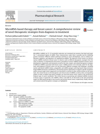
![P.J. Kaboli et al. / Pharmacological Research 97 (2015) 104–121 105
3. miRNA-based therapeutic strategies for breast cancer . . . . . . . . . . . . . . . . . . . . . . . . . . . . . . . . . . . . . . . . . . . . . . . . . . . . . . . . . . . . . . . . . . . . . . . . . . . . . . . . . . . . . . . . . . . . . 111
3.1. Nucleic acid-based strategies. . . . . . . . . . . . . . . . . . . . . . . . . . . . . . . . . . . . . . . . . . . . . . . . . . . . . . . . . . . . . . . . . . . . . . . . . . . . . . . . . . . . . . . . . . . . . . . . . . . . . . . . . . . . . . . . 111
3.1.1. miRNA replacement therapy . . . . . . . . . . . . . . . . . . . . . . . . . . . . . . . . . . . . . . . . . . . . . . . . . . . . . . . . . . . . . . . . . . . . . . . . . . . . . . . . . . . . . . . . . . . . . . . . . . . . . . 111
3.1.2. Anti-miRNA therapy . . . . . . . . . . . . . . . . . . . . . . . . . . . . . . . . . . . . . . . . . . . . . . . . . . . . . . . . . . . . . . . . . . . . . . . . . . . . . . . . . . . . . . . . . . . . . . . . . . . . . . . . . . . . . . . 111
3.2. Drug-based strategies . . . . . . . . . . . . . . . . . . . . . . . . . . . . . . . . . . . . . . . . . . . . . . . . . . . . . . . . . . . . . . . . . . . . . . . . . . . . . . . . . . . . . . . . . . . . . . . . . . . . . . . . . . . . . . . . . . . . . . . 112
3.2.1. Stilbenes . . . . . . . . . . . . . . . . . . . . . . . . . . . . . . . . . . . . . . . . . . . . . . . . . . . . . . . . . . . . . . . . . . . . . . . . . . . . . . . . . . . . . . . . . . . . . . . . . . . . . . . . . . . . . . . . . . . . . . . . . . . 112
3.2.2. Curcumin. . . . . . . . . . . . . . . . . . . . . . . . . . . . . . . . . . . . . . . . . . . . . . . . . . . . . . . . . . . . . . . . . . . . . . . . . . . . . . . . . . . . . . . . . . . . . . . . . . . . . . . . . . . . . . . . . . . . . . . . . . . 113
3.2.3. Flavonoids. . . . . . . . . . . . . . . . . . . . . . . . . . . . . . . . . . . . . . . . . . . . . . . . . . . . . . . . . . . . . . . . . . . . . . . . . . . . . . . . . . . . . . . . . . . . . . . . . . . . . . . . . . . . . . . . . . . . . . . . . . 113
3.2.4. HDAC inhibitors. . . . . . . . . . . . . . . . . . . . . . . . . . . . . . . . . . . . . . . . . . . . . . . . . . . . . . . . . . . . . . . . . . . . . . . . . . . . . . . . . . . . . . . . . . . . . . . . . . . . . . . . . . . . . . . . . . . . 113
3.3. The effects of miRNAs on response to conventional therapy . . . . . . . . . . . . . . . . . . . . . . . . . . . . . . . . . . . . . . . . . . . . . . . . . . . . . . . . . . . . . . . . . . . . . . . . . . . . . . . 113
3.3.1. Increased levels of ABC transporters . . . . . . . . . . . . . . . . . . . . . . . . . . . . . . . . . . . . . . . . . . . . . . . . . . . . . . . . . . . . . . . . . . . . . . . . . . . . . . . . . . . . . . . . . . . . . . 113
3.3.2. Alteration of drug targets . . . . . . . . . . . . . . . . . . . . . . . . . . . . . . . . . . . . . . . . . . . . . . . . . . . . . . . . . . . . . . . . . . . . . . . . . . . . . . . . . . . . . . . . . . . . . . . . . . . . . . . . . . 113
3.3.3. Alterations in DNA repair pathways. . . . . . . . . . . . . . . . . . . . . . . . . . . . . . . . . . . . . . . . . . . . . . . . . . . . . . . . . . . . . . . . . . . . . . . . . . . . . . . . . . . . . . . . . . . . . . . 114
3.3.4. Evasion of apoptosis . . . . . . . . . . . . . . . . . . . . . . . . . . . . . . . . . . . . . . . . . . . . . . . . . . . . . . . . . . . . . . . . . . . . . . . . . . . . . . . . . . . . . . . . . . . . . . . . . . . . . . . . . . . . . . . 114
3.4. Nanoformulas and miRNA-based therapy . . . . . . . . . . . . . . . . . . . . . . . . . . . . . . . . . . . . . . . . . . . . . . . . . . . . . . . . . . . . . . . . . . . . . . . . . . . . . . . . . . . . . . . . . . . . . . . . . . 114
4. miRNA-based therapy and clinical evidence . . . . . . . . . . . . . . . . . . . . . . . . . . . . . . . . . . . . . . . . . . . . . . . . . . . . . . . . . . . . . . . . . . . . . . . . . . . . . . . . . . . . . . . . . . . . . . . . . . . . . . . 116
5. Future directions and challenges . . . . . . . . . . . . . . . . . . . . . . . . . . . . . . . . . . . . . . . . . . . . . . . . . . . . . . . . . . . . . . . . . . . . . . . . . . . . . . . . . . . . . . . . . . . . . . . . . . . . . . . . . . . . . . . . . . . 116
6. Conclusion . . . . . . . . . . . . . . . . . . . . . . . . . . . . . . . . . . . . . . . . . . . . . . . . . . . . . . . . . . . . . . . . . . . . . . . . . . . . . . . . . . . . . . . . . . . . . . . . . . . . . . . . . . . . . . . . . . . . . . . . . . . . . . . . . . . . . . . . . . . 117
Conflicts of interest . . . . . . . . . . . . . . . . . . . . . . . . . . . . . . . . . . . . . . . . . . . . . . . . . . . . . . . . . . . . . . . . . . . . . . . . . . . . . . . . . . . . . . . . . . . . . . . . . . . . . . . . . . . . . . . . . . . . . . . . . . . . . . . . . 117
References. . . . . . . . . . . . . . . . . . . . . . . . . . . . . . . . . . . . . . . . . . . . . . . . . . . . . . . . . . . . . . . . . . . . . . . . . . . . . . . . . . . . . . . . . . . . . . . . . . . . . . . . . . . . . . . . . . . . . . . . . . . . . . . . . . . . . . . . . . . 117
1. Introduction
MicroRNAs (miRNAs or miRs) are small (21–23 nucleotides)
molecules transcribed by type II and type III RNA polymerases. In
general, miRNAs bind the 3 untranslated regions (UTRs) of mRNAs
and suppress mRNA translation [1]. miRNAs play a crucial role in
breast cancer development by promoting uncontrolled cell divi-
sion and blocking apoptosis [2]. Approximately 50% of miRNAs are
located at intergenic regions and the remaining 50% of miRNAs have
been predicted to be located within intragenic (intronic) regions
[3]. It has been estimated that approximately 50% of miRNA genes
are located within genomic regions associated with cancer [4], but
the exact roles of intronic miRNAs are not well understood. To date,
several cancer-associated intronic miRNAs have been discovered in
humans, including miR-10b, miR-15b, miR-16-2, miR-17-92, miR-
26a1, miR-26-a2, miR-26b, and miR-126 [5]. The intronic miRNAs
involved in breast cancer are listed in Tables 1 and 2.
Primary miRNA (pri-miRNA) is transcribed as capped,
polyadenylated, and double-stranded stem-loop containing
structures. The pri-miRNAs are then processed into 70-100
nucleotide-long hairpin structures by the Drosha RNase complex.
The second form of intermediate miRNA is called precursor miRNA
(pre-miRNA), which is transported to the cytoplasm via exportin
5. Cytoplasmic processing of pre-miRNAs occurs when Dicer
(another RNase) cleaves pre-miRNAs into an approximately 22
nucleotide-long double-stranded miRNA duplex. This miRNA
duplex is then incorporated into the miRNA-induced silencing
complex (miRISC). The Argonuate protein located in miRISC
unwinds double-stranded miRNA, and the mature strand is
separated from the passenger [6,7]. According to the currently
accepted hypothesis, the passenger strand (miR-XXX*) is then
released, and the mature strand (miR-XXX) is activated as part
of a powerful cellular regulatory system within miRISC that
targets 3 -UTRs of specific mRNAs; however, it is known that the
passenger strand also has a functional role in post-transcriptional
gene silencing. Therefore, the annotation of miRNA strands has
been changed to miR-XXX-5p (former miR-XXX) and miR-XXX-3p
(former miR-XXX*) for mature strands released from the 5 -end
and 3 -end of mature miRNA hairpin duplex, respectively [8].
miRNAs contain a six to seven nucleotide-long seed sequence
that mediates the interaction with targets [9]. Intronic miRNAs,
which have mostly been predicted by bioinformatics, are only
transcribed by type II RNA polymerase because they bypass the
Drosha dependent step [10]. Instead, a spliceosomal complex is
required for post-transcriptional processing of intronic miRNAs
[11].
It is well-known that miRNAs play a crucial role in cancer
progression, but reports showing the role of miRNAs in cancer
development are rare. Some miRNAs, such as miR-21, miR-221/222,
and miR-182 are known to be involved in cell proliferation (Table 1),
but it cannot be unequivocally concluded that they play roles in
cancer initiation. However, stem cell-associated miRNAs, such as
miR-302 and miR-373-3, may be related to the origin of cancer [12].
It has also been demonstrated that LIN28/let-7, c-MYC-E2F/miR-
17-92, and OCT4/SOX2/miR-302-Cyclin D1 networks play a crucial
role in the pluripotency and self-renewal of cancer stem cells
and embryonic stem cells [13]. During cancer development, pro-
gression, and metastasis, miRNAs are subdivided into two main
categories: tumor suppressor and oncogenic miRNAs. To date, a
number of miRNAs have been found to be associated with breast
cancer; these miRNAs and their molecular targets will be high-
lighted in this review.
According to GLOBACAN 2013, breast cancer is the most fre-
quently diagnosed cancer in women and the second most common
cancer worldwide. In 2012, Twenty-five percent (1.67 million) of all
new cancer cases and 15% (522,000) of all cancer deaths in women
were due to breast cancer [14]. Therefore, advanced therapeutic
strategies are urgently needed for effectively treating breast cancer
patients. However, only a limited number of literature reviews on
the molecular properties and anti-breast cancer effects of miRNAs
with special focus on therapeutic strategies have been published to
date. In addition to our recent review (2014) published on the role
of the natural product, berberine (PubChem CID: 2353), in breast
cancer treatment that briefly focused on miRNA/berberine interac-
tions [15], two other reviews have been recently published on the
role of miRNA in breast cancer. Goh et al. [16] reconstructed and
listed miRNAs involved in breast cancer based on the hallmarks of
cancer, but the review does not discuss the therapeutic importance
of miRNAs in detail. The other review focused on the impact of miR-
NAs on drug resistance in breast cancer with a short discussion on
miRNA targets [17]. Therefore, a comprehensive review discussing
the role of miRNAs in breast cancer development, progression, and
migration as well as miRNA targets and therapeutic strategies has
not been published to date. This review aims to provide an exten-
sive analysis of miRNA effects on various molecular targets (e.g.,
tumor suppressor genes, oncogenes, and other regulators) involved
in breast cancer development as well as miRNA-based therapeutic
opportunities for overcoming breast cancer.](data:image/gif;base64,R0lGODlhAQABAIAAAAAAAP///yH5BAEAAAAALAAAAAABAAEAAAIBRAA7)