Dental panorama part 2
•
3 likes•169 views

Dental Panoramic imaging for dental students Part 2

Recommended
More Related Content
Similar to Dental panorama part 2
Similar to Dental panorama part 2 (20)
Recently uploaded
Recently uploaded (20)
Dental panorama part 2
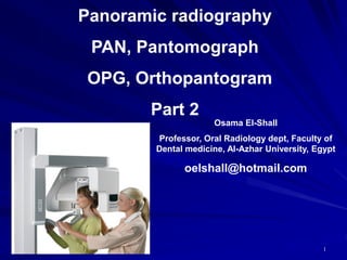
- 1. 5/30/2021 Panoramic radiography PAN, Pantomograph OPG, Orthopantogram Part 2 1 Osama El-Shall Professor, Oral Radiology dept, Faculty of Dental medicine, Al-Azhar University, Egypt oelshall@hotmail.com
- 4. Positioning of teeth anterior to the focal trough: If the patient’s anterior teeth are not positioned in the focal trough indicated by the groove in the bite- block, the teeth appear blurred. If the patients teeth are positioned too far forward on the bite block or anterior to the focal trough the anterior teeth appear skinny and out of focus on the radiograph. Solution: the patient’s anterior teeth must be placed in an end-to end position in the groove on the bite-block.
- 5. teeth anterior to the focal trough
- 6. teeth anterior to the focal trough
- 7. Anterior teeth wider and blurred Teeth too posterior
- 8. Positioning of teeth posterior to the focal trough: if the patient’s anterior teeth are not positioned in the focal trough indicated by the groove in the bite-block, the teeth appear blurred. If the patients teeth are positioned too far back on the bite-block or posterior to the focal trough the anterior teeth appear fat and out of focus on the radiograph. Solution: the patient’s anterior teeth must be placed in an end-to end positioning the groove on the bite-block.
- 9. teeth posterior to the focal trough
- 10. Structures smaller on the side to which head is turned; larger on opposite side.
- 11. Positioning of the midsagital plane: If the patient’s midsagital plane is not perpendicular to the floor, the ramus and posterior teeth appear unequally magnified on the panoramic radiograph. The side farthest from the film appears magnified, and the side closest to the film appears smaller . Solution: the patient’s midsagital plane is perpendicular to the floor. The lateral head supports must then be adjusted to stabilize the patient’s head position .
- 17. Mandibular incisors shortened, V-shaped mandible HEAD TIPPED DOWN
- 18. Positioning of the Frankfort plane-downward: If the patient’s chin is positioned too low or is tipped down the Frankfort plane is angled downward, this results: – The mandibular incisors appear blurred . – There is a loss of detail in the anterior apical regions. – The condyles may not be visible. – An exaggerated smile line (curved up-ward) will appear. – Solution: – the patient’s Frankfort plane must be parallel with the floor.
- 19. Positioning of the Frankfort plane-downward
- 20. Positioning of the Frankfort plane-downward
- 21. HEAD TIPPED UP Squared-off mandible, palate superimposed over maxillary teeth
- 22. Positioning of the Frankfort plane-upward: If the patient’s chin is positioned too high or is tipped up the Frankfort plane is angled upward, and the following results: The hard palate and floor of the nasal cavity appear superimposed over the roots of the maxillary teeth. There is a loss of detail in the maxillary incisor region. The maxillary incisors appear blurred and magnified. A reverse simile line (curved downward) is apparent on the radiograph . Solution: The patient’s Frankfort plane must be parallel with the floor.
- 23. Positioning of the Frankfort plane-upward
- 24. Positioning of the Frankfort plane-upward
- 25. ⚫a ⚫a - g ⚫b ⚫b - g Ghost images of earrings
- 26. Ghost images of earrings
- 27. 5/30/2021 27
- 28. 5/30/2021 28
- 29. Ghost image of metal used to restore left angle of mandible
- 33. Ghost image of thyroid collar
- 34. Shadow of vertebral column, usually from patient not standing straight
- 36. Palatoglossal air space (black area above) caused by failure to keep tongue against palate during exposure. Makes it difficult to diagnose periapical pathology in maxillary area.
- 38. Static electricity; caused by removing film from box or cassette too quickly, creating static discharge
- 39. Partial dentures
- 40. Glasses
- 41. Patient movement
- 42. Double exposure
- 43. Ossama El-Shall നന്ദി धन्यवाद hvala ti Salamat