
Recommended
More Related Content
Similar to 2-Lower Respiratory Tract (3).pptx
Similar to 2-Lower Respiratory Tract (3).pptx (20)
Recently uploaded
Recently uploaded (20)
2-Lower Respiratory Tract (3).pptx
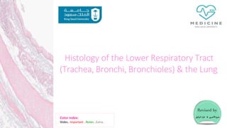
- 1. Histology of the Lower Respiratory Tract (Trachea, Bronchi, Bronchioles) & the Lung Color index: Slides.. Important ..Notes ..Extra..
- 2. Objectives : The microscopic structures of the wall of: Trachea. Primary or extrapulmonary bronchi. Intrapulmonary (secondary and tertiary) bronchi. Bronchioles The microscopic structures of : Interalveolar septum. Alveolar phagocytes. Pleura.
- 3. TRACHEA The wall of trachea is formed of Mucosa Epithelium: Respiratory epithelium Lamina Propria Elastic Lamina ( membrane) Submucosa C.T Numerous Mucous and seromucous glands Lymphoid elements (cells) Adventitia* Fibroelastic C.T C-shaped rings (12-16) of hyaline cartilage (incomplete) Trachealis muscle (bundle of smooth muscle fibers ) connects the 2 ends of each C-shaped(incomplete) rings of cartilage In the trachea the elastic fibers is so dense and form a membrane , this membrane separates the lamina propria form the submucosa. Note : you MUST differentiate between the elastic cartilage and elastic fibers ! The mucosa (MAINLY) composed of 2 things : • epithelium • Lamina propria (connective tissue) * Adventitia is the outermost fibroblastic connective tissue+cartilage covering of an organ, vessel, or other structure The trachea is highly humidified because not only does the mucosa has glands but also the submucosaleading to a high humidity
- 4. Pictures of the different layers of mucosa As you can see here the trachealis muscle enclosed the c-shapedhayline cartilage The elastic lamina is not visible because it needs a special stain C-shapedcartilage
- 5. 1-Mucosa: 2 Layers: A- Epithelium: Respiratory epithelium (pseudo-stratified Ciliated columnar Epithelium with goblet cell). B- Lamina propria. (It’s narrow so it doesn’t contain 1- glands 2- lymphoid follicles.) N.B.No elasticlamina. 2-Musclecoat (complete)not like trachea: Two distinct layers of smooth muscle fibers spirally arranged in opposite direction (2 spiral shape crisscrossing layers one is clockwise and the other is anti- clockwise). Adventitia: Sub-mucosa: C.T . contains: A- Seromucous glands. B- Lymphoid elements. A- Loose C.T. B- Irregular plates of hyaline cartilage (complete layer) hallmark . C- Solitary lymphoid nodules. 1-EXTRAPULMONARY BRONCHUS(1ry BRONCHUS): Generally have the same histological appearance as the trachea. 2-INTRAPULMONARY BRONCHI(2ry & 3ry BRONCHI): BRONCHI
- 6. less than 0.5mm in diameter . Similar structure to preterminal bronchioles, but: Epithelium: Simple cuboidal partially ciliated epithelium With Clara cells ( With NO goblet cells). CLARA CELLS: Structure: columnar cells (non ciliated). Function: 1- Degrade toxins in inhaled air . (immune cell likefunction) 2- Divideto regenerate the bronchiolar epithelium. 3- Produce surfactant-like material. Location: Terminal bronchioles and respiratory bronchioles. *Important notes : •The Terminal Bronchioles are the last part of the conduction zone . •The RespiratoryBronchiolesare the first part of the respiratory zone. •The main differencebetween the bronchioles and the bronchus is the absent of: 1- Cartilages 2- Seromucous glands 3- Lymph nodules 4- Goblet cells 5- Sub-mucosa B- Lamina propria : C.T . rich in elastic fibers. (although it’s rich in elastic fiber but there’s no membrane) Muscle coat: 2 helically arranged smooth muscle layers. Adventitia: C.T. No cartilage at all, No seromucous glands, No lymph nodules. Preterminal Bronchioles(1ry): less than 1mm in diameter . Mucosa: (has longitudinal folds ) A- Epithelium: Simple ciliated Columnar Epithelium with occasional goblet cells. (The doctor said usually you will not find goblet cells ) their walls are interrupted by the presence of few pulmonary alveoli. Why is the mucosa folded ? To give larger surface area for dilatation. BRONCHIOLES (DOESN’T CONTAIN CARTILAGE) Respiratory Bronchioles(3ry): Terminal Bronchioles(2ry): Similarstructure to terminal bronchioles,but:
- 7. EXTRA بع ش ال ت ف ر ع د ا ز ا م ل ك و تق م ع ت ا م ل ك هنا يف ك ك ل حضوي انه يوكتنها ف ي ةلخادال ءايشالا و تالراكيب كدنع ت ن ق ص ةيئاوهال Note-team435-: Special features of everyone of them: Pretermenal > Goblet cells Terminal > NO goblet cells , Rispiratory > Alveoli Bronchioles Terminal bronchiole = end of conducting portion + end of goblet cells, Goblet cells are replaced by Clara cells
- 8. ALVEOLAR DUCTS The wall of alveolar ducts consist almost of pulmonaryalveoli. N.B. Alveolar duct → ends by: atrium → communicates with: 2-3 alveolar sacs PULMONARY ALVEOLI Definition: They are small out-pouching of respiratory bronchioles, alveolar ducts & alveolar sacs. INTERALVEOLAR SEPTA Definition: The region between 2 adjacentalveoli. the alveoli are like rooms and the septas are like the walls in between Components: (A) Alveolar Epithelium: lines both sides of interalveolar septum. (B) Interstitium. PULMONARY ALVEOLI Alveolar epithelium. Interalveolar septa. Alveolar phagocytes (Lung macrophages).
- 9. (1)Alveolarepithelium consist of twomajor cells : Type I Pneumocytes Type II Pneumocytes Type I Pneumocytes Type IIPneumocytes line 95% of the alveolarsurface. Line 5% of the alveolarsurfaces. Count: less numerous than type II pneumocytes. Are more numerousthan type I pneumocytes. L/M: Are cuboidal or roundedcells, With Foamy cytoplasm. Nucleus:central & rounded. - The cytoplasm contains membrane- boundLamellar bodies (contain pulmonary surfactant). Function: Exchange ofgases. 1- Synthesis & secretionof pulmonarysurfactant. 2- Renewalof alveolar epithelial cells )STEM CELL( : TypeII cells can divide to regenerate both type I & type II pneumocytes. ALVEOLAR EPITHELIUM Lining: simple squamousepithelium. The number of type I pneumocyte in alveolar epithelium is less then type 2 but its lining surface is greater , why ? Simply because the epithelium of pneumocyte type 1 is SIMPLE SAQUAMOUS and since it’s SQUAMOUS it can fill the space with less number of cells .
- 10. * Interstitium of interalveolar septa Continuous Pulmonary Capillaries. InterstitialC.T . C. T .Fibers: elastic fibers type III collagen (reticular fibers). C. T .Cells: Fibroblasts Macrophages (Alveolar macrophage) Mast cells Lymphocytes Interstitium of interalveolar septa
- 11. BLOOD-GAS BARRIER (BLOOD-AIR BARRIER) Definition: It is the region of the interalveolar septum that is traversed by O2 & CO2 . Components: 1- Thin layer of surfactant. (frompneumocyte type II ) 2- Type I pneumocyte . (Exchange of gases) 3- Fused basal laminae of typeI pneumocytes& endothelial cells of the pulmonary capillary. 4- Endothelial cells of the pulmonary capillary. * The wall of blood capillaries is continues. Alveolar phagocytes (Alveolar Macrophages) (Dust Cells) Sites: 1- In the lumenof pulmonary alveoli. 2- In the interstitium of interalveolar septa. Function: Phagocytoseparticulate matter (e.g. dust) & bacteria in the lumen of pulmonary alveoli and in the interstitium of interalveolar septa. When the O2 molecules diffuse to the capillaries it they pass the following structures respectively : 1. surfactant ( surface lining ) 2. Alveolar epithelium 3. Fuesed basel lamnie (base) 4. Endothelium
- 12. Pleura Is formed of two layers: 1 Parietal and visceral. 2 It is formed of simple squamous mesothelium . ( Mesothelium is the epithelium of serous membranes e.g. Pleura ) 3 The two layers are separated by serous fluid . Visceral Layer : has sub-epithelium loose C.T that extends into the lung tissue .
- 13. Mind Map Thanks to our dear fellow student Norah Alshabib for sharing! FULL MIND MAP
- 14. Conducting Portion Comparison Trachea Extra-pulmonary bronchi ( 1ryBronchus) Intrapulmonary bronchi (2ry &3ryBronchi) Preterminal Bronchioles TerminalBronchioles Mucosa 1 - Respiratoryepithelium 2- Laminapropria 3- ElasticLamina 1 -Respiratory Epithelium 2 - Laminapropria (No elasticlamina) 1- Simple ciliated epithelium with occasional gobletcells 2- Laminapropria ( C.T. is rich in elsatic fibers) 1 Simplecuboidal partiallyciliated epithelium With Clara cells (NO gobletcells). 2 Laminapropria Submucosa 1 -C.T. 2 - Numerous mucous & seromuscousglands 3 - Lymphoid elements C.T.contains: A- Seromucousglands B - Lymphoid elements here there’s no submucosa , why ? Simply because the elastic fibres do not form a elastic lamina ( membrane ) and therefore ,there’ll be no submucosallayer . Adventitia 1 - FibroelasticC.T. 2 - C -shaped rings of hayalinecartilage (incomplete rings of cartilage) A-LooseC.T. B.Irregular platesof hyaline cartilage (completelayer). C.Solitarylymphoid nodules Nocartilage No seromucousglands No lymphnodes Muscle coat Only trachealis muscle to compete the C-shapedrings (complete) ---------------------------------------------------------------
- 15. EXTRA NOTE –team435- For the 1st & 2nd lectures
- 16. Videos : We recommend watching this 5min video: https://www.youtube.com/watch?v=UDIgNteqVag Full playlist for respiratory system : https://www.youtube.com/watch?v=23_aHo4X2Vs&list=PLEf8wmJpS_1HmywDPF1Ve0zRhWfHCH COy Links to help you !
- 17. MCQ : 1 which one of the following has an elastic lamina? A-trachea B-Intrapulmonay bronchi C-preterminal bronchioles D- Terminal bronchioles 2the incomplete ring of hyaline cartilage in trachea completed by : A-Trachealis muscle B-Tendon C- ligament D-elastic cartilage 3 type of muscle coat found in intrapulmonary bronchus is : A-cardiac muscle B-smooth muscle C- skeletal D- All of them 4 -The preterminal bronchioles have no … A-Mucosa ,cartilage and seromucous glands B-lymph nodes, smooth muscle and cartilage C-cartilage,seromucous gland and lymph nodes 5- which one of the following has clara cells? A-trachea B-extrapulmonary bronchi C-terminal bronchioles D-alveolar sacs 6what is the type of epithelium found in both type I and II pneumocytes ? A-simple columnar – simple squamous B pseudo stratified columnar - simple squamous C-Stratified squamous - simple cuboidal D-simple squamous – simple cuboidal 7which one of the following is responsible for the secretion and synthesis of pulmonary surfactant ? A-type I pneumocytes B-type II pneumocytes C- alveolar ducts D-trachea
- 18. Thank you & good luck - Histology team Done by : Ahmed Badahdah Mutasem Alhasani Omar Turkistani Nawaf Aldarweesh Mohammed Khojah Shahad Alanzan Team leaders: Reema Alotaibi Faisal Alrabaii Please if you need anything or even further explanation contact us on : HistologyTeam436@gmail.com @histology436