intravascular lymphoma. Common computed tomography brian findings
•Download as PPT, PDF•
0 likes•1 view
CT Brain of a patient with intravascular lymphoma
Report
Share
Report
Share
Recommended
Recommended
❤️Ludhiana Call Girls ☎️98157-77685☎️ Call Girl service in Ludhiana☎️Ludhiana Call Girls Service ☎️ Call Girls In Ludhiana Ludhiana City Centre, Park Plaza Ludhiana, Windsor Fountain, G.T Road Ludhiana escort all Ludhiana service Russian available model female girls in Ludhiana VIP Lo price personal Ludhiana off class call girls payment high profile model and female escort 70% Off On Your First Booking Ludhiana Call Girls Service Cash Payment
Welcome to DILPREET Ludhiana Call Girl Service, the Trusted call girl agency around. We Offer 70% Discount On Your First Booking For Ludhiana Call Girls Service Cash Payment is available.❤️Ludhiana Call Girls ☎️98157-77685☎️ Call Girl service in Ludhiana☎️Ludhiana...
❤️Ludhiana Call Girls ☎️98157-77685☎️ Call Girl service in Ludhiana☎️Ludhiana...dilpreetentertainmen
🍑👄Ludhiana Escorts Service☎️98157-77685🍑👄 Call Girl service in Ludhiana☎️Ludhiana Call Girls Service 🍑👄 Call Girls In Ludhiana Book Now :- 98157-77685
Our agency presents a selection of young, charming call girls available for bookings at Oyo Hotels. Experience high-class escort services at pocket-friendly rates, with our female escorts exuding both beauty and a delightful personality, ready to meet your desires. Whether it's Housewives, College girls, Russian girls, Muslim girls, or any other preference, we offer a diverse range of options to cater to your tastes.
We provide both in-call and out-call services for your convenience. Our in-call location in Delhi ensures cleanliness, hygiene, and 100% safety, while our out-call services offer doorstep delivery for added ease.
We value your time and money, hence we kindly request pic collectors, time-passers, and bargain hunters to refrain from contacting us. l Ludhiana, Majestic Grand Hotel, Ramada by Wyndham Ludhiana City Centre, Park Plaza Ludhiana, Windsor Fountain, G.T Road Ludhiana escort all Ludhiana service Russian available model female girls in Ludhiana VIP Lo price personal Ludhiana off class call girls payment high profile model and female escort 70% Off On Your First Booking Ludhiana Call Girls Service Cash Payment
Welcome to DILPREET Ludhiana Call Girl Service, the Trusted call girl agency around. We Offer 70% Discount On Your First Booking For Ludhiana Call Girls Service Cash Payment is available.🍑👄Ludhiana Escorts Service☎️98157-77685🍑👄 Call Girl service in Ludhiana☎️Ludh...
🍑👄Ludhiana Escorts Service☎️98157-77685🍑👄 Call Girl service in Ludhiana☎️Ludh...dilpreetentertainmen
Independent Call Girls Hyderabad 💋 9352988975 💋 Genuine WhatsApp Number for Real Meet
WHATSAPP On Here: 9352988975
Today call girl service available 24X7*▬█⓿▀█▀ 𝐈𝐍𝐃𝐄𝐏𝐄𝐍𝐃𝐄𝐍𝐓 CALL 𝐆𝐈𝐑𝐋 𝐕𝐈𝐏 𝐄𝐒𝐂𝐎𝐑𝐓 SERVICE ✅
⭐➡️HOT & SEXY MODELS // COLLEGE GIRLS
AVAILABLE FOR COMPLETE ENJOYMENT WITH HIGH PROFILE INDIAN MODEL AVAILABLE HOTEL & HOME
★ SAFE AND SECURE HIGH CLASS SERVICE AFFORDABLE RATE
★ 100% SATISFACTION,UNLIMITED ENJOYMENT.
★ All Meetings are confidential and no information is provided to any one at any cost.
★ EXCLUSIVE PROFILes Are Safe and Consensual with Most Limits Respected
★ Service Available In: - HOME & HOTEL 24x7 :: 3 * 5 *7 *Star Hotel Service .In Call & Out call SeRvIcEs :
★ A-Level (5 star escort)
★ Strip-tease
★ BBBJ (Bareback Blowjob)Receive advanced sexual techniques in different mode make their life more pleasurable #G05.
★ Spending time in hotel rooms
★ BJ (Blowjob Without a Condom)
★ Completion (Oral to completion)
★ Covered (Covered blowjob Without condom
100% SAFE AND SECURE 24 HOURS SERVICE AVAILABLE HOME AND HOTEL SERVICESIndependent Call Girls Hyderabad 💋 9352988975 💋 Genuine WhatsApp Number for R...
Independent Call Girls Hyderabad 💋 9352988975 💋 Genuine WhatsApp Number for R...Ahmedabad Call Girls
More Related Content
Recently uploaded
❤️Ludhiana Call Girls ☎️98157-77685☎️ Call Girl service in Ludhiana☎️Ludhiana Call Girls Service ☎️ Call Girls In Ludhiana Ludhiana City Centre, Park Plaza Ludhiana, Windsor Fountain, G.T Road Ludhiana escort all Ludhiana service Russian available model female girls in Ludhiana VIP Lo price personal Ludhiana off class call girls payment high profile model and female escort 70% Off On Your First Booking Ludhiana Call Girls Service Cash Payment
Welcome to DILPREET Ludhiana Call Girl Service, the Trusted call girl agency around. We Offer 70% Discount On Your First Booking For Ludhiana Call Girls Service Cash Payment is available.❤️Ludhiana Call Girls ☎️98157-77685☎️ Call Girl service in Ludhiana☎️Ludhiana...
❤️Ludhiana Call Girls ☎️98157-77685☎️ Call Girl service in Ludhiana☎️Ludhiana...dilpreetentertainmen
🍑👄Ludhiana Escorts Service☎️98157-77685🍑👄 Call Girl service in Ludhiana☎️Ludhiana Call Girls Service 🍑👄 Call Girls In Ludhiana Book Now :- 98157-77685
Our agency presents a selection of young, charming call girls available for bookings at Oyo Hotels. Experience high-class escort services at pocket-friendly rates, with our female escorts exuding both beauty and a delightful personality, ready to meet your desires. Whether it's Housewives, College girls, Russian girls, Muslim girls, or any other preference, we offer a diverse range of options to cater to your tastes.
We provide both in-call and out-call services for your convenience. Our in-call location in Delhi ensures cleanliness, hygiene, and 100% safety, while our out-call services offer doorstep delivery for added ease.
We value your time and money, hence we kindly request pic collectors, time-passers, and bargain hunters to refrain from contacting us. l Ludhiana, Majestic Grand Hotel, Ramada by Wyndham Ludhiana City Centre, Park Plaza Ludhiana, Windsor Fountain, G.T Road Ludhiana escort all Ludhiana service Russian available model female girls in Ludhiana VIP Lo price personal Ludhiana off class call girls payment high profile model and female escort 70% Off On Your First Booking Ludhiana Call Girls Service Cash Payment
Welcome to DILPREET Ludhiana Call Girl Service, the Trusted call girl agency around. We Offer 70% Discount On Your First Booking For Ludhiana Call Girls Service Cash Payment is available.🍑👄Ludhiana Escorts Service☎️98157-77685🍑👄 Call Girl service in Ludhiana☎️Ludh...
🍑👄Ludhiana Escorts Service☎️98157-77685🍑👄 Call Girl service in Ludhiana☎️Ludh...dilpreetentertainmen
Independent Call Girls Hyderabad 💋 9352988975 💋 Genuine WhatsApp Number for Real Meet
WHATSAPP On Here: 9352988975
Today call girl service available 24X7*▬█⓿▀█▀ 𝐈𝐍𝐃𝐄𝐏𝐄𝐍𝐃𝐄𝐍𝐓 CALL 𝐆𝐈𝐑𝐋 𝐕𝐈𝐏 𝐄𝐒𝐂𝐎𝐑𝐓 SERVICE ✅
⭐➡️HOT & SEXY MODELS // COLLEGE GIRLS
AVAILABLE FOR COMPLETE ENJOYMENT WITH HIGH PROFILE INDIAN MODEL AVAILABLE HOTEL & HOME
★ SAFE AND SECURE HIGH CLASS SERVICE AFFORDABLE RATE
★ 100% SATISFACTION,UNLIMITED ENJOYMENT.
★ All Meetings are confidential and no information is provided to any one at any cost.
★ EXCLUSIVE PROFILes Are Safe and Consensual with Most Limits Respected
★ Service Available In: - HOME & HOTEL 24x7 :: 3 * 5 *7 *Star Hotel Service .In Call & Out call SeRvIcEs :
★ A-Level (5 star escort)
★ Strip-tease
★ BBBJ (Bareback Blowjob)Receive advanced sexual techniques in different mode make their life more pleasurable #G05.
★ Spending time in hotel rooms
★ BJ (Blowjob Without a Condom)
★ Completion (Oral to completion)
★ Covered (Covered blowjob Without condom
100% SAFE AND SECURE 24 HOURS SERVICE AVAILABLE HOME AND HOTEL SERVICESIndependent Call Girls Hyderabad 💋 9352988975 💋 Genuine WhatsApp Number for R...
Independent Call Girls Hyderabad 💋 9352988975 💋 Genuine WhatsApp Number for R...Ahmedabad Call Girls
Recently uploaded (20)
Rishikesh Call Girls Service 6398383382 Real Russian Girls Looking Models
Rishikesh Call Girls Service 6398383382 Real Russian Girls Looking Models
Premium Call Girls Bangalore {7304373326} ❤️VVIP POOJA Call Girls in Bangalor...
Premium Call Girls Bangalore {7304373326} ❤️VVIP POOJA Call Girls in Bangalor...
Budhwar Peth ( Call Girls ) Pune 6297143586 Hot Model With Sexy Bhabi Ready...
Budhwar Peth ( Call Girls ) Pune 6297143586 Hot Model With Sexy Bhabi Ready...
Sexy Call Girl Palani Arshi 💚9058824046💚 Palani Escort Service
Sexy Call Girl Palani Arshi 💚9058824046💚 Palani Escort Service
💞 Safe And Secure Call Girls Coimbatore 🧿 9332606886 🧿 High Class Call Girl S...
💞 Safe And Secure Call Girls Coimbatore 🧿 9332606886 🧿 High Class Call Girl S...
Kolkata Call Girls Miss Inaaya ❤️ at @30% discount Everyday Call girl
Kolkata Call Girls Miss Inaaya ❤️ at @30% discount Everyday Call girl
❤️Ludhiana Call Girls ☎️98157-77685☎️ Call Girl service in Ludhiana☎️Ludhiana...
❤️Ludhiana Call Girls ☎️98157-77685☎️ Call Girl service in Ludhiana☎️Ludhiana...
Vip Call Girls Makarba 👙 6367187148 👙 Genuine WhatsApp Number for Real Meet
Vip Call Girls Makarba 👙 6367187148 👙 Genuine WhatsApp Number for Real Meet
Sexy Call Girl Nagercoil Arshi 💚9058824046💚 Nagercoil Escort Service
Sexy Call Girl Nagercoil Arshi 💚9058824046💚 Nagercoil Escort Service
Call Now ☎ 8868886958 || Call Girls in Chandigarh Escort Service Chandigarh
Call Now ☎ 8868886958 || Call Girls in Chandigarh Escort Service Chandigarh
Top 20 Famous Indian Female Pornstars Name List 2024
Top 20 Famous Indian Female Pornstars Name List 2024
🍑👄Ludhiana Escorts Service☎️98157-77685🍑👄 Call Girl service in Ludhiana☎️Ludh...
🍑👄Ludhiana Escorts Service☎️98157-77685🍑👄 Call Girl service in Ludhiana☎️Ludh...
Sexy Call Girl Villupuram Arshi 💚9058824046💚 Villupuram Escort Service
Sexy Call Girl Villupuram Arshi 💚9058824046💚 Villupuram Escort Service
AECS Layout Escorts (Bangalore) 9352852248 Women seeking Men Real Service
AECS Layout Escorts (Bangalore) 9352852248 Women seeking Men Real Service
Call Girls in Udaipur Girija Udaipur Call Girl ✔ VQRWTO ❤️ 100% offer with...
Call Girls in Udaipur Girija Udaipur Call Girl ✔ VQRWTO ❤️ 100% offer with...
Independent Call Girls Hyderabad 💋 9352988975 💋 Genuine WhatsApp Number for R...
Independent Call Girls Hyderabad 💋 9352988975 💋 Genuine WhatsApp Number for R...
Call Girl in Indore 8827247818 {Low Price}👉 Meghna Indore Call Girls * DXZ...
Call Girl in Indore 8827247818 {Low Price}👉 Meghna Indore Call Girls * DXZ...
👉Bangalore Call Girl Service👉📞 7304373326 👉📞 Just📲 Call Rajveer Call Girls Se...
👉Bangalore Call Girl Service👉📞 7304373326 👉📞 Just📲 Call Rajveer Call Girls Se...
Gorgeous Call Girls In Pune {9xx000xx09} ❤️VVIP ANKITA Call Girl in Pune Maha...
Gorgeous Call Girls In Pune {9xx000xx09} ❤️VVIP ANKITA Call Girl in Pune Maha...
Ernakulam Call Girls 👙 6297143586 👙 Genuine WhatsApp Number for Real Meet
Ernakulam Call Girls 👙 6297143586 👙 Genuine WhatsApp Number for Real Meet
Featured
Featured (20)
Product Design Trends in 2024 | Teenage Engineerings
Product Design Trends in 2024 | Teenage Engineerings
How Race, Age and Gender Shape Attitudes Towards Mental Health
How Race, Age and Gender Shape Attitudes Towards Mental Health
AI Trends in Creative Operations 2024 by Artwork Flow.pdf
AI Trends in Creative Operations 2024 by Artwork Flow.pdf
Content Methodology: A Best Practices Report (Webinar)
Content Methodology: A Best Practices Report (Webinar)
How to Prepare For a Successful Job Search for 2024
How to Prepare For a Successful Job Search for 2024
Social Media Marketing Trends 2024 // The Global Indie Insights
Social Media Marketing Trends 2024 // The Global Indie Insights
Trends In Paid Search: Navigating The Digital Landscape In 2024
Trends In Paid Search: Navigating The Digital Landscape In 2024
5 Public speaking tips from TED - Visualized summary
5 Public speaking tips from TED - Visualized summary
Google's Just Not That Into You: Understanding Core Updates & Search Intent
Google's Just Not That Into You: Understanding Core Updates & Search Intent
The six step guide to practical project management
The six step guide to practical project management
Beginners Guide to TikTok for Search - Rachel Pearson - We are Tilt __ Bright...
Beginners Guide to TikTok for Search - Rachel Pearson - We are Tilt __ Bright...
Unlocking the Power of ChatGPT and AI in Testing - A Real-World Look, present...
Unlocking the Power of ChatGPT and AI in Testing - A Real-World Look, present...
intravascular lymphoma. Common computed tomography brian findings
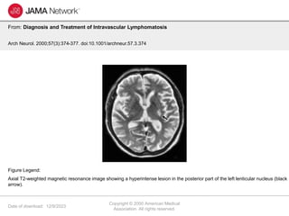
- 1. Date of download: 12/9/2023 Copyright © 2000 American Medical Association. All rights reserved. From: Diagnosis and Treatment of Intravascular Lymphomatosis Arch Neurol. 2000;57(3):374-377. doi:10.1001/archneur.57.3.374 Axial T2-weighted magnetic resonance image showing a hyperintense lesion in the posterior part of the left lenticular nucleus (black arrow). Figure Legend: