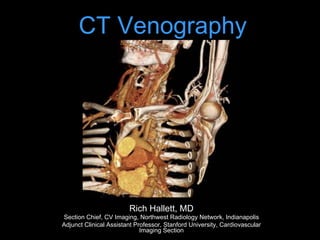
2. Introduction
• CT venography (CTV) is a technique targeted to
assess venous anatomy, determine venous
patency & delineate collateral circulation
• Non-invasive, simple protocols, wide anatomic
coverage, short acquisition time, and ability to be
combined with arterial-phase CTA
5. Doppler US
• Well established clinical utility
• No ionizing radiation
• Portable
• Inexpensive
• Flow direction information
• Operator / Patient dependent
• Some areas inaccessible (pelvis, SVC)
• Collateral pathways not well delineated
7. Performance of Doppler vs. CTV in ICU
patients – LE DVT
Sens Spec
Indiect CTV 70 96
Doppler US 70 100
Taffoni, AJR, 2004
8. MR Venography - Positives
• Excellent for pelvic venous system, CNS
• May not require contrast
• SI ratio thrombus:blood higher for MRV vs. CTV
– 3.7-8:1 vs 1.8-3.2 *
• For PE: Sens 80-95%, Spec 95%, depends on
technique (Perf imaging best)+
• For DVT: Sens ~92%, Spec ~95%
• 0.25 mmol/kg Gd better than 0.125 mmol/kg
+Sampson, Eur Radiol 2007; 17:175-181
* Kluge, AJR, 2006
9. Combo MR-PA / Indirect MRV
• MRA: TRuFISP, perfusion, MRA
(0.25mmol/Kg)
• MRV: 3D FLASH w/ PV coil, voxel size of
1.2x0.8x1.1 mm
– High agreement w/ CTA/CTV but requires a
change in coil and pt. position to obtain MRV
after chest MRA
• Good agreement w/ Doppler in legs,
moderate in pelvis
Kluge, AJR, 2006
10. MR Venography - Negatives
• Expensive, availability sometimes limited
• Exam may be lengthy
• Pt. cooperation?
• Spatial resolution (vs other choices)
• Limited anatomic coverage
11. Radionuclide Venography
• 99mTc-labeled MAA
• 99mTc-labeled RBC
• 99mTc-human serum albumin
• 99mTc-labeled platelets
– Direct evidence of acute / active DVT
– BUT: Arduous prep, false positives – pts on heparin
• 99mTc-apcitide (GIIb/IIIa receptor binding)
– Can tell acute (+) vs. chronic (-) clot
– Interpreter dependent?
Anatomic agents,
indirect evidence
12. Catheter Venography
• Considered the gold standard
• Invasive (but can treat lesions)
• You only see what you can fill
• Risks:
– Minor Complications: 18%
– Thrombosis: 2%
– Bronchospasm, Contrast reactions, etc
13. CTV: Challenges
• Goal: visualize all venous structures, with good
opacification, but without artifacts
Direct CTV
• good opacification (too good; needs dilution)
• but difficult to show all venous structures or full
extent of collateral circulation
Indirect CTV
• shows all veins; but difficult to achieve strong
enhancement; timing
14. CTV: Challenges
• Goal: visualize all venous structures, with good
opacification, but without artifacts
Direct CTV
• good opacification (too good; needs dilution)
• but difficult to visualize all venous structures or
full extent of collateral circulation
Indirect CTV
• visualizes all veins (recirculation of CM)
• but difficult to achieve strong enhancement;
timing difficulties
15. 60M smoker, r/o lung cancer
Routine chest with contrast: 100cc contrast @ 2cc/sec, 40 sec diagnostic delay
16. CTV: Challenges
• Goal: visualize all venous structures, with good
opacification, but without artifacts
Direct CTV
• good opacification (too good; needs dilution)
• but difficult to visualize all venous structures or
full extent of collateral circulation
Indirect CTV
• visualizes all veins (recirculation of CM)
• but difficult to achieve strong enhancement;
timing difficulties
18. CTV: Imaging Techniques
• Direct Venography (first pass):
– Dilute contrast (1:5 - 1:10)
– Fill veins of interest (50cc or more)
– Slow infusion, 1-2cc/sec
– Start acquisition towards end of infusion
• Indirect Venography (recirculation)
– 100-150cc contrast needed for adequate
venous opacification
– Empiric imaging delay
• 60 seconds: upper extremity and pelvic veins
• 3 to 3.5 min: lower extremity veins
– Smart prep off vein of interest
Baldt MM, et al. Radiology 1996;200:423-428
19. 40M prior left arm DVT. Acute pain and swelling of
the left upper arm, rule out DVT.
1:5 dilution (20cc contrast + 80cc NS) @ 3cc/sec. Tourniquet around biceps
region, released 15 sec before initiation of scan.
basilic
Brachial artery
basilic
cephalic
cephalic
20. CTV: Imaging Techniques
• Direct Venography (first pass):
– Dilute contrast (1:5 or 1:6)
– Fill veins of interest (50cc or more)
– Slow infusion, 1-2cc/sec
– Start acquisition towards end of infusion
• Indirect Venography (recirculation)
– 100-150cc contrast needed for adequate
venous opacification
– Empiric imaging delay
• 60 seconds: upper extremity and pelvic veins
• 3 to 3.5 min: lower extremity veins
– Smart prep off vein of interest
Baldt MM, et al. Radiology 1996;200:423-428
21. 65M with metastatic lung ca and recent PEs. An IVC filter was placed but did
not fully deploy. A second IVC filter was placed above the first one.
120cc contrast, diagnostic delay = 70sec
22. CTV: Imaging Techniques
• Direct Venography (first pass):
–Dilute contrast medium (1:5 or 1:6)
–Fill veins of interest (50cc or more)
–Slow infusion, 1-2cc/sec
–Start acquisition towards end of
infusion
Baldt MM, et al. Radiology 1996;200:423-428
24. • Large bolus of contrast followed by a delay to
image the recirculation phase
– 150 mL (2 mL/kg BW)
• Empiric Delay (depends on venous territory)
• 60 seconds: thoracic
• 70-80 seconds: upper extremity
• 110 seconds: abdomen & pelvis
• 180 seconds: lower extremity
• NO Bolus Trigger
• Not an exact science, no target HU
INDIRECT CT VENOGRAPHY
25. Combo Direct / Indirect CTV
• R/O LUE venous
malformation; L hand
and arm swelling
• 120 mL @ 5 mL/s
followed by 100 mL
1:10 dilution at 2.5
mL/s via L hand IV
• Caudocranial
acquisition
26. Combo Direct / Indirect CTV
Protocol and dataset courtesy of Scott Alexander, MD
29. CTA for TOS:
Combo Direct / Indirect CTA
• Ipsilateral IV, arm over head w/ palm taped up
• Bolus: 120 mL full-strength @ 4ml/s
• Chase: 100 mL dilute (10%) contrast @2.5 ml/s
• Can inject contralateral arm at same time (dilute)
• 65 sec empiric delay, scan caudo-cranial
• Arm down, immediate re-scan cranio-caudal
• Volumetric Review
30. MRA for TOS: Blood Pool MRA
• Anatomic imaging: Oblique sag and cor T1/T2
• Relaxed and Challenged imaging:
§ Gadofosveset (blood pool agent)
§ Breath-hold FSPGR, ECG-gated, high resolution (1.8
mm ST, 448 x 448 matrix) CORONAL acquisition
§ Challenged: Arm Abducted
§ Relaxed: Arm Down
33. SVC Obstruction
• Stanford, et al.: Venography series with
4 main collateral pathways
I. Partial SVC occlusion w/ patent Azygous v.
II. Near complete obstruction SVC w/ antegrade
flow azygous à RA
III. Near complete obstruction SVC w/
retrograde flow azygous
IV. Complete obstruction SVC + one or more
major tributaries (e.g. azygous v.)
Stanford W, et al AJR 1987:148. 259-62.
NOT A COMPREHENSIVE SYSTEM!
34. SVC Occlusion
• Mass / Adenopathy
• Catheter / Device (pacer / ICD leads)
• Fibrosing Mediastinitis
• Catheter + Mass
• Catheter + pleural effusion
• Thrombus
• Catheter + lymph nodes
More common
Less common
36. Classification of all collateral pathways one series
From: Cihangiroglu: J Comput Assist Tomogr, Volume 25(1).January/February 2001.1-8
37. Most common venous collaterals listed in order of frequency (n = 21).
From: Cihangiroglu: J Comput Assist Tomogr, Volume 25(1).January/February 2001.1-8
48. Thoracic Outlet Syndrome (TOS)
• Symptoma(c compression/entrapment
of neurovascular structures by bone
and/or so7 (ssue as they pass through
the cervicoaxillary canal
• 90% Neurogenic (PT, postural Tx, NSAIDs)
• 10% Vascular
• Venous > Arterial
49. Linda D D et al. Radiographics 2010;30:1373-1400
Components of Cervico-Axillary
Canal
• Interscalene Triangle:
#1 site of compression
• Costoclavicular Space:
#1 site for vascular
TOS
• Retro-pectoralis minor
space: #1 site for
masses
50. CTA for TOS:
Combo Direct / Indirect CTA
• Ipsilateral IV, arm over head w/ palm taped up
• Bolus: 120 mL full-strength @ 4ml/s
• Chase: 100 mL dilute (10%) contrast @2.5 ml/s
• Can inject contralateral arm at same time (dilute)
• 65 sec empiric delay, scan caudo-cranial
• Arm down, immediate re-scan cranio-caudal
• Volumetric Review
61. 35M hx thigh sarcoma.
Facial swelling & chest
wall varicosities when he
bent over to tie his shoes.
Documented central
venous obstruction.
Treatment planning:
Assess vascular access,
particularly axillary &
subclavian veins B/L.
Simultaneous bilateral arm
injection:
1: 6 dilution (30cc contrast
+ 170 cc NS, each arm) @
2cc/sec. Courtesy of Anne Chin, MD
62. 90cc contrast, 60 sec diagnostic delay.
Imaging range: angle of mandible to lesser trochanters.
LT IJV
68. LT IJ injection 1:2 dilution (12cc contrast
+ 12cc NS @ 2cc/sec) acquired on flat-
panel detector Dyna-CT.
SVC
RIMV
L innominate Occlusion - C-Arm CT
Courtesy of Anne Chin, MD
69.
70.
71.
72.
73. IV cannula in left arm. 100cc contrast + 20cc
NS flush, diagnostic delay = 60sec.
60F ESRD, 3 overlapping stents placed for venous
stenosis from previous catheters.
Courtesy of Anne Chin, MD
91. • 120 cc contrast
• Monitoring delay = 40sec
• Smart prep at infrarenal IVC
28F May-Thurner syndrome,
CIV/EIV stent placement
3 years ago
Courtesy of Anne Chin, MD
F/U stenting for May Thurner
95. IVC Aneurysm
• Rare
• Saccular > fusiform
• Cause unknown, may be related to anomalous
connections in embryologic venous systems
– Acquired (trauma, AV fistulae)
– May be associated with other congenital CV
anomalies
• Sx: Thrombosis (7/16), pain, rupture, leg
swelling
– Massive penile bleeding (1/16)
– PE if thrombus
96. • CTV is a robust, non-invasive technique to visualize venous
anatomy, and can be combined with arterial phase CTA
• Direct CTV: better opacification, less CM needed, but only the
injected and downstream veins will be visualized
• Indirect CTV: all venous anatomy is delineated, empiric delay
or smart-prep at ROI, opacification occasionally unpredictable
• Combo CTV : Perhaps the best choice for excellent and
consistent venous opacification
• Provides accurate 3D visualization of venous anatomy for
treatment planning
Conclusions