

Recommended
More Related Content
Similar to liver trauma.pptx
Similar to liver trauma.pptx (20)
Recently uploaded
Recently uploaded (20)
liver trauma.pptx
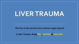
- 1. LIVERTRAUMA The liver is the second most common organ injured in abd.Trauma. being big , central & vascular
- 2. Background Largest solid abdominal organ,fixed position Second most common injured, but most common cause of death after abdominal trauma Blunt MVA most common 80% adults, 97% children-conservative rx
- 3. Pathophysiology Friable parenchyma, thin capsule, fixed position in relation to spine. Right lobe gets hit more since its larger, and closer to ribs. 85% injuries involve segments 6,7,8 from compressioin against ribs, spine, abd wall. Shear forces at attachments to diaphragm Transmission thru right hemithorax.
- 4. Liver injured easily in children since ribs are compliant, force transmitted. Liver not as developed in children, with weaker connective tissue framework. Iatrogenic injuries by biopsies, biliary drainage, TIPS, can cause capsular tears and bile leaks, fistulas, hemoperitoneum.
- 8. (3) Injury to blood vessels and biliary structure : May occur in both type of trauma The liver have two vascular pedicle
- 9. A- Inflow pedicel : Portal vein , H. artery. easy accessible in hepato duod. Lig & controlled by Pringle: maneuver B – Out flow pedicle Hepatic vein , retro hepatic v. cava inaccessible .Total vascular isolation of the liver may be needed ! (4) Spontaneous rupture e.g primary carcinoma , haemangioma , blunt trauma
- 10. Liver injury scale (AMercian ASSocitation of the surgery of trauma (AAST)
- 11. 1. Superficial parenchymal wound: simple suture , diathermy 2. Deep or extensive parenchymal injury : → Paking 3. Deep injury involving major vascular structure → Require special maneuver to achieve hemostasis ( poor prognosis) AAST SUMMURIZESTHE LIVER INJURY SCALE INTO 3TYPES
- 12. Diagnosis A. Clinical : 1. Pt with blunt abd. trauma & Hypotension 2. Contusion over the lower chest 3. Fracture of Rt Lower ribs (9-12 ribs) 4. Penetrating injuries to the Rt lower chest (below the fourth intercostal space 5. Sign of hoemoperitonium
- 13. B. Taping Diagnostic Negative tape does not rule out intra peritoneal organ damage C. Laboratory investigation Blood picture leuko cytosis HB concentration Liver function Of little value in first 24h D. Imaging X Ray abd. And chest Elevated RT copula of the diaphragm Fracture lower sibs E. Ultra sound Nan invasive & can be repeated Highly sensitive in detecting >200ml of intra peritoneal fluid
- 14. F. Computed tomography (CT Scan ) Delineate the anatomy of injured sold organ & grading of the injuries Used in haemodynamically stable Pt. suitable for non operative management )
- 15. G . Selective angiography Should be considered in Pt with active contrast extravasations into the peritoneum seen on CT scan
- 16. Management Hemodynamic stability of the pt. Types of injury Blunt Stable Conservative non operative regardless the grade or extent of injury Un stable Urgent laparotomy penetrating Stap wound stable Unstable Gun shot Usually operative conservative ( careful selected case)
- 17. Complication of Non operative management 1- Ongoing Bleeding Stable Repeat CT Extravasations Selective angiographic embolization Unstable Urgent laparotomy Hypotension ↓ HB ↑ liver size
- 18. 2- Biloma 3- Biliary fistula T. Drainage under U/S guide Failed ERCP ( Endoscopic papillotomy & stending
- 19. 4. Intra Abd. abscess 5. Hepatic abscess 6. Haemobilia • Delayed rupture of false aneurysm of branch of H. artery into biliary tree • c/p → jaundice / biliary colic, upper G.I. Hge • Treatment angiographic Embolization (treat. of choice) t. drainage under U/S guide
- 20. 7. Liver necrosis Small area Large area No treatment Difficult to diagnose May become infected May need resectional debridement & drainage
- 21. 8.Missed Hollow viscus injury • Rare, difficult in diagnosis • RepeatedCT • Laparotomy • Failure to achieve the source ⇛ Death
- 22. laparotomy Superficial Simple suture Local Homeostasis Deep injuries Packing Control Removed after 48h POST operative angiography for selective embolization Uncontrolled Inflow occlusion Bringle maneuver Unstable
- 24. Pringle maneuver for reducing bleeding in the liver surgery has stood the test of time for over a century.The medial free edge of the hepatoduodenal ligament contains the portal vein, hepatic artery, and the common duct. By inserting one’s index finger through the foramen of Winslow and compressing the portal triad between the thumb and index finger, all of the inflow to the liver can be occluded with the rare exception of an anomalous hepatic artery coursing outside the pinched area.This occlusion can alternatively be performed with a nontraumatic instrument or touniquet. C caudate lobe of the liver, P portal vein, hepatic artery, and common duct, D duodenum, IVC inferior vena cava
- 25. Bringle maneuver Bleeding stop Not stop Consider injuries H. vein Retro hepatic IVC Abberant H artery . Special technique .Total Hepatic isolation . Mobilization of the liver . Mortality high
- 26. Summary 1. Blunt hepatic injury in stable Pt ⇛non op. management with adjunctive measures to manage sequelae & complication 2. Missed Hollow Viscous is unusual & carry high mortality with the conservative strategy
- 27. 3. Haemodynamically unstable patient ⇛ emergency laparotomy & managed first by Paking 4. Rarely Pt . With injury to hepatic vein or retro hepatic vena cava ⇛ needs proximal & distal vascular control & even in most experienced hand the mortality rate is high.