SlideShare a Scribd company logo
1 of 76
- Dr. Miliya Parveen
AIRWAY ANALYSIS AND ITS
RELEVANCE IN ORTHODONTICS
CONTENTS
• Introduction
• Anatomy
• Naso – respiratory function and craniofacial
growth
• Methods of analysis
- Clinical examination
- Otorhinolaryngology tests for upper airway
- Supplementary examinations
• LC
• CBCT
• Airway and skeletal patterns
• Obstructive Sleep Apnoea
• Mouth breathing
• Effect of orthodontics on airway
• Extraction cases
• Expansion
• Mandibular advancement
• Orthognathic surgery
• Adenoidectomy or tonsillectomy
• Role of orthodontist
• Conclusion
INTRODUCTION
• Upper and lower airway has always been an area of interest because
the oropharyngeal and nasopharyngeal structures play important roles
in the growth and development of the craniofacial complex.
• In particular, upper airway assessment and its interactions with
craniofacial growth and development are studied by ENT specialists,
laryngologists, speech therapists, pediatricians and dentists.
• The airway, mode of breathing, and craniofacial formation are inter-
related during growth and development that form can follow function
and function can follow form .
• So, it is imperative to normalize form and function as early as
possible, so that function is optimized for life.
• Airway obstruction impairs respiration which leads to craniofacial
malformation, malocclusion and jaw deformation.
• Research also shows that abnormal craniofacial formation can lead to
airway obstruction, impaired respiration, impaired nasal breathing,
chronic mouth breathing, sleep apnea, sleep disorders and lifelong ill-
health.
ANATOMY
• The essential organs of the respiratory
system are the lungs, located on either
side of the thorax, on each side of the
heart, and the great vessels.
• To reach the lungs, atmospheric air
follows the airway, which comprises of
the nasal cavity, the mouth, the pharynx,
larynx, trachea, and bronchi. The upper
airway is formed by the nasal cavity and
the pharynx.
A. Nasal cavity
• The normal airway starts, from the functional perspective, in the
nostrils. The nasal cavity includes the nose, the nasal cavities, and
extend to the back with the nasopharynx.
• A deviated septum, a narrow nasal cavity, and turbinate hypertrophy are
some of the signs that cause mouth breathing and OSAS.
• In allergic rhinitis, which is also related to upper airway obstruction, the
nasal mucous membrane swell with dust particles, pollen or even cold,
also affecting the eyes and nose and causing a decrease in air flow
B. Oral cavity
• It is an essential organ for mastication, phonation, taste, deglutition and
breathing, formed by the maxillary, palatal and mandible bones, the
tongue, lips and cheeks, and the oropharynx at the back.
• The tongue plays many important function but as its muscle tone
decreases during sleep, the tongue can block the upper airway.
• Jointly with the loss of muscle tone of the pharyngeal walls and the soft
palate, it contributes to the collapse of the airway, one of the main causes
of obstructive sleep apnea syndrome.
C. Pharynx
• The pharynx is a tube-like structure formed by muscles and
membranes. It measures approximately 12-14 cm and is divided into
three parts: nasopharynx, oropharynx and laringopharynx.
• The nasopharynx is the upper part of the respiratory system - located
behind the nasal cavity and on the soft palate.
• In the roof sub-mucosa, there is a collection of lymphoid tissue called
pharyngeal tonsil (adenoids) - when large in size, is the main
obstruction to the passage of air through the nasopharynx.
• The oropharynx extends from the 2nd to 4th
vertebra and opens into the oral cavity
through an isthmus. The upper end - soft
palate & lower end - lingual side of the
epiglottis.
• The tongue is the main blocking element
in the oropharynx- contraction of the
genioglossus muscle moves the tongue
forward during inspiration, and thereby
acts as a pharyngeal dilator.
• The laringopharynx joins the oropharynx
at the pharyngoepiglottic fold and hyoid
bone, and continues up to the sixth
vertebra. It is behind the opening in the
larynx.
NASO – REPIRATORY FUNCTION AND
CRANIOFACIAL GROWTH
• Dentofacial morphology can be altered by naso-respiratory obstruction
depending on the magnitude, duration & time of occurrence.
• Quinn in 1978 reiterated that mouth breathing is one of the early
symptoms of unnatural acts of breathing and that dramatic deformities
of the face, jaws and dentition can be caused by inability to breathe
through the nose properly.
• Ricketts in 1979 stated that when there is a lack of function in the
nose, there can be a growth inhibition. Under normal function in
breathing, a pressure develops. In the abnormal, this pressure is
changed to vacuum and the maxillary complex is sucked inward,
restricting the basal cone.
• In 1918, Norlund introduced the ‘compression theory’ which stated
that constriction of the maxillary arch is related to the absence of the
lateralizing pressure of the tongue against the palate. In response to
nasal obstruction, the tongue drops and the medializing effects of the
buccal musculature is left unopposed. The effect is further enhanced
by a pressure differential across the hard palate in the absence of
nasal airflow, leading to a narrow, high- arched palate.
• Norlund also put forward the ‘theory of inactivity’, according to
which there is reduced growth of the nasal cavity, due to its inactivity,
as suggested by Korner (1891) and Bentzen (1903).
• The air pressure theory described by Kantorowicz (1916) and James
& Hastings (1932) holds that a change to mouth breathing causes the
normal negative pressure in the anteriorly sealed oral cavity to be
lost, and the palate thus is not carried downward with the growth of
the maxillary alveolar process.
• The excavation theory proposed by Bloch (1903) and Michel (1908)
states that an upward stream of oral airflow presses on the palate
leading to higher palatal vault.
• Although the literature is replete with statements that airway
impairment alters facial and dental growth, there is substantial
evidence to the contrary.
• Bushey found no relationship between nasal respiration and linear
measurements of the adenoids on lateral skull cephalograms before
and after surgical removal of the tonsils and adenoids.
• Kingsley (1989) noted normal craniofacial development in children
with severe nasal obstruction and Whitaker described severe palate
malformations in patients who had undergone adenoidectomy at an
early age.
• More recent findings suggest that nasal-oral breathing per se is not
necessarily harmful to craniofacial growth. However, in instances
where the nasopharyngeal or oropharyngeal airspace is small,
exaggerated postural responses in mouthbreathers may be detrimental
to craniofacial growth.
METHODS OF ANALYSIS
Clinical examination
• The general well-being of the child, including growth and development,
must be determined.
• Pertinent past history include birth trauma, early childhood trauma,
previous hospitalizations, medications, and surgical history.
• Physical assessment includes facial morphology, skeletal jaw
relationships, functional assessment of nostrils, the size and function of
the tongue and the anatomy of the soft palate, uvula and tonsils.
• Regarding facial morphology, Class II patterns due to mandibular
retrusion have smaller upper airway volumes, which is usually
associated with adenoid hypertrophy with a very short upper lip and a
thick and everted bottom lip.
• Swallowing difficulties may be noted.
• Voice quality (degree of nasality) and clarity, daytime
hypersomnolence, and school/behavioral difficulties should be
evaluated.
• Sleep history may often reveal loud irregular snoring, restless sleep,
abnormal sleep position and nocturnal mouth breathing.
• Resting mouth position is noted.
• "Adenoid facies" is characterized by an open mouth, dull facial
appearance, and short upper lip. This is nonspecific for chronic nasal
obstruction.
• Other craniofacial anomalies may be associated with these symptoms
including cleft palate, Down syndrome, etc.
• Tonsillar hypertrophy, macroglossia and oropharyngeal masses should
be evaluated.
• The nasal cavity was inspected for the presence of secretions, edema
and erythema of the nasal mucosa.
• The ears should be evaluated as otitis media certainly is associated
with nasal obstruction problems.
• Bony nasal anomalies, external masses, pits, etc. should be evaluated.
a) Functional assessment of nostrils (Duran V.) :
To do this we observe nostril response to intense inspiration, paying
special attention to the degree of collapse during the maneuver. This is
the classification obtained -
• Value 0: Dilated nostrils both at rest and in deep inspiration
• Value 1: Narrowed nostrils at rest, without functional collapse
• Value 2: Functional partial unilateral collapse
• Value 3: Functional total unilateral or bilateral partial collapse
• Value 4: Functional partial collapse of one nostril and total collapse of
the other one
• Value 5: Total functional collapse in both nostrils
b) Intraoral evaluation of tonsils
The tonsils are assessed according to the degree of obstruction of the
oropharynx - a reliable clinical evaluation method.
• Grade 1 - the tonsils are within their cavity
• Grade 2 - do not exceed the midline between the uvula and the anterior
pillar of the soft palate
• Grade 3 - go over the midline between the uvula and the anterior pillar
• Grade 4 - the tonsils are less than 4 mm between them.
Grades 3 or 4 represents a decrease in airway permeability
c) Upper airway assessment with the Mallampati score - evaluates the
risk of obstruction of the airway.
• Based on the visual assessment of the oropharyngeal structures, mainly
the distance between the tip of the uvula and the tongue base.
• Class 1 - full visibility of the tonsils, uvula and soft palate
• Class 2 - hard and soft palate, the upper section of the tonsils and uvula
• Class 3 - hard and soft palate, and the base of the uvula
• Class 4 - only the hard palate. Classes 3 and 4 are commonly present in
breathing-related sleep disorders, even after an adenotonsillectomy
Most Commonly Used Otorhinolaryngology
Tests For Upper Airway Assessment
a) Rhinomanometry - evaluate nasal obstruction.
• There are different types of rhinomanometry (RMM), active anterior
RMM being the one most frequently used. This evaluates nasal
airflow in inspiration and expiration by detecting potential
obstructions and/or resistance. This can be done with a face mask and
it requires full patient cooperation.
• After placing the mask, airflows are measured with the
rhinomanometer, and the data are analyzed computationally and then
graphs are designed in pressure/volume curves.
• The recording is repeated under the effect of a topical vasoconstrictor,
which will differentiate mechanical obstructions (which do not vary
with the vasoconstrictor), vasomotor obstructions (which fully
improve with the vasoconstrictor) and mixed obstructions (which
improve partially with the vasoconstrictor).
b) Acoustic rhinometry - study of the geometry of the nasal cavity.
• Based on the analysis of sound reflection and provides a calculation
of cross-sectional areas of the nasal cavity and of certain nasal
volumes.
• Done by generating an audible sound in the nostril with an adapter,
taking care not to deform the nasal vestibule. The sound wave
penetrates the cavities and is reflected on the different nasal structures
or their irregularities.
• MCA1 corresponds anatomically to the area at the nasal valve level
which has the greatest resistance in the normal nose. MCA2
corresponds to the area at the level of the head of the inferior
turbinate.
• As in active anterior RMM, the study can be performed before and
after applying a vasoconstrictor for the same purpose and with a
similar interpretation of the results.
• Incident wave signals are measured and
reflected according to time, which makes
it possible to determine the distance, from
the nostril, where there is a change in
acoustic impedance.
• The most interesting data are the
“minimum cross-sectional areas 1 and 2
(MCA1 and MCA2)”.
c) C) Nasopharyngolaryngoscopy - evaluates the anatomy of the upper
airway, soft palate, the movement of the vocal cords and the process
of deglutition.
• Performed with a flexible fiberscope which is inserted through the
nasal cavities to observe both pharynx and larynx.
• The patient is usually awake, and topical lidocaine is applied on the
nostrils.
• During the test, the patient may be asked to talk, cough or swallow,
depending on what is being evaluated.
• The following anatomical elements should be evaluated:
i. deviations of the nasal septum
ii. size of inferior turbinates
iii. presence and size of the adenoid tissue
iv. quantity and quality of nasal secretion
v. size of palatine tonsils & base of the tongue
vi. its relationship with the oropharyngeal cavity
vii. abduction of the vocal cords
viii. subglottic diameter
ix. presence of masses or pathological deformities at any levels
d) Functional Nasal Permeability (PeNaF) - assesses the independent
functional nasal permeability of each cavity.
• The performance is recorded as negative (-) when the patient
maintains nasal breathing for six inspirations at rest, and positive (+)
when the patient fails to maintain it for six inspirations.
• A study validated in Chile recommends orthodontists implement this
simple examination to rule out a possible nasal obstruction. If this is
not the case, they should request an objective assessment to check the
increase in nasal resistance
e) SNORT - Simultaneous Nasal Oral Respirometric Technique by
Gurley and Vig reported a enabled the direct and simultaneous
measurement of inspired and expired air, both orally and nasally.
• Using a custom fitted face mask with separate valves attached to the
nose and mouth and attached to a flow meter, air pressure transducer,
recorder and computer, it can give the nasal versus the oral
inspiration, expiration and their ratios.
• SNORT permits the objective quantification of the ratio of oral to
nasal airflow and permits a numerical determination of both normal
and pathological states of breathing mode.
Supplementary Examinations
a) Upper airway assessment in lateral cephalograms
• Lateral cephalometry is commonly used in clinical practice given its
relative simplicity, accessibility, low cost and low exposure to
radiation.
• Lateral cephalograms provide reliable linear measurements, can
measure the dimensions of the nasopharyngeal and retropalatal
regions, but have not proven to be reliable to measure the airway in
the back of the tongue.
• Though multiple authors have tried to standardize a method of airway
analysis on lateral cephalogram, very few methods have been widely
accepted.
McNamara’s Analysis (1984) –
• Two measurements are used to examine the possibility of an airway
impairment.
• The upper pharyngeal width is measured from a point on the posterior
outline of the soft palate to the closest point on the posterior pharyngeal
wall.
• This measurement is taken on the anterior half of the soft palate outline
because the area immediately adjacent to the posterior opening of the
nose is critical in determining upper respiratory patency.
• Apparent airway obstruction, as indicated by an opening of 5 mm or
less in the upper pharyngeal measurement - only an indicator of
possible airway impairment.
• The average upper airway measurement for adults of both sexes is
17.4 mm.
• Lower pharyngeal width is measured from the intersection of the
posterior border of the tongue and the inferior border of the mandible
to the closest point on the posterior pharyngeal wall.
• The average value for this measurement is 10 to 12 mm and does not
change appreciably with age.
• In contrast to the upper pharynx, a smaller than average value for the
lower pharynx is not remarkable. A lower pharyngeal width of greater
than 15 mm suggests anterior positioning of the tongue, either as a
result of habitual posture or due to an enlargement of the tonsils.
Martin’s Analysis (2006) –
The following cephalometric measurements
were made –
1. PNS-AD1: lower airway thickness;
distance between PNS and the nearest
adenoid tissue measured through the PNS-
Ba line (AD1).
2. AD1-Ba: lower adenoid thickness; defined
as the soft-tissue thickness at the posterior
nasopharynx wall through the PNS-Ba line.
3. PNS-Ba: lower airway thickness; distance
between PNS and Ba—the sum of
variables 1 and 2.
4. PNS-AD2: upper airway thickness;
distance between PNS and the nearest
adenoid tissue measured through a
perpendicular line to S-Ba from PNS
(AD2).
5. AD2-H: upper adenoid thickness;
defined as the soft-tissue thickness at the
posterior nasopharynx wall through the
PNS-H line.
● Hormion (H): cephalometric point located
near the adenoidal tissue at cranial base
localized where a perpendicular to S-Ba line
crosses the sphenoid bone. The variations of
this point are minimal because it is located
far from growing sites.
6. PNS-H: upper airway thickness; distance
between PNS and H—the sum of variables
1 and 2.
7. N-H: nasal fossa length; distance between
N and H.
8. S-N: anterior cranial base.
9. McNamara’s upper pharynx dimension
10.McNamara’s lower pharynx dimension
11 and 12. Total adenoidal, and aerial areas: the Ba-N plane, the bispinal
plane, and 2 perpendicular lines to the bispinal plane: 1 that crosses the
more anterior point at atlas vertebra and other that crosses the PNS. The
resulting trapezoid is divided in 2 spaces (aerial and adenoid).
Fujioka et al. (1979) - described the adenoidal-nasopharyngeal ratio
(AN ratio).
• Relates the length of the line perpendicular to the sphenoid bone (A)
by the thickest portion of the adenoids with the distance between the
posterior nasal spine and the anterior edge of the sphenobasioccipital
synchondrosis (N).
• An AN< 0.8 is considered normal and an AN > 0.8 is considered
enlarged
• In addition, Feres, Murilo et al. in 2012 found that both parameters
had good reproducibility and a variability which was not clinically
significant.
• One of the most common reasons for upper airway obstruction is
hypertrophic adenoids. Before planning an orthodontic treatment, this
area is usually observed in the lateral cephalometry.
• A study was conducted to assess whether adenoidal ratio on lateral
cephalograms can be used to estimate airway volumes, using CBCT
as the validation method. They concluded that the lateral cephalogram
can provide some information about the nasopharyngeal space,
particularly in patients over 15.
• This is due to the stability reached by the tissue at this age; however,
it cannot be used as a diagnostic procedure to determine the volume
of the total airway, but rather as an assessment tool to determine the
need for a more comprehensive ENT examination.
b) Upper airway assessment using CBCT
• Several studies have shown that CBCT is accurate and reliable for
upper airway assessment.
• Volumetric reconstructions that may be obtained from CBCTs help
clinicians make a correct diagnosis and indicate a better treatment
plan for some pathologies of the maxillofacial area, especially those
related to the airway.
• For the volumetric reconstruction and visualization of the upper
airway, software programs must allow us to find the correct location
of the boundaries of the pharynx and nasal cavity (segmentation)
through a process that can be manual, automatic or semi-automatic.
• Studies comparing the commercial software programs for the study of
the airway were analyzed. They were found to have reliable
reproducible and accurate results of linear measurements, but they
lost accuracy when calculating the volume of the airway. This could
be due to the automatic segmentation of the nasal cavity, the
nasopharynx and oropharynx.
• The head position and the position of the patient when the CBCT is
taken must be considered to obtain accurate and repeatable upper
airway measurements and volumes.
• The position of the hyoid bone and tongue, and the dimension of the
airway would be highly reproducible using the natural position of the
head when taking lateral cephalograms.
• It has been found that individuals would be approximately 40% more
affected by the width of the airway in an upright position.
• Solow et al. determined that in an upright position or by increasing
the cervical skull angle, there is an increase in upper airway
diameters.
• Alsufyani et al. states that images must be obtained with the patient in
a sitting position so as not to affect airway diameter.
• Aboudara et al. stated that CBCT is a simple and effective method to
evaluate upper airway. They compared the volumetric measurements
from CBCT with known physical airway phantoms and found that the
errors ranged from 0 to 5%. They also reported a moderately high
correlation (r = 0.75) between the sagittal area and the volume when
correlating lateral cephalograms measurements with CBCT data.
• Two systematic literature reviews concluded that although major
progress has been made in the capture and management of CBCT
images, there is no optimized evidence-based protocol to obtain
images to analyze the upper airway.
• Several obstacles must still be overcome, such as the influence of the
position of the tongue, mandible morphology, the impact of the
respiratory phase and the definition of the anatomical boundaries of
the upper airway, as well as the lack of consistency in the
configuration of the equipment and in how images and volumetric
reconstructions are obtained.
AIRWAY AND SKELETAL PATTERNS
• A study conducted in New Delhi compared the reliability of lateral
cephalograms and computed tomographies to assess airways. They
compared three skeletal patterns determined by the different values of
the ANB angle, and related their linear values taken from the
cephalometries to volumetric values delivered by CT, and concluded
that the skeletal pattern had a strong association with pharyngeal
volume and its linear dimensions. They also noted that the S-shaped
soft palate can be considered high-risk for sleep apnea compared to
the leaf shape, which is more common.
• In contrast, Dalmau et al., found no statistically significant
differences that correlate airway with skeletal patterns or facial
biotypes.
• The recent results of Lucas Castro-Silva et al., in Brazil, found a
positive correlation of higher values of pharyngeal airway for Class
III patients.
• A new study by El and Palomo found that oropharyngeal airway
volumes were lower in Class II patients compared to Class I and
Class III patients. They also state that the mandibular position with
respect to the skull base has a strong impact on oropharyngeal
volume.
OBSTRUCTIVE SLEEP APNEA
• Definition: condition caused either by complete occlusion or partial
collapse of the upper airway despite the presence of simultaneous
respiratory effort. Cessation occurs at the level of nostrils and mouth.
• Condition is considered pathologic when the episodes last for at least
ten seconds and at a frequency of 30 times or more during 7 hrs. of
nocturnal sleep in REM and especially in non REM stages of sleep.
• Diagnosis is by polysomnography - Measurements are made to assess
sleep stages of breathing and gas exchange to detect sleep stages.
PSG ensures the no. of apnic episodes per hour of sleep expressed by
respiratory(Disturbance Index)measurements of chest and abdominal
efforts and oxygen saturation.
• Airway measurement by cephalometric 3D imaging – lateral
pharyngeal dimension.
• Treatment Options -
− Enlarging the upper airway
− Continuous positive airway pressure
− Weight loss
− Oral appliances - mandibular advancement devices
− Upper airway surgery
− Combination CPAP/ dental sleep device therapy
MOUTH BREATHING
• Clinical examination – ask patient to hold water in the mouth, use
double sided mouth mirror or cotton wisps
• Cephalometric Analysis: -
1. McNamara airway analysis –
• Upper pharyngeal width – the point on posterior outline on soft palate
to closest point on pharyngeal wall – 15 to 20 mm in width. Values
2mm or less indicate airway impairment. Lower pharyngeal width
from point of intersection of posterior border of tongue and inferior
border of mandible to the closet point on posterior pharyngeal wall –
11 to 14mm.
2. Vertical growth pattern, increased ANB, increased gonial angle,
decreased mandibular length, steep MP angle, over erupted upper
posterior segments
EFFECT OF ORTHODONTICS ON AIRWAY
1. Extraction and airway changes:
• A systematic review on the effects of bicuspid extractions and incisor
retraction on upper airway of Asian adults and late adolescents
showed that Linear airway response to incisor retraction measured on
lateral cephalograms varied substantially, while linear, cross‐sectional
and volumetric measurements of posterior airway space using CT
showed larger effect sizes and smaller variations, providing evidence
of airway narrowing with bicuspid extractions and incisor retraction.
• An article published in Orthodontics and craniofacial research journal
on three-dimensional pharyngeal airway changes in orthodontic
patients treated with and without extractions analysed the pharyngeal
airway for subjects treated with extractions and without extractions.
• Nasopharyngeal (NP) and oropharyngeal (OP) volumes, area of
maximum pharyngeal constriction (AMPC), and upper arch perimeter
were measured on CBCT scans.
• There were no statistically significant differences in the pharyngeal
airway values between the extraction and non-extraction groups.
• The extraction group showed a statistically significant increase for NP
and OP volumes and AMPC values. Such increase was also noted in
the non-extraction group, without statistical significance for AMPC
values.
• A retrospective study evaluating the airway space changes after
extraction of four second premolars and orthodontic space closure in
adult female patients with bimaxillary protrusion showed orthodontic
treatment increases the vertical airway length, which is the amount of
distance between base of the tongue and posterior nasal spine.
• A study conducted on pharyngeal airway dimensional changes after
premolar extraction in skeletal class II and class III, growing and
adult orthodontic patients showed significant increase in
nasopharyngeal airway dimension in class II and class III patients,
whereas palatopharyngeal and glossopharyngeal dimensions were
insignificantly decreased in both groups.
2. Rapid Maxillary Expansion
• RME is successful in increasing the nasal permeability and reducing
airway resistance.
• Reduced airway resistance reduces negative pressure during
ventilation, with promising results in the treatment of paediatric sleep
disordered breathing, including obstructive sleep apnoea
• The effects on more distant structures include stretching of the tensor
palatine muscles by the expanding maxilla with subsequent
improvement in drainage of the Eustachian tubes, aiding in reducing
otitis media and conductive hearing loss
• Enlarged palatal space may also allow for an improved tongue
posture, which could facilitate increased airway space in the
oropharynx
• Donald Timms, in a study with Rapid Maxillary Expansion, reported
a 37 percent mean drop in nasal resistance with 7 mm of expansion.
• In another larger study of children with a previous history respiratory
disease treated with Rapid Maxillary Expansion, 82 percent reported
improvement in number of upper respiratory tract infections and 60
percent reported improvement of allergic rhinitis.
• According to Bishara and Staley -
Anatomically, there is an increase in the
width of the nasal cavity immediately
following expansion, particularly at the
floor of the nose adjacent to the
midpalatal suture. As the maxillae
separate, the outer walls of the nasal
cavity move laterally. The total effect is
an increase in the intranasal capacity. The
nasal cavity width gain averages 1.9 mm
but can widen as much as 8 to 10 mm at
the level of the inferior turbinates, while
the more superior areas might move
medially.
• A number of rhinologists indicate that RME, in addition to its
widening procedure, results in correction of septal deformity as a
result of the lowering of the floor of the nasal cavity.
• Hershey, Stewart, and Warren, and Turbyfill reported a reduction of
nasal airway resistance by an average of 45% to 53% with RME.
• Wertz concluded that opening the midpalatal suture for the purpose of
increasing nasal permeability cannot be justified unless the
obstruction is shown to be in the lower anterior portion of the nasal
cavity and accompanied by a relative maxillary arch width deficiency.
• Graber believes that the claims of improved nasal breathing
apparently as a result of RME are most likely only temporary.
Spontaneous regression of lymphoid tissues during growth
automatically improves nasal breathing, even if nothing is done to the
palate.
• Therefore, it can be concluded that the effect of RME on the nasal
airway will to a great extent depend on the cause, location, and the
severity of the nasal obstruction
3. Mandibular advancement
• Mandibular repositioning is most effective in mild to moderate
obstructive sleep apnea.
• It enhances retro pharyngeal air space thereby increasing nasal airway
patency.
1. Mandibular repositioners - open the airway by moving the mandible
forward.
As the jaw is moved forward, the collapsible part of your airway is held
open by the forward movement of the tongue and other airway muscles.
• 2. Mandibular Advancement Devices (MADs) also improve the
strength and rigidity of the airway by increasing the muscle activity
of the tongue and other muscles of the airway.
3. Elastic Mandibular Advancement (EMA ) Appliance
4. Tongue retaining devices (with airway tubes)
• Like MADs, Tongue Retaining Devices (TRDs) also work by holding the
tongue in a forward position.
• These devices pull the tongue forward, but instead of moving the jaw
forward like a Mandibular Advancement Device (MAD), TRDs directly
control the tongue itself.
• In some cases, Tongue Retaining Devices (TRDs) have decreased
therapeutic complications compared to MADs, but TRDs can also be less
comfortable and generally take several weeks or months to be worn
comfortably.
EFFECT OF ORTHOGNATHIC SURGERY ON
AIRWAY
• If the difference between the maxillary and the mandibular unit length
is greater than 16mm at the age of 12 in class II malocclusion and
greater than 29mm at the age of 12 in class III dysplasias, surgical
intervention is necessary.
• Surgical superior impaction of the maxilla has become an accepted
treatment for the correction of vertical maxillary excess and it does
reduce nasal resistance, but it does not increase the percentage of
nasal airflow.
• It has been speculated that this change may be associated with the
common postoperative increase in interalar width and widening of the
external nares, which result in an opening of the liminal valve.
• At the present time,
maxillomandibular advancement
(MMA) remains the most
effective surgical procedure in
treating patients with moderate
to severe OSA.
• Advancement of maxillofacial
skeletal structures opens the
oropharyngeal airway and places
the oropharyngeal musculature
on tension.
• MMA surgery results in a significant increase in the volume and a
morphologic airway change from a round to an elliptical f shape in the
upper airway space in patients with OSA.
• These changes are stated to reduce the collapsibility of the upper airway
during deep stages of sleep.
ROLE OF ORTHODONTIST
• In 1987, Weimert published a study of 1360 patients referred to
otolaryngologists by orthodontists because of suspicion of nasal
obstruction. Although it was not a solid scientific study, the findings
suggest that orthodontists can effectively screen for nasal obstruction.
• The most common reasons for referral were: dentofacial characteristics
suggestive of upper airway obstruction, inability to retain a dental
appliance, and unsatisfactory results from an orthodontic program.
• Dentists are important as both referrers and treating doctors.
• Allergies generally must be treated first, before consideration of any
surgery. The greatest number of surgical failures are in allergic patients.
• The greatest number of orthodontic relapses are in patients who have not
had their breathing problems successfully treated.
• Orthodontic therapy is affected by the function of the lips, tongue,
and masticatory musculature, all of which may accommodate to nasal
obstruction in ways which can effect occlusion.
• Effective orthodontic therapy may require the elimination of the nasal
obstruction to allow for normalization of the facial musculature
surrounding the dentition.
• According to Meredith, the growth of the face (excluding the
mandible) is completed at a relatively early age. 60% of craniofacial
development takes place during the first 4 years of life and 90% by
age 12. Based on these observations, any intervention to open the
airway must take place at an early age.
• As a referring doctor, one has to decide between allergist, ENT,
pediatrician or sleep lab.
CONCLUSION
• In spite of the long history of research between respiration and
malocclusion, only now is this aspect of diagnosis and treatment
given its due importance.
• Evaluation of children with nasal obstruction and dental abnormalities
requires a multidisciplinary approach between the orthodontists,
pediatricians, and otolaryngologists.
• The orthodontists must be familiar with the dental literature so as to
provide optimal care for pediatric patients as they have an opportunity
to examine and institute treatment to the patients at a very early age.
REFERENCES
• Rojas E, Corvalán R, Messen E, Sandoval P. Upper airway assessment in Orthodontics: a review. Odontoestomatologia. 2017 Dec
1;19(30).
• Martins LS, Liedke GS, Heraldo LD, Da Silveira PF, Arus NA, Ongkosuwito EM, Vizzotto MB. Airway volume analysis: is there a
correlation between two and three-dimensions?. European journal of orthodontics. 2018 May 25;40(3):262-7.
• Huynh NT, Desplats E, Almeida FR. Orthodontics treatments for managing obstructive sleep apnea syndrome in children: A
systematic review and meta-analysis. Sleep medicine reviews. 2016 Feb 1;25:84-94.
• Indriksone I, Jakobsone G. The upper airway dimensions in different sagittal craniofacial patterns: a systematic review.
Stomatologija. 2014 Jan 1;16(3):109-7.
• Lenza MG, Lenza MD, Dalstra M, Melsen B, Cattaneo PM. An analysis of different approaches to the assessment of upper airway
morphology: a CBCT study. Orthodontics & craniofacial research. 2010 May;13(2):96-105
• Preston CB, Lampasso JD, Tobias PV. Cephalometric evaluation and measurement of the upper airway. InSeminars in Orthodontics
2004 Mar 1 (Vol. 10, No. 1, pp. 3-15). WB Saunders.
• Ng JH, Song YL, Yap AU. Effects of bicuspid extractions and incisor retraction on upper airway of Asian adults and late
adolescents: A systematic review. Journal of oral rehabilitation. 2019 Nov;46(11):1071-87.
• Becking BE, Verweij JP, Kalf-Scholte SM, Valkenburg C, Bakker EW, van Merkesteyn JP. Impact of adenotonsillectomy on the
dentofacial development of obstructed children: a systematic review and meta-analysis. European journal of orthodontics. 2017 Oct
1;39(5):509-18.
• Banerjee D, Shrivastav D, Kamble D, Dhannawat D, Purva V. Lateral Cephalogram and CBCT as a diagnostic aid for analysis of
airway-Review Article. European Journal of Molecular & Clinical Medicine. 2020 Dec 22;7(7):1802-8
• Bidjan D, Sallmann R, Eliades T, Papageorgiou SN. Orthopedic Treatment for Class II Malocclusion with Functional Appliances and
Its Effect on Upper Airways: A Systematic Review with Meta-Analysis. Journal of clinical medicine. 2020 Dec;9(12):3806.
• Stefanovic N, El HA, Chenin DL, Glisic B, Palomo JM. Three‐dimensional pharyngeal airway changes in orthodontic patients treated
with and without extractions. Orthodontics & craniofacial research. 2013 May;16(2):87-96.
• Schendel SA, Broujerdi JA, Jacobson RL. Three-dimensional upper-airway changes with maxillomandibular advancement for
obstructive sleep apnea treatment. American Journal of Orthodontics and Dentofacial Orthopedics. 2014 Sep 1;146(3):385-93.
• Jayan B, Kadu A. Airway-focused orthodontics. Journal of Indian Orthodontic Society. 2018 Apr;52(4_suppl1):23-8.
Airway analysis and its relevance in orthodontics

More Related Content

What's hot

Traditional begg technique stage 1 and stage 2
Traditional begg technique stage 1 and stage 2Traditional begg technique stage 1 and stage 2
Traditional begg technique stage 1 and stage 2Indian dental academy
 
Tongue and its importance in orthodontic treatment /certified fixed orthodont...
Tongue and its importance in orthodontic treatment /certified fixed orthodont...Tongue and its importance in orthodontic treatment /certified fixed orthodont...
Tongue and its importance in orthodontic treatment /certified fixed orthodont...Indian dental academy
 
Natural head position /certified fixed orthodontic courses by Indian dental a...
Natural head position /certified fixed orthodontic courses by Indian dental a...Natural head position /certified fixed orthodontic courses by Indian dental a...
Natural head position /certified fixed orthodontic courses by Indian dental a...Indian dental academy
 
Begg’s philosophy and technique
Begg’s philosophy and techniqueBegg’s philosophy and technique
Begg’s philosophy and techniqueDr Susna Paul
 
COGS analysis (Cephelometrics for orthognathic surgery) / fixed orthodontics ...
COGS analysis (Cephelometrics for orthognathic surgery) / fixed orthodontics ...COGS analysis (Cephelometrics for orthognathic surgery) / fixed orthodontics ...
COGS analysis (Cephelometrics for orthognathic surgery) / fixed orthodontics ...Indian dental academy
 
Condyle secondary cartilage-a misnomer /certified fixed orthodontic courses ...
Condyle  secondary cartilage-a misnomer /certified fixed orthodontic courses ...Condyle  secondary cartilage-a misnomer /certified fixed orthodontic courses ...
Condyle secondary cartilage-a misnomer /certified fixed orthodontic courses ...Indian dental academy
 
Molar distalisation
Molar distalisationMolar distalisation
Molar distalisationTony Pious
 
Functional matrix Hypothesis- Revisited
Functional matrix Hypothesis- RevisitedFunctional matrix Hypothesis- Revisited
Functional matrix Hypothesis- RevisitedDr Susna Paul
 
Postero anterior cephalometric analysis /certified fixed orthodontic courses ...
Postero anterior cephalometric analysis /certified fixed orthodontic courses ...Postero anterior cephalometric analysis /certified fixed orthodontic courses ...
Postero anterior cephalometric analysis /certified fixed orthodontic courses ...Indian dental academy
 

What's hot (20)

Traditional begg technique stage 1 and stage 2
Traditional begg technique stage 1 and stage 2Traditional begg technique stage 1 and stage 2
Traditional begg technique stage 1 and stage 2
 
Sassouni's analysis
Sassouni's analysisSassouni's analysis
Sassouni's analysis
 
Pitchfork Analysis
Pitchfork AnalysisPitchfork Analysis
Pitchfork Analysis
 
Tongue and its importance in orthodontic treatment /certified fixed orthodont...
Tongue and its importance in orthodontic treatment /certified fixed orthodont...Tongue and its importance in orthodontic treatment /certified fixed orthodont...
Tongue and its importance in orthodontic treatment /certified fixed orthodont...
 
Natural head position /certified fixed orthodontic courses by Indian dental a...
Natural head position /certified fixed orthodontic courses by Indian dental a...Natural head position /certified fixed orthodontic courses by Indian dental a...
Natural head position /certified fixed orthodontic courses by Indian dental a...
 
Tweeds
TweedsTweeds
Tweeds
 
Begg’s philosophy and technique
Begg’s philosophy and techniqueBegg’s philosophy and technique
Begg’s philosophy and technique
 
Occlusograms
OcclusogramsOcclusograms
Occlusograms
 
Intrusion arches
Intrusion archesIntrusion arches
Intrusion arches
 
COGS analysis (Cephelometrics for orthognathic surgery) / fixed orthodontics ...
COGS analysis (Cephelometrics for orthognathic surgery) / fixed orthodontics ...COGS analysis (Cephelometrics for orthognathic surgery) / fixed orthodontics ...
COGS analysis (Cephelometrics for orthognathic surgery) / fixed orthodontics ...
 
Pa ceph analysis
Pa ceph analysisPa ceph analysis
Pa ceph analysis
 
Cephalometrics / for orthodontists by Almuzian
Cephalometrics / for orthodontists by AlmuzianCephalometrics / for orthodontists by Almuzian
Cephalometrics / for orthodontists by Almuzian
 
Growth rotations in orthodontics
Growth rotations  in orthodonticsGrowth rotations  in orthodontics
Growth rotations in orthodontics
 
Condyle secondary cartilage-a misnomer /certified fixed orthodontic courses ...
Condyle  secondary cartilage-a misnomer /certified fixed orthodontic courses ...Condyle  secondary cartilage-a misnomer /certified fixed orthodontic courses ...
Condyle secondary cartilage-a misnomer /certified fixed orthodontic courses ...
 
Soft tissue analysis
Soft tissue analysisSoft tissue analysis
Soft tissue analysis
 
Growth prediction (2)
Growth prediction (2)Growth prediction (2)
Growth prediction (2)
 
Vertical maxillary excess
Vertical maxillary excessVertical maxillary excess
Vertical maxillary excess
 
Molar distalisation
Molar distalisationMolar distalisation
Molar distalisation
 
Functional matrix Hypothesis- Revisited
Functional matrix Hypothesis- RevisitedFunctional matrix Hypothesis- Revisited
Functional matrix Hypothesis- Revisited
 
Postero anterior cephalometric analysis /certified fixed orthodontic courses ...
Postero anterior cephalometric analysis /certified fixed orthodontic courses ...Postero anterior cephalometric analysis /certified fixed orthodontic courses ...
Postero anterior cephalometric analysis /certified fixed orthodontic courses ...
 

Similar to Airway analysis and its relevance in orthodontics

Obstructive sleep apnea (osa)The relationship of airway obstruction and dento...
Obstructive sleep apnea (osa)The relationship of airway obstruction and dento...Obstructive sleep apnea (osa)The relationship of airway obstruction and dento...
Obstructive sleep apnea (osa)The relationship of airway obstruction and dento...奇卿 黃
 
Importance of respiration, deglutition and speech in development of head and ...
Importance of respiration, deglutition and speech in development of head and ...Importance of respiration, deglutition and speech in development of head and ...
Importance of respiration, deglutition and speech in development of head and ...Kunaal Agrawal
 
Upper airway and cranial morphology
Upper airway and cranial morphologyUpper airway and cranial morphology
Upper airway and cranial morphologyDr.shifaya nasrin
 
NASOPHARYNGEAL SPACE AND ORTHODONTICS.pptx
NASOPHARYNGEAL SPACE AND ORTHODONTICS.pptxNASOPHARYNGEAL SPACE AND ORTHODONTICS.pptx
NASOPHARYNGEAL SPACE AND ORTHODONTICS.pptxDr. Zeba Siddiqui
 
Orthodontic diagnostic procedures part 3
Orthodontic diagnostic procedures                part 3 Orthodontic diagnostic procedures                part 3
Orthodontic diagnostic procedures part 3 Maher Fouda
 
FUNCTIONAL INFLUENCES ON OROFACIAL DEVELOPMENT
FUNCTIONAL INFLUENCES ON OROFACIAL DEVELOPMENTFUNCTIONAL INFLUENCES ON OROFACIAL DEVELOPMENT
FUNCTIONAL INFLUENCES ON OROFACIAL DEVELOPMENTShehnaz Jahangir
 
hard and soft palate in prosthodontics
 hard and soft palate in prosthodontics hard and soft palate in prosthodontics
hard and soft palate in prosthodonticsmadhushreepatil5
 
3.NASO-RESPIRATORY OBSTRUCTION.ppt
3.NASO-RESPIRATORY OBSTRUCTION.ppt3.NASO-RESPIRATORY OBSTRUCTION.ppt
3.NASO-RESPIRATORY OBSTRUCTION.pptIRONMASTER96
 
Obstructive sleep apnea /certified fixed orthodontic courses by Indian denta...
Obstructive sleep apnea  /certified fixed orthodontic courses by Indian denta...Obstructive sleep apnea  /certified fixed orthodontic courses by Indian denta...
Obstructive sleep apnea /certified fixed orthodontic courses by Indian denta...Indian dental academy
 
Velopharyngeal insufficiency parag
Velopharyngeal insufficiency paragVelopharyngeal insufficiency parag
Velopharyngeal insufficiency paragParag Deshmukh
 
Group a presentation 20th feb 2012
Group a presentation 20th feb 2012Group a presentation 20th feb 2012
Group a presentation 20th feb 2012Sana Anwari
 
mouth-breathing-habit-in-children-pedo
 mouth-breathing-habit-in-children-pedo mouth-breathing-habit-in-children-pedo
mouth-breathing-habit-in-children-pedoParth Thakkar
 
H.A. MOUTH, THROAT, ETC.pptx
H.A. MOUTH, THROAT, ETC.pptxH.A. MOUTH, THROAT, ETC.pptx
H.A. MOUTH, THROAT, ETC.pptxMcRamilBPradero1
 
development & growth of tongue
development & growth of tonguedevelopment & growth of tongue
development & growth of tonguedrdishashah
 
3-140604041741-phpapp01 (2).pdf
3-140604041741-phpapp01 (2).pdf3-140604041741-phpapp01 (2).pdf
3-140604041741-phpapp01 (2).pdfAderawAlemie
 

Similar to Airway analysis and its relevance in orthodontics (20)

Obstructive sleep apnea (osa)The relationship of airway obstruction and dento...
Obstructive sleep apnea (osa)The relationship of airway obstruction and dento...Obstructive sleep apnea (osa)The relationship of airway obstruction and dento...
Obstructive sleep apnea (osa)The relationship of airway obstruction and dento...
 
Importance of respiration, deglutition and speech in development of head and ...
Importance of respiration, deglutition and speech in development of head and ...Importance of respiration, deglutition and speech in development of head and ...
Importance of respiration, deglutition and speech in development of head and ...
 
Upper airway and cranial morphology
Upper airway and cranial morphologyUpper airway and cranial morphology
Upper airway and cranial morphology
 
NASOPHARYNGEAL SPACE AND ORTHODONTICS.pptx
NASOPHARYNGEAL SPACE AND ORTHODONTICS.pptxNASOPHARYNGEAL SPACE AND ORTHODONTICS.pptx
NASOPHARYNGEAL SPACE AND ORTHODONTICS.pptx
 
Malocclusion caused by nasal airway
Malocclusion caused by nasal airwayMalocclusion caused by nasal airway
Malocclusion caused by nasal airway
 
Nasal airway and malocclusion
Nasal airway and malocclusionNasal airway and malocclusion
Nasal airway and malocclusion
 
Orthodontic diagnostic procedures part 3
Orthodontic diagnostic procedures                part 3 Orthodontic diagnostic procedures                part 3
Orthodontic diagnostic procedures part 3
 
FUNCTIONAL INFLUENCES ON OROFACIAL DEVELOPMENT
FUNCTIONAL INFLUENCES ON OROFACIAL DEVELOPMENTFUNCTIONAL INFLUENCES ON OROFACIAL DEVELOPMENT
FUNCTIONAL INFLUENCES ON OROFACIAL DEVELOPMENT
 
hard and soft palate in prosthodontics
 hard and soft palate in prosthodontics hard and soft palate in prosthodontics
hard and soft palate in prosthodontics
 
hard and soft palate
 hard and soft palate hard and soft palate
hard and soft palate
 
3.NASO-RESPIRATORY OBSTRUCTION.ppt
3.NASO-RESPIRATORY OBSTRUCTION.ppt3.NASO-RESPIRATORY OBSTRUCTION.ppt
3.NASO-RESPIRATORY OBSTRUCTION.ppt
 
Obstructive sleep apnea /certified fixed orthodontic courses by Indian denta...
Obstructive sleep apnea  /certified fixed orthodontic courses by Indian denta...Obstructive sleep apnea  /certified fixed orthodontic courses by Indian denta...
Obstructive sleep apnea /certified fixed orthodontic courses by Indian denta...
 
Velopharyngeal insufficiency parag
Velopharyngeal insufficiency paragVelopharyngeal insufficiency parag
Velopharyngeal insufficiency parag
 
Group a presentation 20th feb 2012
Group a presentation 20th feb 2012Group a presentation 20th feb 2012
Group a presentation 20th feb 2012
 
mouth-breathing-habit-in-children-pedo
 mouth-breathing-habit-in-children-pedo mouth-breathing-habit-in-children-pedo
mouth-breathing-habit-in-children-pedo
 
H.A. MOUTH, THROAT, ETC.pptx
H.A. MOUTH, THROAT, ETC.pptxH.A. MOUTH, THROAT, ETC.pptx
H.A. MOUTH, THROAT, ETC.pptx
 
Obstructive sleep apnea
Obstructive sleep apneaObstructive sleep apnea
Obstructive sleep apnea
 
development & growth of tongue
development & growth of tonguedevelopment & growth of tongue
development & growth of tongue
 
Cleft Palate.pptx
Cleft Palate.pptxCleft Palate.pptx
Cleft Palate.pptx
 
3-140604041741-phpapp01 (2).pdf
3-140604041741-phpapp01 (2).pdf3-140604041741-phpapp01 (2).pdf
3-140604041741-phpapp01 (2).pdf
 

More from Miliya Parveen

Molar distalisation in Orthodontics
Molar distalisation in OrthodonticsMolar distalisation in Orthodontics
Molar distalisation in OrthodonticsMiliya Parveen
 
Bio progressive therapy
Bio progressive therapyBio progressive therapy
Bio progressive therapyMiliya Parveen
 
Management of impacted canines
Management of impacted caninesManagement of impacted canines
Management of impacted caninesMiliya Parveen
 
Molecular and ultracellular basis of orthodontic tooth movement
Molecular and ultracellular basis of orthodontic tooth movementMolecular and ultracellular basis of orthodontic tooth movement
Molecular and ultracellular basis of orthodontic tooth movementMiliya Parveen
 
Class III treatment mbt
Class III treatment   mbtClass III treatment   mbt
Class III treatment mbtMiliya Parveen
 
Esthetics in orthodontics
Esthetics in orthodonticsEsthetics in orthodontics
Esthetics in orthodonticsMiliya Parveen
 
Study models in Orthodontics
Study models in OrthodonticsStudy models in Orthodontics
Study models in OrthodonticsMiliya Parveen
 
Bows and retractors for Undergraduates
Bows and retractors for UndergraduatesBows and retractors for Undergraduates
Bows and retractors for UndergraduatesMiliya Parveen
 
Embryology In Orthodontics
Embryology In OrthodonticsEmbryology In Orthodontics
Embryology In OrthodonticsMiliya Parveen
 

More from Miliya Parveen (14)

Roth philosophy
Roth philosophyRoth philosophy
Roth philosophy
 
Molar distalisation in Orthodontics
Molar distalisation in OrthodonticsMolar distalisation in Orthodontics
Molar distalisation in Orthodontics
 
Bio progressive therapy
Bio progressive therapyBio progressive therapy
Bio progressive therapy
 
Tip edge mechanics
Tip edge mechanicsTip edge mechanics
Tip edge mechanics
 
Management of impacted canines
Management of impacted caninesManagement of impacted canines
Management of impacted canines
 
Molecular and ultracellular basis of orthodontic tooth movement
Molecular and ultracellular basis of orthodontic tooth movementMolecular and ultracellular basis of orthodontic tooth movement
Molecular and ultracellular basis of orthodontic tooth movement
 
Class III treatment mbt
Class III treatment   mbtClass III treatment   mbt
Class III treatment mbt
 
Esthetics in orthodontics
Esthetics in orthodonticsEsthetics in orthodontics
Esthetics in orthodontics
 
Study models in Orthodontics
Study models in OrthodonticsStudy models in Orthodontics
Study models in Orthodontics
 
Bows and retractors for Undergraduates
Bows and retractors for UndergraduatesBows and retractors for Undergraduates
Bows and retractors for Undergraduates
 
Orthodontic diagnosis
Orthodontic diagnosisOrthodontic diagnosis
Orthodontic diagnosis
 
Bone metabolism
Bone metabolismBone metabolism
Bone metabolism
 
Growth rotations
Growth rotations Growth rotations
Growth rotations
 
Embryology In Orthodontics
Embryology In OrthodonticsEmbryology In Orthodontics
Embryology In Orthodontics
 

Recently uploaded

High Profile Call Girls Jaipur Vani 8445551418 Independent Escort Service Jaipur
High Profile Call Girls Jaipur Vani 8445551418 Independent Escort Service JaipurHigh Profile Call Girls Jaipur Vani 8445551418 Independent Escort Service Jaipur
High Profile Call Girls Jaipur Vani 8445551418 Independent Escort Service Jaipurparulsinha
 
Bangalore Call Girls Marathahalli 📞 9907093804 High Profile Service 100% Safe
Bangalore Call Girls Marathahalli 📞 9907093804 High Profile Service 100% SafeBangalore Call Girls Marathahalli 📞 9907093804 High Profile Service 100% Safe
Bangalore Call Girls Marathahalli 📞 9907093804 High Profile Service 100% Safenarwatsonia7
 
Vip Call Girls Anna Salai Chennai 👉 8250192130 ❣️💯 Top Class Girls Available
Vip Call Girls Anna Salai Chennai 👉 8250192130 ❣️💯 Top Class Girls AvailableVip Call Girls Anna Salai Chennai 👉 8250192130 ❣️💯 Top Class Girls Available
Vip Call Girls Anna Salai Chennai 👉 8250192130 ❣️💯 Top Class Girls AvailableNehru place Escorts
 
Aspirin presentation slides by Dr. Rewas Ali
Aspirin presentation slides by Dr. Rewas AliAspirin presentation slides by Dr. Rewas Ali
Aspirin presentation slides by Dr. Rewas AliRewAs ALI
 
Call Girls Hsr Layout Just Call 7001305949 Top Class Call Girl Service Available
Call Girls Hsr Layout Just Call 7001305949 Top Class Call Girl Service AvailableCall Girls Hsr Layout Just Call 7001305949 Top Class Call Girl Service Available
Call Girls Hsr Layout Just Call 7001305949 Top Class Call Girl Service Availablenarwatsonia7
 
VIP Call Girls Mumbai Arpita 9910780858 Independent Escort Service Mumbai
VIP Call Girls Mumbai Arpita 9910780858 Independent Escort Service MumbaiVIP Call Girls Mumbai Arpita 9910780858 Independent Escort Service Mumbai
VIP Call Girls Mumbai Arpita 9910780858 Independent Escort Service Mumbaisonalikaur4
 
Low Rate Call Girls Pune Esha 9907093804 Short 1500 Night 6000 Best call girl...
Low Rate Call Girls Pune Esha 9907093804 Short 1500 Night 6000 Best call girl...Low Rate Call Girls Pune Esha 9907093804 Short 1500 Night 6000 Best call girl...
Low Rate Call Girls Pune Esha 9907093804 Short 1500 Night 6000 Best call girl...Miss joya
 
Call Girls In Andheri East Call 9920874524 Book Hot And Sexy Girls
Call Girls In Andheri East Call 9920874524 Book Hot And Sexy GirlsCall Girls In Andheri East Call 9920874524 Book Hot And Sexy Girls
Call Girls In Andheri East Call 9920874524 Book Hot And Sexy Girlsnehamumbai
 
Bangalore Call Girls Majestic 📞 9907093804 High Profile Service 100% Safe
Bangalore Call Girls Majestic 📞 9907093804 High Profile Service 100% SafeBangalore Call Girls Majestic 📞 9907093804 High Profile Service 100% Safe
Bangalore Call Girls Majestic 📞 9907093804 High Profile Service 100% Safenarwatsonia7
 
Call Girls Whitefield Just Call 7001305949 Top Class Call Girl Service Available
Call Girls Whitefield Just Call 7001305949 Top Class Call Girl Service AvailableCall Girls Whitefield Just Call 7001305949 Top Class Call Girl Service Available
Call Girls Whitefield Just Call 7001305949 Top Class Call Girl Service Availablenarwatsonia7
 
Call Girls Service Chennai Jiya 7001305949 Independent Escort Service Chennai
Call Girls Service Chennai Jiya 7001305949 Independent Escort Service ChennaiCall Girls Service Chennai Jiya 7001305949 Independent Escort Service Chennai
Call Girls Service Chennai Jiya 7001305949 Independent Escort Service ChennaiNehru place Escorts
 
Call Girls Hosur Just Call 7001305949 Top Class Call Girl Service Available
Call Girls Hosur Just Call 7001305949 Top Class Call Girl Service AvailableCall Girls Hosur Just Call 7001305949 Top Class Call Girl Service Available
Call Girls Hosur Just Call 7001305949 Top Class Call Girl Service Availablenarwatsonia7
 
Call Girls Electronic City Just Call 7001305949 Top Class Call Girl Service A...
Call Girls Electronic City Just Call 7001305949 Top Class Call Girl Service A...Call Girls Electronic City Just Call 7001305949 Top Class Call Girl Service A...
Call Girls Electronic City Just Call 7001305949 Top Class Call Girl Service A...narwatsonia7
 
Kesar Bagh Call Girl Price 9548273370 , Lucknow Call Girls Service
Kesar Bagh Call Girl Price 9548273370 , Lucknow Call Girls ServiceKesar Bagh Call Girl Price 9548273370 , Lucknow Call Girls Service
Kesar Bagh Call Girl Price 9548273370 , Lucknow Call Girls Servicemakika9823
 
College Call Girls Pune Mira 9907093804 Short 1500 Night 6000 Best call girls...
College Call Girls Pune Mira 9907093804 Short 1500 Night 6000 Best call girls...College Call Girls Pune Mira 9907093804 Short 1500 Night 6000 Best call girls...
College Call Girls Pune Mira 9907093804 Short 1500 Night 6000 Best call girls...Miss joya
 
Call Girls ITPL Just Call 7001305949 Top Class Call Girl Service Available
Call Girls ITPL Just Call 7001305949 Top Class Call Girl Service AvailableCall Girls ITPL Just Call 7001305949 Top Class Call Girl Service Available
Call Girls ITPL Just Call 7001305949 Top Class Call Girl Service Availablenarwatsonia7
 
Call Girl Indore Vrinda 9907093804 Independent Escort Service Indore
Call Girl Indore Vrinda 9907093804 Independent Escort Service IndoreCall Girl Indore Vrinda 9907093804 Independent Escort Service Indore
Call Girl Indore Vrinda 9907093804 Independent Escort Service IndoreRiya Pathan
 
Artifacts in Nuclear Medicine with Identifying and resolving artifacts.
Artifacts in Nuclear Medicine with Identifying and resolving artifacts.Artifacts in Nuclear Medicine with Identifying and resolving artifacts.
Artifacts in Nuclear Medicine with Identifying and resolving artifacts.MiadAlsulami
 

Recently uploaded (20)

High Profile Call Girls Jaipur Vani 8445551418 Independent Escort Service Jaipur
High Profile Call Girls Jaipur Vani 8445551418 Independent Escort Service JaipurHigh Profile Call Girls Jaipur Vani 8445551418 Independent Escort Service Jaipur
High Profile Call Girls Jaipur Vani 8445551418 Independent Escort Service Jaipur
 
Bangalore Call Girls Marathahalli 📞 9907093804 High Profile Service 100% Safe
Bangalore Call Girls Marathahalli 📞 9907093804 High Profile Service 100% SafeBangalore Call Girls Marathahalli 📞 9907093804 High Profile Service 100% Safe
Bangalore Call Girls Marathahalli 📞 9907093804 High Profile Service 100% Safe
 
Vip Call Girls Anna Salai Chennai 👉 8250192130 ❣️💯 Top Class Girls Available
Vip Call Girls Anna Salai Chennai 👉 8250192130 ❣️💯 Top Class Girls AvailableVip Call Girls Anna Salai Chennai 👉 8250192130 ❣️💯 Top Class Girls Available
Vip Call Girls Anna Salai Chennai 👉 8250192130 ❣️💯 Top Class Girls Available
 
Aspirin presentation slides by Dr. Rewas Ali
Aspirin presentation slides by Dr. Rewas AliAspirin presentation slides by Dr. Rewas Ali
Aspirin presentation slides by Dr. Rewas Ali
 
Call Girls Hsr Layout Just Call 7001305949 Top Class Call Girl Service Available
Call Girls Hsr Layout Just Call 7001305949 Top Class Call Girl Service AvailableCall Girls Hsr Layout Just Call 7001305949 Top Class Call Girl Service Available
Call Girls Hsr Layout Just Call 7001305949 Top Class Call Girl Service Available
 
VIP Call Girls Mumbai Arpita 9910780858 Independent Escort Service Mumbai
VIP Call Girls Mumbai Arpita 9910780858 Independent Escort Service MumbaiVIP Call Girls Mumbai Arpita 9910780858 Independent Escort Service Mumbai
VIP Call Girls Mumbai Arpita 9910780858 Independent Escort Service Mumbai
 
Low Rate Call Girls Pune Esha 9907093804 Short 1500 Night 6000 Best call girl...
Low Rate Call Girls Pune Esha 9907093804 Short 1500 Night 6000 Best call girl...Low Rate Call Girls Pune Esha 9907093804 Short 1500 Night 6000 Best call girl...
Low Rate Call Girls Pune Esha 9907093804 Short 1500 Night 6000 Best call girl...
 
Call Girls In Andheri East Call 9920874524 Book Hot And Sexy Girls
Call Girls In Andheri East Call 9920874524 Book Hot And Sexy GirlsCall Girls In Andheri East Call 9920874524 Book Hot And Sexy Girls
Call Girls In Andheri East Call 9920874524 Book Hot And Sexy Girls
 
Bangalore Call Girls Majestic 📞 9907093804 High Profile Service 100% Safe
Bangalore Call Girls Majestic 📞 9907093804 High Profile Service 100% SafeBangalore Call Girls Majestic 📞 9907093804 High Profile Service 100% Safe
Bangalore Call Girls Majestic 📞 9907093804 High Profile Service 100% Safe
 
Call Girls Whitefield Just Call 7001305949 Top Class Call Girl Service Available
Call Girls Whitefield Just Call 7001305949 Top Class Call Girl Service AvailableCall Girls Whitefield Just Call 7001305949 Top Class Call Girl Service Available
Call Girls Whitefield Just Call 7001305949 Top Class Call Girl Service Available
 
Call Girls Service Chennai Jiya 7001305949 Independent Escort Service Chennai
Call Girls Service Chennai Jiya 7001305949 Independent Escort Service ChennaiCall Girls Service Chennai Jiya 7001305949 Independent Escort Service Chennai
Call Girls Service Chennai Jiya 7001305949 Independent Escort Service Chennai
 
Call Girls Hosur Just Call 7001305949 Top Class Call Girl Service Available
Call Girls Hosur Just Call 7001305949 Top Class Call Girl Service AvailableCall Girls Hosur Just Call 7001305949 Top Class Call Girl Service Available
Call Girls Hosur Just Call 7001305949 Top Class Call Girl Service Available
 
Call Girls Electronic City Just Call 7001305949 Top Class Call Girl Service A...
Call Girls Electronic City Just Call 7001305949 Top Class Call Girl Service A...Call Girls Electronic City Just Call 7001305949 Top Class Call Girl Service A...
Call Girls Electronic City Just Call 7001305949 Top Class Call Girl Service A...
 
sauth delhi call girls in Bhajanpura 🔝 9953056974 🔝 escort Service
sauth delhi call girls in Bhajanpura 🔝 9953056974 🔝 escort Servicesauth delhi call girls in Bhajanpura 🔝 9953056974 🔝 escort Service
sauth delhi call girls in Bhajanpura 🔝 9953056974 🔝 escort Service
 
Kesar Bagh Call Girl Price 9548273370 , Lucknow Call Girls Service
Kesar Bagh Call Girl Price 9548273370 , Lucknow Call Girls ServiceKesar Bagh Call Girl Price 9548273370 , Lucknow Call Girls Service
Kesar Bagh Call Girl Price 9548273370 , Lucknow Call Girls Service
 
College Call Girls Pune Mira 9907093804 Short 1500 Night 6000 Best call girls...
College Call Girls Pune Mira 9907093804 Short 1500 Night 6000 Best call girls...College Call Girls Pune Mira 9907093804 Short 1500 Night 6000 Best call girls...
College Call Girls Pune Mira 9907093804 Short 1500 Night 6000 Best call girls...
 
Call Girls ITPL Just Call 7001305949 Top Class Call Girl Service Available
Call Girls ITPL Just Call 7001305949 Top Class Call Girl Service AvailableCall Girls ITPL Just Call 7001305949 Top Class Call Girl Service Available
Call Girls ITPL Just Call 7001305949 Top Class Call Girl Service Available
 
Russian Call Girls in Delhi Tanvi ➡️ 9711199012 💋📞 Independent Escort Service...
Russian Call Girls in Delhi Tanvi ➡️ 9711199012 💋📞 Independent Escort Service...Russian Call Girls in Delhi Tanvi ➡️ 9711199012 💋📞 Independent Escort Service...
Russian Call Girls in Delhi Tanvi ➡️ 9711199012 💋📞 Independent Escort Service...
 
Call Girl Indore Vrinda 9907093804 Independent Escort Service Indore
Call Girl Indore Vrinda 9907093804 Independent Escort Service IndoreCall Girl Indore Vrinda 9907093804 Independent Escort Service Indore
Call Girl Indore Vrinda 9907093804 Independent Escort Service Indore
 
Artifacts in Nuclear Medicine with Identifying and resolving artifacts.
Artifacts in Nuclear Medicine with Identifying and resolving artifacts.Artifacts in Nuclear Medicine with Identifying and resolving artifacts.
Artifacts in Nuclear Medicine with Identifying and resolving artifacts.
 

Airway analysis and its relevance in orthodontics

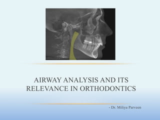
  • 1. - Dr. Miliya Parveen AIRWAY ANALYSIS AND ITS RELEVANCE IN ORTHODONTICS
  • 2. CONTENTS • Introduction • Anatomy • Naso – respiratory function and craniofacial growth • Methods of analysis - Clinical examination - Otorhinolaryngology tests for upper airway - Supplementary examinations • LC • CBCT • Airway and skeletal patterns • Obstructive Sleep Apnoea • Mouth breathing • Effect of orthodontics on airway • Extraction cases • Expansion • Mandibular advancement • Orthognathic surgery • Adenoidectomy or tonsillectomy • Role of orthodontist • Conclusion
  • 3. INTRODUCTION • Upper and lower airway has always been an area of interest because the oropharyngeal and nasopharyngeal structures play important roles in the growth and development of the craniofacial complex. • In particular, upper airway assessment and its interactions with craniofacial growth and development are studied by ENT specialists, laryngologists, speech therapists, pediatricians and dentists. • The airway, mode of breathing, and craniofacial formation are inter- related during growth and development that form can follow function and function can follow form . • So, it is imperative to normalize form and function as early as possible, so that function is optimized for life.
  • 4. • Airway obstruction impairs respiration which leads to craniofacial malformation, malocclusion and jaw deformation. • Research also shows that abnormal craniofacial formation can lead to airway obstruction, impaired respiration, impaired nasal breathing, chronic mouth breathing, sleep apnea, sleep disorders and lifelong ill- health.
  • 5. ANATOMY • The essential organs of the respiratory system are the lungs, located on either side of the thorax, on each side of the heart, and the great vessels. • To reach the lungs, atmospheric air follows the airway, which comprises of the nasal cavity, the mouth, the pharynx, larynx, trachea, and bronchi. The upper airway is formed by the nasal cavity and the pharynx.
  • 6. A. Nasal cavity • The normal airway starts, from the functional perspective, in the nostrils. The nasal cavity includes the nose, the nasal cavities, and extend to the back with the nasopharynx. • A deviated septum, a narrow nasal cavity, and turbinate hypertrophy are some of the signs that cause mouth breathing and OSAS. • In allergic rhinitis, which is also related to upper airway obstruction, the nasal mucous membrane swell with dust particles, pollen or even cold, also affecting the eyes and nose and causing a decrease in air flow
  • 7. B. Oral cavity • It is an essential organ for mastication, phonation, taste, deglutition and breathing, formed by the maxillary, palatal and mandible bones, the tongue, lips and cheeks, and the oropharynx at the back. • The tongue plays many important function but as its muscle tone decreases during sleep, the tongue can block the upper airway. • Jointly with the loss of muscle tone of the pharyngeal walls and the soft palate, it contributes to the collapse of the airway, one of the main causes of obstructive sleep apnea syndrome.
  • 8. C. Pharynx • The pharynx is a tube-like structure formed by muscles and membranes. It measures approximately 12-14 cm and is divided into three parts: nasopharynx, oropharynx and laringopharynx. • The nasopharynx is the upper part of the respiratory system - located behind the nasal cavity and on the soft palate. • In the roof sub-mucosa, there is a collection of lymphoid tissue called pharyngeal tonsil (adenoids) - when large in size, is the main obstruction to the passage of air through the nasopharynx.
  • 9. • The oropharynx extends from the 2nd to 4th vertebra and opens into the oral cavity through an isthmus. The upper end - soft palate & lower end - lingual side of the epiglottis. • The tongue is the main blocking element in the oropharynx- contraction of the genioglossus muscle moves the tongue forward during inspiration, and thereby acts as a pharyngeal dilator. • The laringopharynx joins the oropharynx at the pharyngoepiglottic fold and hyoid bone, and continues up to the sixth vertebra. It is behind the opening in the larynx.
  • 10. NASO – REPIRATORY FUNCTION AND CRANIOFACIAL GROWTH • Dentofacial morphology can be altered by naso-respiratory obstruction depending on the magnitude, duration & time of occurrence. • Quinn in 1978 reiterated that mouth breathing is one of the early symptoms of unnatural acts of breathing and that dramatic deformities of the face, jaws and dentition can be caused by inability to breathe through the nose properly.
  • 11. • Ricketts in 1979 stated that when there is a lack of function in the nose, there can be a growth inhibition. Under normal function in breathing, a pressure develops. In the abnormal, this pressure is changed to vacuum and the maxillary complex is sucked inward, restricting the basal cone.
  • 12. • In 1918, Norlund introduced the ‘compression theory’ which stated that constriction of the maxillary arch is related to the absence of the lateralizing pressure of the tongue against the palate. In response to nasal obstruction, the tongue drops and the medializing effects of the buccal musculature is left unopposed. The effect is further enhanced by a pressure differential across the hard palate in the absence of nasal airflow, leading to a narrow, high- arched palate.
  • 13. • Norlund also put forward the ‘theory of inactivity’, according to which there is reduced growth of the nasal cavity, due to its inactivity, as suggested by Korner (1891) and Bentzen (1903). • The air pressure theory described by Kantorowicz (1916) and James & Hastings (1932) holds that a change to mouth breathing causes the normal negative pressure in the anteriorly sealed oral cavity to be lost, and the palate thus is not carried downward with the growth of the maxillary alveolar process. • The excavation theory proposed by Bloch (1903) and Michel (1908) states that an upward stream of oral airflow presses on the palate leading to higher palatal vault.
  • 14. • Although the literature is replete with statements that airway impairment alters facial and dental growth, there is substantial evidence to the contrary. • Bushey found no relationship between nasal respiration and linear measurements of the adenoids on lateral skull cephalograms before and after surgical removal of the tonsils and adenoids.
  • 15. • Kingsley (1989) noted normal craniofacial development in children with severe nasal obstruction and Whitaker described severe palate malformations in patients who had undergone adenoidectomy at an early age. • More recent findings suggest that nasal-oral breathing per se is not necessarily harmful to craniofacial growth. However, in instances where the nasopharyngeal or oropharyngeal airspace is small, exaggerated postural responses in mouthbreathers may be detrimental to craniofacial growth.
  • 16. METHODS OF ANALYSIS Clinical examination • The general well-being of the child, including growth and development, must be determined. • Pertinent past history include birth trauma, early childhood trauma, previous hospitalizations, medications, and surgical history. • Physical assessment includes facial morphology, skeletal jaw relationships, functional assessment of nostrils, the size and function of the tongue and the anatomy of the soft palate, uvula and tonsils.
  • 17. • Regarding facial morphology, Class II patterns due to mandibular retrusion have smaller upper airway volumes, which is usually associated with adenoid hypertrophy with a very short upper lip and a thick and everted bottom lip.
  • 18. • Swallowing difficulties may be noted. • Voice quality (degree of nasality) and clarity, daytime hypersomnolence, and school/behavioral difficulties should be evaluated. • Sleep history may often reveal loud irregular snoring, restless sleep, abnormal sleep position and nocturnal mouth breathing.
  • 19. • Resting mouth position is noted. • "Adenoid facies" is characterized by an open mouth, dull facial appearance, and short upper lip. This is nonspecific for chronic nasal obstruction. • Other craniofacial anomalies may be associated with these symptoms including cleft palate, Down syndrome, etc.
  • 20. • Tonsillar hypertrophy, macroglossia and oropharyngeal masses should be evaluated. • The nasal cavity was inspected for the presence of secretions, edema and erythema of the nasal mucosa. • The ears should be evaluated as otitis media certainly is associated with nasal obstruction problems. • Bony nasal anomalies, external masses, pits, etc. should be evaluated.
  • 21. a) Functional assessment of nostrils (Duran V.) : To do this we observe nostril response to intense inspiration, paying special attention to the degree of collapse during the maneuver. This is the classification obtained - • Value 0: Dilated nostrils both at rest and in deep inspiration • Value 1: Narrowed nostrils at rest, without functional collapse • Value 2: Functional partial unilateral collapse • Value 3: Functional total unilateral or bilateral partial collapse • Value 4: Functional partial collapse of one nostril and total collapse of the other one • Value 5: Total functional collapse in both nostrils
  • 22. b) Intraoral evaluation of tonsils The tonsils are assessed according to the degree of obstruction of the oropharynx - a reliable clinical evaluation method. • Grade 1 - the tonsils are within their cavity • Grade 2 - do not exceed the midline between the uvula and the anterior pillar of the soft palate • Grade 3 - go over the midline between the uvula and the anterior pillar • Grade 4 - the tonsils are less than 4 mm between them. Grades 3 or 4 represents a decrease in airway permeability
  • 23. c) Upper airway assessment with the Mallampati score - evaluates the risk of obstruction of the airway. • Based on the visual assessment of the oropharyngeal structures, mainly the distance between the tip of the uvula and the tongue base. • Class 1 - full visibility of the tonsils, uvula and soft palate • Class 2 - hard and soft palate, the upper section of the tonsils and uvula • Class 3 - hard and soft palate, and the base of the uvula • Class 4 - only the hard palate. Classes 3 and 4 are commonly present in breathing-related sleep disorders, even after an adenotonsillectomy
  • 24. Most Commonly Used Otorhinolaryngology Tests For Upper Airway Assessment a) Rhinomanometry - evaluate nasal obstruction. • There are different types of rhinomanometry (RMM), active anterior RMM being the one most frequently used. This evaluates nasal airflow in inspiration and expiration by detecting potential obstructions and/or resistance. This can be done with a face mask and it requires full patient cooperation. • After placing the mask, airflows are measured with the rhinomanometer, and the data are analyzed computationally and then graphs are designed in pressure/volume curves.
  • 25. • The recording is repeated under the effect of a topical vasoconstrictor, which will differentiate mechanical obstructions (which do not vary with the vasoconstrictor), vasomotor obstructions (which fully improve with the vasoconstrictor) and mixed obstructions (which improve partially with the vasoconstrictor).
  • 26. b) Acoustic rhinometry - study of the geometry of the nasal cavity. • Based on the analysis of sound reflection and provides a calculation of cross-sectional areas of the nasal cavity and of certain nasal volumes. • Done by generating an audible sound in the nostril with an adapter, taking care not to deform the nasal vestibule. The sound wave penetrates the cavities and is reflected on the different nasal structures or their irregularities.
  • 27. • MCA1 corresponds anatomically to the area at the nasal valve level which has the greatest resistance in the normal nose. MCA2 corresponds to the area at the level of the head of the inferior turbinate. • As in active anterior RMM, the study can be performed before and after applying a vasoconstrictor for the same purpose and with a similar interpretation of the results. • Incident wave signals are measured and reflected according to time, which makes it possible to determine the distance, from the nostril, where there is a change in acoustic impedance. • The most interesting data are the “minimum cross-sectional areas 1 and 2 (MCA1 and MCA2)”.
  • 28. c) C) Nasopharyngolaryngoscopy - evaluates the anatomy of the upper airway, soft palate, the movement of the vocal cords and the process of deglutition. • Performed with a flexible fiberscope which is inserted through the nasal cavities to observe both pharynx and larynx. • The patient is usually awake, and topical lidocaine is applied on the nostrils.
  • 29. • During the test, the patient may be asked to talk, cough or swallow, depending on what is being evaluated. • The following anatomical elements should be evaluated: i. deviations of the nasal septum ii. size of inferior turbinates iii. presence and size of the adenoid tissue iv. quantity and quality of nasal secretion v. size of palatine tonsils & base of the tongue vi. its relationship with the oropharyngeal cavity vii. abduction of the vocal cords viii. subglottic diameter ix. presence of masses or pathological deformities at any levels
  • 30. d) Functional Nasal Permeability (PeNaF) - assesses the independent functional nasal permeability of each cavity. • The performance is recorded as negative (-) when the patient maintains nasal breathing for six inspirations at rest, and positive (+) when the patient fails to maintain it for six inspirations. • A study validated in Chile recommends orthodontists implement this simple examination to rule out a possible nasal obstruction. If this is not the case, they should request an objective assessment to check the increase in nasal resistance
  • 31. e) SNORT - Simultaneous Nasal Oral Respirometric Technique by Gurley and Vig reported a enabled the direct and simultaneous measurement of inspired and expired air, both orally and nasally. • Using a custom fitted face mask with separate valves attached to the nose and mouth and attached to a flow meter, air pressure transducer, recorder and computer, it can give the nasal versus the oral inspiration, expiration and their ratios. • SNORT permits the objective quantification of the ratio of oral to nasal airflow and permits a numerical determination of both normal and pathological states of breathing mode.
  • 32. Supplementary Examinations a) Upper airway assessment in lateral cephalograms • Lateral cephalometry is commonly used in clinical practice given its relative simplicity, accessibility, low cost and low exposure to radiation. • Lateral cephalograms provide reliable linear measurements, can measure the dimensions of the nasopharyngeal and retropalatal regions, but have not proven to be reliable to measure the airway in the back of the tongue. • Though multiple authors have tried to standardize a method of airway analysis on lateral cephalogram, very few methods have been widely accepted.
  • 33. McNamara’s Analysis (1984) – • Two measurements are used to examine the possibility of an airway impairment. • The upper pharyngeal width is measured from a point on the posterior outline of the soft palate to the closest point on the posterior pharyngeal wall. • This measurement is taken on the anterior half of the soft palate outline because the area immediately adjacent to the posterior opening of the nose is critical in determining upper respiratory patency.
  • 34. • Apparent airway obstruction, as indicated by an opening of 5 mm or less in the upper pharyngeal measurement - only an indicator of possible airway impairment. • The average upper airway measurement for adults of both sexes is 17.4 mm. • Lower pharyngeal width is measured from the intersection of the posterior border of the tongue and the inferior border of the mandible to the closest point on the posterior pharyngeal wall. • The average value for this measurement is 10 to 12 mm and does not change appreciably with age. • In contrast to the upper pharynx, a smaller than average value for the lower pharynx is not remarkable. A lower pharyngeal width of greater than 15 mm suggests anterior positioning of the tongue, either as a result of habitual posture or due to an enlargement of the tonsils.
  • 35. Martin’s Analysis (2006) – The following cephalometric measurements were made – 1. PNS-AD1: lower airway thickness; distance between PNS and the nearest adenoid tissue measured through the PNS- Ba line (AD1). 2. AD1-Ba: lower adenoid thickness; defined as the soft-tissue thickness at the posterior nasopharynx wall through the PNS-Ba line. 3. PNS-Ba: lower airway thickness; distance between PNS and Ba—the sum of variables 1 and 2.
  • 36. 4. PNS-AD2: upper airway thickness; distance between PNS and the nearest adenoid tissue measured through a perpendicular line to S-Ba from PNS (AD2). 5. AD2-H: upper adenoid thickness; defined as the soft-tissue thickness at the posterior nasopharynx wall through the PNS-H line. ● Hormion (H): cephalometric point located near the adenoidal tissue at cranial base localized where a perpendicular to S-Ba line crosses the sphenoid bone. The variations of this point are minimal because it is located far from growing sites.
  • 37. 6. PNS-H: upper airway thickness; distance between PNS and H—the sum of variables 1 and 2. 7. N-H: nasal fossa length; distance between N and H. 8. S-N: anterior cranial base. 9. McNamara’s upper pharynx dimension 10.McNamara’s lower pharynx dimension
  • 38. 11 and 12. Total adenoidal, and aerial areas: the Ba-N plane, the bispinal plane, and 2 perpendicular lines to the bispinal plane: 1 that crosses the more anterior point at atlas vertebra and other that crosses the PNS. The resulting trapezoid is divided in 2 spaces (aerial and adenoid).
  • 39.
  • 40. Fujioka et al. (1979) - described the adenoidal-nasopharyngeal ratio (AN ratio). • Relates the length of the line perpendicular to the sphenoid bone (A) by the thickest portion of the adenoids with the distance between the posterior nasal spine and the anterior edge of the sphenobasioccipital synchondrosis (N). • An AN< 0.8 is considered normal and an AN > 0.8 is considered enlarged • In addition, Feres, Murilo et al. in 2012 found that both parameters had good reproducibility and a variability which was not clinically significant.
  • 41. • One of the most common reasons for upper airway obstruction is hypertrophic adenoids. Before planning an orthodontic treatment, this area is usually observed in the lateral cephalometry. • A study was conducted to assess whether adenoidal ratio on lateral cephalograms can be used to estimate airway volumes, using CBCT as the validation method. They concluded that the lateral cephalogram can provide some information about the nasopharyngeal space, particularly in patients over 15. • This is due to the stability reached by the tissue at this age; however, it cannot be used as a diagnostic procedure to determine the volume of the total airway, but rather as an assessment tool to determine the need for a more comprehensive ENT examination.
  • 42. b) Upper airway assessment using CBCT • Several studies have shown that CBCT is accurate and reliable for upper airway assessment. • Volumetric reconstructions that may be obtained from CBCTs help clinicians make a correct diagnosis and indicate a better treatment plan for some pathologies of the maxillofacial area, especially those related to the airway.
  • 43. • For the volumetric reconstruction and visualization of the upper airway, software programs must allow us to find the correct location of the boundaries of the pharynx and nasal cavity (segmentation) through a process that can be manual, automatic or semi-automatic. • Studies comparing the commercial software programs for the study of the airway were analyzed. They were found to have reliable reproducible and accurate results of linear measurements, but they lost accuracy when calculating the volume of the airway. This could be due to the automatic segmentation of the nasal cavity, the nasopharynx and oropharynx.
  • 44. • The head position and the position of the patient when the CBCT is taken must be considered to obtain accurate and repeatable upper airway measurements and volumes. • The position of the hyoid bone and tongue, and the dimension of the airway would be highly reproducible using the natural position of the head when taking lateral cephalograms. • It has been found that individuals would be approximately 40% more affected by the width of the airway in an upright position. • Solow et al. determined that in an upright position or by increasing the cervical skull angle, there is an increase in upper airway diameters. • Alsufyani et al. states that images must be obtained with the patient in a sitting position so as not to affect airway diameter.
  • 45. • Aboudara et al. stated that CBCT is a simple and effective method to evaluate upper airway. They compared the volumetric measurements from CBCT with known physical airway phantoms and found that the errors ranged from 0 to 5%. They also reported a moderately high correlation (r = 0.75) between the sagittal area and the volume when correlating lateral cephalograms measurements with CBCT data.
  • 46. • Two systematic literature reviews concluded that although major progress has been made in the capture and management of CBCT images, there is no optimized evidence-based protocol to obtain images to analyze the upper airway. • Several obstacles must still be overcome, such as the influence of the position of the tongue, mandible morphology, the impact of the respiratory phase and the definition of the anatomical boundaries of the upper airway, as well as the lack of consistency in the configuration of the equipment and in how images and volumetric reconstructions are obtained.
  • 47. AIRWAY AND SKELETAL PATTERNS • A study conducted in New Delhi compared the reliability of lateral cephalograms and computed tomographies to assess airways. They compared three skeletal patterns determined by the different values of the ANB angle, and related their linear values taken from the cephalometries to volumetric values delivered by CT, and concluded that the skeletal pattern had a strong association with pharyngeal volume and its linear dimensions. They also noted that the S-shaped soft palate can be considered high-risk for sleep apnea compared to the leaf shape, which is more common.
  • 48. • In contrast, Dalmau et al., found no statistically significant differences that correlate airway with skeletal patterns or facial biotypes. • The recent results of Lucas Castro-Silva et al., in Brazil, found a positive correlation of higher values of pharyngeal airway for Class III patients.
  • 49. • A new study by El and Palomo found that oropharyngeal airway volumes were lower in Class II patients compared to Class I and Class III patients. They also state that the mandibular position with respect to the skull base has a strong impact on oropharyngeal volume.
  • 50. OBSTRUCTIVE SLEEP APNEA • Definition: condition caused either by complete occlusion or partial collapse of the upper airway despite the presence of simultaneous respiratory effort. Cessation occurs at the level of nostrils and mouth. • Condition is considered pathologic when the episodes last for at least ten seconds and at a frequency of 30 times or more during 7 hrs. of nocturnal sleep in REM and especially in non REM stages of sleep.
  • 51. • Diagnosis is by polysomnography - Measurements are made to assess sleep stages of breathing and gas exchange to detect sleep stages. PSG ensures the no. of apnic episodes per hour of sleep expressed by respiratory(Disturbance Index)measurements of chest and abdominal efforts and oxygen saturation. • Airway measurement by cephalometric 3D imaging – lateral pharyngeal dimension.
  • 52. • Treatment Options - − Enlarging the upper airway − Continuous positive airway pressure − Weight loss − Oral appliances - mandibular advancement devices − Upper airway surgery − Combination CPAP/ dental sleep device therapy
  • 53.
  • 54. MOUTH BREATHING • Clinical examination – ask patient to hold water in the mouth, use double sided mouth mirror or cotton wisps • Cephalometric Analysis: - 1. McNamara airway analysis – • Upper pharyngeal width – the point on posterior outline on soft palate to closest point on pharyngeal wall – 15 to 20 mm in width. Values 2mm or less indicate airway impairment. Lower pharyngeal width from point of intersection of posterior border of tongue and inferior border of mandible to the closet point on posterior pharyngeal wall – 11 to 14mm. 2. Vertical growth pattern, increased ANB, increased gonial angle, decreased mandibular length, steep MP angle, over erupted upper posterior segments
  • 55. EFFECT OF ORTHODONTICS ON AIRWAY 1. Extraction and airway changes: • A systematic review on the effects of bicuspid extractions and incisor retraction on upper airway of Asian adults and late adolescents showed that Linear airway response to incisor retraction measured on lateral cephalograms varied substantially, while linear, cross‐sectional and volumetric measurements of posterior airway space using CT showed larger effect sizes and smaller variations, providing evidence of airway narrowing with bicuspid extractions and incisor retraction.
  • 56. • An article published in Orthodontics and craniofacial research journal on three-dimensional pharyngeal airway changes in orthodontic patients treated with and without extractions analysed the pharyngeal airway for subjects treated with extractions and without extractions. • Nasopharyngeal (NP) and oropharyngeal (OP) volumes, area of maximum pharyngeal constriction (AMPC), and upper arch perimeter were measured on CBCT scans. • There were no statistically significant differences in the pharyngeal airway values between the extraction and non-extraction groups. • The extraction group showed a statistically significant increase for NP and OP volumes and AMPC values. Such increase was also noted in the non-extraction group, without statistical significance for AMPC values.
  • 57. • A retrospective study evaluating the airway space changes after extraction of four second premolars and orthodontic space closure in adult female patients with bimaxillary protrusion showed orthodontic treatment increases the vertical airway length, which is the amount of distance between base of the tongue and posterior nasal spine. • A study conducted on pharyngeal airway dimensional changes after premolar extraction in skeletal class II and class III, growing and adult orthodontic patients showed significant increase in nasopharyngeal airway dimension in class II and class III patients, whereas palatopharyngeal and glossopharyngeal dimensions were insignificantly decreased in both groups.
  • 58. 2. Rapid Maxillary Expansion • RME is successful in increasing the nasal permeability and reducing airway resistance. • Reduced airway resistance reduces negative pressure during ventilation, with promising results in the treatment of paediatric sleep disordered breathing, including obstructive sleep apnoea • The effects on more distant structures include stretching of the tensor palatine muscles by the expanding maxilla with subsequent improvement in drainage of the Eustachian tubes, aiding in reducing otitis media and conductive hearing loss • Enlarged palatal space may also allow for an improved tongue posture, which could facilitate increased airway space in the oropharynx
  • 59. • Donald Timms, in a study with Rapid Maxillary Expansion, reported a 37 percent mean drop in nasal resistance with 7 mm of expansion. • In another larger study of children with a previous history respiratory disease treated with Rapid Maxillary Expansion, 82 percent reported improvement in number of upper respiratory tract infections and 60 percent reported improvement of allergic rhinitis.
  • 60. • According to Bishara and Staley - Anatomically, there is an increase in the width of the nasal cavity immediately following expansion, particularly at the floor of the nose adjacent to the midpalatal suture. As the maxillae separate, the outer walls of the nasal cavity move laterally. The total effect is an increase in the intranasal capacity. The nasal cavity width gain averages 1.9 mm but can widen as much as 8 to 10 mm at the level of the inferior turbinates, while the more superior areas might move medially.
  • 61. • A number of rhinologists indicate that RME, in addition to its widening procedure, results in correction of septal deformity as a result of the lowering of the floor of the nasal cavity. • Hershey, Stewart, and Warren, and Turbyfill reported a reduction of nasal airway resistance by an average of 45% to 53% with RME. • Wertz concluded that opening the midpalatal suture for the purpose of increasing nasal permeability cannot be justified unless the obstruction is shown to be in the lower anterior portion of the nasal cavity and accompanied by a relative maxillary arch width deficiency.
  • 62. • Graber believes that the claims of improved nasal breathing apparently as a result of RME are most likely only temporary. Spontaneous regression of lymphoid tissues during growth automatically improves nasal breathing, even if nothing is done to the palate. • Therefore, it can be concluded that the effect of RME on the nasal airway will to a great extent depend on the cause, location, and the severity of the nasal obstruction
  • 63. 3. Mandibular advancement • Mandibular repositioning is most effective in mild to moderate obstructive sleep apnea. • It enhances retro pharyngeal air space thereby increasing nasal airway patency. 1. Mandibular repositioners - open the airway by moving the mandible forward. As the jaw is moved forward, the collapsible part of your airway is held open by the forward movement of the tongue and other airway muscles.
  • 64. • 2. Mandibular Advancement Devices (MADs) also improve the strength and rigidity of the airway by increasing the muscle activity of the tongue and other muscles of the airway. 3. Elastic Mandibular Advancement (EMA ) Appliance
  • 65. 4. Tongue retaining devices (with airway tubes) • Like MADs, Tongue Retaining Devices (TRDs) also work by holding the tongue in a forward position. • These devices pull the tongue forward, but instead of moving the jaw forward like a Mandibular Advancement Device (MAD), TRDs directly control the tongue itself. • In some cases, Tongue Retaining Devices (TRDs) have decreased therapeutic complications compared to MADs, but TRDs can also be less comfortable and generally take several weeks or months to be worn comfortably.
  • 66. EFFECT OF ORTHOGNATHIC SURGERY ON AIRWAY • If the difference between the maxillary and the mandibular unit length is greater than 16mm at the age of 12 in class II malocclusion and greater than 29mm at the age of 12 in class III dysplasias, surgical intervention is necessary. • Surgical superior impaction of the maxilla has become an accepted treatment for the correction of vertical maxillary excess and it does reduce nasal resistance, but it does not increase the percentage of nasal airflow. • It has been speculated that this change may be associated with the common postoperative increase in interalar width and widening of the external nares, which result in an opening of the liminal valve.
  • 67. • At the present time, maxillomandibular advancement (MMA) remains the most effective surgical procedure in treating patients with moderate to severe OSA. • Advancement of maxillofacial skeletal structures opens the oropharyngeal airway and places the oropharyngeal musculature on tension. • MMA surgery results in a significant increase in the volume and a morphologic airway change from a round to an elliptical f shape in the upper airway space in patients with OSA. • These changes are stated to reduce the collapsibility of the upper airway during deep stages of sleep.
  • 68.
  • 69.
  • 70. ROLE OF ORTHODONTIST • In 1987, Weimert published a study of 1360 patients referred to otolaryngologists by orthodontists because of suspicion of nasal obstruction. Although it was not a solid scientific study, the findings suggest that orthodontists can effectively screen for nasal obstruction. • The most common reasons for referral were: dentofacial characteristics suggestive of upper airway obstruction, inability to retain a dental appliance, and unsatisfactory results from an orthodontic program. • Dentists are important as both referrers and treating doctors. • Allergies generally must be treated first, before consideration of any surgery. The greatest number of surgical failures are in allergic patients. • The greatest number of orthodontic relapses are in patients who have not had their breathing problems successfully treated.
  • 71. • Orthodontic therapy is affected by the function of the lips, tongue, and masticatory musculature, all of which may accommodate to nasal obstruction in ways which can effect occlusion. • Effective orthodontic therapy may require the elimination of the nasal obstruction to allow for normalization of the facial musculature surrounding the dentition. • According to Meredith, the growth of the face (excluding the mandible) is completed at a relatively early age. 60% of craniofacial development takes place during the first 4 years of life and 90% by age 12. Based on these observations, any intervention to open the airway must take place at an early age. • As a referring doctor, one has to decide between allergist, ENT, pediatrician or sleep lab.
  • 72.
  • 73.
  • 74. CONCLUSION • In spite of the long history of research between respiration and malocclusion, only now is this aspect of diagnosis and treatment given its due importance. • Evaluation of children with nasal obstruction and dental abnormalities requires a multidisciplinary approach between the orthodontists, pediatricians, and otolaryngologists. • The orthodontists must be familiar with the dental literature so as to provide optimal care for pediatric patients as they have an opportunity to examine and institute treatment to the patients at a very early age.
  • 75. REFERENCES • Rojas E, Corvalán R, Messen E, Sandoval P. Upper airway assessment in Orthodontics: a review. Odontoestomatologia. 2017 Dec 1;19(30). • Martins LS, Liedke GS, Heraldo LD, Da Silveira PF, Arus NA, Ongkosuwito EM, Vizzotto MB. Airway volume analysis: is there a correlation between two and three-dimensions?. European journal of orthodontics. 2018 May 25;40(3):262-7. • Huynh NT, Desplats E, Almeida FR. Orthodontics treatments for managing obstructive sleep apnea syndrome in children: A systematic review and meta-analysis. Sleep medicine reviews. 2016 Feb 1;25:84-94. • Indriksone I, Jakobsone G. The upper airway dimensions in different sagittal craniofacial patterns: a systematic review. Stomatologija. 2014 Jan 1;16(3):109-7. • Lenza MG, Lenza MD, Dalstra M, Melsen B, Cattaneo PM. An analysis of different approaches to the assessment of upper airway morphology: a CBCT study. Orthodontics & craniofacial research. 2010 May;13(2):96-105 • Preston CB, Lampasso JD, Tobias PV. Cephalometric evaluation and measurement of the upper airway. InSeminars in Orthodontics 2004 Mar 1 (Vol. 10, No. 1, pp. 3-15). WB Saunders. • Ng JH, Song YL, Yap AU. Effects of bicuspid extractions and incisor retraction on upper airway of Asian adults and late adolescents: A systematic review. Journal of oral rehabilitation. 2019 Nov;46(11):1071-87. • Becking BE, Verweij JP, Kalf-Scholte SM, Valkenburg C, Bakker EW, van Merkesteyn JP. Impact of adenotonsillectomy on the dentofacial development of obstructed children: a systematic review and meta-analysis. European journal of orthodontics. 2017 Oct 1;39(5):509-18. • Banerjee D, Shrivastav D, Kamble D, Dhannawat D, Purva V. Lateral Cephalogram and CBCT as a diagnostic aid for analysis of airway-Review Article. European Journal of Molecular & Clinical Medicine. 2020 Dec 22;7(7):1802-8 • Bidjan D, Sallmann R, Eliades T, Papageorgiou SN. Orthopedic Treatment for Class II Malocclusion with Functional Appliances and Its Effect on Upper Airways: A Systematic Review with Meta-Analysis. Journal of clinical medicine. 2020 Dec;9(12):3806. • Stefanovic N, El HA, Chenin DL, Glisic B, Palomo JM. Three‐dimensional pharyngeal airway changes in orthodontic patients treated with and without extractions. Orthodontics & craniofacial research. 2013 May;16(2):87-96. • Schendel SA, Broujerdi JA, Jacobson RL. Three-dimensional upper-airway changes with maxillomandibular advancement for obstructive sleep apnea treatment. American Journal of Orthodontics and Dentofacial Orthopedics. 2014 Sep 1;146(3):385-93. • Jayan B, Kadu A. Airway-focused orthodontics. Journal of Indian Orthodontic Society. 2018 Apr;52(4_suppl1):23-8.

Editor's Notes

  1. These anomalies require early detection, and it has been shown that the early diagnosis and treatment of obstructive sleep apnea-hypopnea syndrome allows for an almost complete normalization of dentofacial morphology
  2. Modified – without tongue protruding
  3. continuous positive airway pressure therapy, is a treatment method for patients who have sleep apnea. CPAP machines use mild air pressure to keep the airways open, and are typically used by patients who have breathing problems during sleep. More specifically, what CPAP therapy helps accomplish is making sure that your airway doesn't collapse when you breathe while asleep.
  4. The Journal of Craniomandibular & Sleep Practice
  5. ‘‘Mewing’’ is an eponym of Dr. John Mew (1929), a professor at his own London School of Facial Orthotropics, who was recently stripped of his dental license by the General Dental Council. The dismissal was on grounds of misconduct for publicly denigrating the traditional practices of orthodontic tooth movement, in conjunction with exodontia and orthognathic surgery, as treatment for malocclusion2,3 and boldly heralding his own etiologic concepts of malocclusion, somewhat based on Moss’s functional matric hypothesis.4 Specifically, Mew’s theory postulates that genetic control of skeletal growth is not precise, but rather, that the articulation of the jaws and teeth is dependent on environmental guidance from the orofacial musculature.5 He and his followers further stipulate that the ideal facial esthetic (that is, ideal nose-chin relationship, well-defined jawline, and appropriately prominent malar eminences) can be realized by posturing the tongue against the palate with the lips sealed and the teeth in or near contact.2