More Related Content
Similar to Micromotor-based on–off fluorescence detection of sarin and soman simulants (20)
Micromotor-based on–off fluorescence detection of sarin and soman simulants
- 1. 11190 | Chem. Commun., 2015, 51, 11190--11193 This journal is ©The Royal Society of Chemistry 2015
Cite this: Chem. Commun., 2015,
51, 11190
Micromotor-based on–off fluorescence detection
of sarin and soman simulants†
Virendra V. Singh, Kevin Kaufmann, Jahir Orozco, Jinxing Li, Michael Galarnyk,
Gaurav Arya and Joseph Wang*
Self-propelled micromotor-based fluorescent ‘‘On–Off’’ detection
of nerve agents is described. The motion-based assay utilizes Si/Pt
Janus micromotors coated with fluoresceinamine toward real-time
‘‘on-the-fly’’ field detection of sarin and soman simulants.
Chemical warfare agents (CWA) are some of the most nefarious
weapons of mass destruction.1
The ease of manufacturing and
dispensability, ease of availability, and inexpensive starting materials
make them an eminent global threat.1
Nerve agents are a particularly
dangerous class of CWA that continue to be a threat in spite of
controls imposed by the chemical weapon convention (CWC) which
prohibits the production and use of chemicals on enemy forces.2,3
Their rapid and severe effects on human health stem from their
ability to irreversibly inhibit acetylcholinesterase activity by phos-
phorylation and leads to neuromuscular paralysis and eventually
death.4–6
A particularly dangerous class of organophosphorus nerve
agents is the phosphoryl fluoride containing species such as sarin
and soman,7–9
referred to as GB and GD agents, respectively.
Unfortunately, these highly reactive and volatile nerve agents are
colorless, odorless, and tasteless, making their detection very diffi-
cult. Therefore, a reliable and rapid nerve agent detection system is
highly desirable amidst the current climate of terrorism awareness.
Among a variety of detection methods that have been developed for
these nerve agents,9
fluorescent detection offers the unrivaled merits
of high sensitivity, low-cost and operational simplicity.10–12
The
chromo-fluorogenic detection is especially attractive because it allows
simple visual detection in situ, or on-site, without any sample
pretreatment or the use of complex equipment.10–12
As a result,
the development of highly sensitive, selective, ready-to-use, and rapid
field screening methods for these nerve agents has become an
increasingly imperative research goal.
Herein we demonstrate an extremely fast (B10 s) fluores-
cent ‘‘On–Off ’’ detection of sarin and soman simulants based
on self-propelled dye-coated micromotors. Artificial/synthetic
micromotors have recently attracted considerable interest due to
their attractive capabilities and diverse potential applications, ran-
ging from directed drug delivery to environmental detection.13–15
The self-propulsion capability of micromotors induces efficient fluid
mixing, a very useful property for environments that would not allow
mechanical agitation (i.e., bodies of water, stealthy national defense
operations, etc.) or at the microscale level. This motor-induced
mixing has shown to be extremely effective in accelerating both
target–receptor interaction,13–15
detection,16,17
and detoxification
reactions.18–20
Recently, there has been a considerable interest in
micromotor-based defenses against chemical and biological warfare
agents;18–20
these include the potential use of functionalized micro-
motors as efficient screening tools for the real time and on-site
detection of chemical threats in environmental matrices.
Fluorescence quenching methods have been emerging as an
effective route for sensitive and low cost field detection.21–23
In this
regard, fluorescent ‘turn-off’ based sensors have been developed for
nitrated explosive compounds22
and Hg2+
.23
Such ‘turn-off’ sensors
have shown to be more sensitive than their ‘turn-on’ sensor counter-
part.22,23
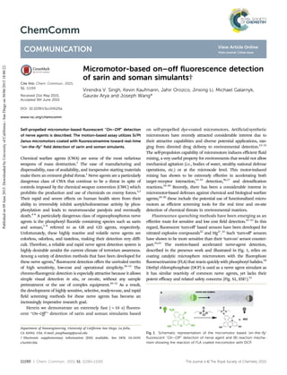
The motion-based accelerated nerve-agent detection,
described in the presence work and illustrated in Fig. 1, relies on
coating catalytic microsphere micromotors with the fluorophore
fluoresceinamine (FLA) that reacts quickly with phosphoryl halides.24
Diethyl chlorophosphate (DCP) is used as a nerve agent simulant as
it has similar reactivity of common nerve agents, yet lacks their
potent efficacy and related safety concerns (Fig. S1, ESI†).12
Fig. 1 Schematic representation of the micromotor based ‘on-the-fly’
fluorescent ‘‘On–Off’’ detection of nerve agent and (B) reaction mecha-
nism showing the reaction of FLA coated micromotor with DCP.
Department of Nanoengineering, University of California San Diego, La Jolla,
CA 92093, USA. E-mail: josephwang@ucsd.edu
† Electronic supplementary information (ESI) available. See DOI: 10.1039/
c5cc04120a
Received 21st May 2015,
Accepted 9th June 2015
DOI: 10.1039/c5cc04120a
www.rsc.org/chemcomm
ChemComm
COMMUNICATION
Publishedon09June2015.DownloadedbyUniversityofCalifornia-SanDiegoon30/06/201518:00:22.
View Article Online
View Journal | View Issue
- 2. This journal is ©The Royal Society of Chemistry 2015 Chem. Commun., 2015, 51, 11190--11193 | 11191
Microbead chemical switches were shown to be useful for
detecting reactive organophosphate CWA vapors using buffers and
appropriate proton scavengers.25
Moreover, it was also found that
fluorescence intensity increased first and then decreased over time
which may lead to false positive fluorescent ‘‘Off–On’’ detection.
However, since in the field conditions, the concentration of CWA
and other metrological conditions are often unknown, the detection
of CWA in natural water matrices is more challenging compared to
vapor measurements since proper buffering and a suitable proton
scavenger are very important. To date, this has made the detection
process unfeasible. As will be illustrated below, the new micromotor
strategy addresses these challenges toward rapid fluorescent
‘‘On–Off’’ route for instantaneous detection of sarin and soman
simulants.
The self-propulsion capability of the dye-coated micromotors,
along with the corresponding bubble tail, are shown to generate
effective fluid convection (without external force) that results in an
increased rate of collision between the micromotors and the target
nerve agent, thereby offering a rapid ‘on-the-fly’ fluorescent ‘‘On–Off’’
detection method. The fluid mixing capability of micromotors26
has
been used recently for accelerated environmental remediation and
detoxification processes.18–20
Similarly, the efficient movement of the
FLA-coated microsphere, along with the substantial fluid transport,
are used in the following sections for developing an attractive
‘‘On–Off ’’ CWA detection approach.
As illustrated in Fig. 1A, self-propelled FLA/silica–NH2/Pt
micromotors perform ‘‘on-the-fly’’ fluorescent detection of
nerve agents in environmental matrices. Fig. 1B displays the
reaction mechanism involving the FLA/silica–NH2/Pt micromotor
and nerve agent. These micromotors, along with inherent efficient
solution mixing, resulted in the dramatic quenching of FLA fluores-
cence upon encountering nerve agents in solution.
Fig. 2A illustrates the fabrication steps involved in the preparation
of FLA/silica–NH2/Pt micromotors for the detection of nerve agents.
The new micromotors were prepared by impregnation of FLA into
silica microparticles using the incipient wetness technique27
(refer to
Experimental section) followed by asymmetric deposition (for the
dynamic movement of micromotors) of Pt by sputtering (Fig. 2A).
This technique allows the FLA solution to just wet the adsorbent and
be completely adsorbed on solid silica–NH2 particles.
Scanning electron microscopy (SEM) images of silica–NH2 particle
before and after the dye loading are shown in the ESI.† The surface of
FLA coated silica particles displays a rough, shiny, and bright white
morphology when compared to unmodified silica particles, indicat-
ing that the FLA molecules are adsorbed on the surface of silica–NH2
microparticles. Energy dispersive spectroscopic (EDX) analysis of
impregnated and un-impregnated samples qualitatively confirms
the impregnation as indicated by the presence of the respective
element in the EDX spectra (refer to ESI†). The presence of Janus
micromotors is also confirmed by the corresponding EDX mapping
of Pt and carbon displayed in Fig. 2C and D. Fig. 2E and F illustrate
the initial movement of the dye coated micromotors in pure water
and the movement after 5 minute propulsion, respectively. The
tracking trajectories in Fig. 2E and F display the motion over a 5 s
period, with an average speed of B145 mm sÀ1
.
To demonstrate the practical utility of the new micromotor-based
fluorescent ‘‘On–Off’’ nerve-agent detection in environmental
matrices, we investigated the ability to detect the sarin and soman
simulants (Fig. S1, ESI†). In order to mimic the natural environment,
experiments were carried out in water without buffer for the real
time detection of nerve agents. Fig. 3 shows a FLA-coated silica–NH2
particle that exhibits fluorescence counts of B40000. Fig. 3B and C
displays the comparative fluorescence intensity before and after FLA/
silica–NH2/Pt micromotors navigated a DCP contaminated solution
(10À3
M), respectively. There is an instant fluorescence quenching of
the moving micromotor compared to static particles that did not
show any fluorescence quenching under the same conditions, even
after 5 min of interaction (Fig. 3D and E). Fig. 3F shows the crucial
role of the movement of micromotors in the contaminated solution
for the rapid screening of nerve agents when compared to the static
coated particles under similar conditions. The unique movement of
multiple micromotors with bubble generation across the contami-
nated samples results in a continuous mixing (without external
agitation), which increases the likelihood of collision with nerve
agent and leads to an increase in the rate of reaction, with
the concomitant fluorescence quenching within a minute, as per
collision theory.28
This dramatic fluorescence quenching is attribu-
ted to the reaction of reactive phosphonates with the –NH2 group of
Fig. 2 (A) Schematic detailing the fabrication steps in the preparation of
FLA/silica–NH2/Pt micromotors; (B–D) energy-dispersive X-ray spectro-
scopy images illustrating the distribution of Si, dye and Pt; (E) tracking line
(taken from ESI,† Video S1) illustrating the typical trajectories traveled by a
micromotor over 5 second intervals at (E) t = 0 min and (F) t = 5 min.
Fig. 3 (A) Fluorescent intensity of FLA coated silica–NH2 particles;
(B) fluorescent intensity of FLA/silica–NH2/Pt micromotors before expo-
sure to DCP; (C) after exposure; (D) fluorescent intensity of FLA/silica–
NH2/Pt static particles before exposure of DCP; (E) after exposure; and
(F) graph showing the movement of micromotors leading to rapid quenching
of fluorescence compared to static. Reaction conditions: conc. of DCP =
10À3
M, H2O2 (2%), SDS (1%), and 2 Â 104
micromotors; lex, 490 nm;
lem, 510 nm. Scale bar 45 mm.
Communication ChemComm
Publishedon09June2015.DownloadedbyUniversityofCalifornia-SanDiegoon30/06/201518:00:22.
View Article Online
- 3. 11192 | Chem. Commun., 2015, 51, 11190--11193 This journal is ©The Royal Society of Chemistry 2015
dye-coated micromotors which leads to the respective phos-
phoramide while releasing HCl which causes the interruption
of the fluorophore’s conjugation.29
Unlike the fluorescence
intensity increase expected from the formation of phosphora-
mide,25
we observed a dramatic quenching in the fluorescence
intensity of the FLPA adduct, attributed to the local decrease in
pH due to the HCl release.
To expand the practical utility of the dye-coated micromo-
tors towards diverse environmental applications, the fluores-
cence intensity of micromotors were also tested in sea water,
pool water, and lake water. The resultant fluorescence intensity
profile was consistent with that found in water experiments,
which reflects its applicability in diverse natural environments.
The specificity of this micromotor based detection was con-
firmed by monitoring the fluorescence with non-reactive phos-
phonates, such as dimethyl methyl phosphonate (DMMP;
10À3
M), and no fluorescence change was observed. The efficiency
of the new micromotor-based on-the-fly fluorescent ‘‘On–Off’’ detec-
tion strongly depends on the concentration of nerve agents. Fig. 4A
displays the time dependent quenching of FLA-coated micro-
motors with respect to varying DCP concentrations ranging
from 10À1
to 10À6
M using a fixed number of micromotors. As
expected, the fluorescence quenching is very fast (within a
minute) for concentrations as low as 10À3
M DCP. Moreover,
this micromotors based detection platform is able to sense
concentrations as low as 10À6
M DCP within 3 minutes.
The detection efficiency at different concentrations of DCP
with micromotor and static dye-coated particles was also com-
pared (Fig. 4B). As expected, the reaction between the FLA-
coated micromotors and the target DCP was very fast. For
example, quenching of the motors was observed after only
120 s of interaction with a 10À4
M DCP contaminated solution.
In contrast, when the contaminated solution is in quiescent
conditions, the reaction between static FLA-coated silica parti-
cles and DCP is inefficient, thereby requiring around 30 min to
exhibit a quenching of the fluorescence.
In order to optimize the density of micromotors for the rapid
detection of 10À6
M DCP solution, different numbers of FLA-coated
silica micromotors (1 Â 104
to 2 Â 104
micromotors per ml), were
propelled in 600 ml contaminated solution for 3 min. It was found
that an increase in the micromotor density increases the rate of CWA
detection. Fig. S4 (ESI†) displays the effect of micromotor density on
the detection rate (rapid fluorescence quenching) of DCP in a
contaminated solution. We have also performed micromotor-based
fluorescent enhancement detection of DCP in a phosphate buffer
(pH 7.2) medium based on the conversion of dye in to phosphor-
amide. This resulted in a rapid and intense initial fluorescence
increase in the presence of different DCP concentrations (Fig. S5,
ESI†). However, this data illustrates that at longer times (41.5 min)
this enhancement may decay, depending on the DCP concentration.
Such decrease is expected for this specific phosphoramide reaction
since there is also a release of hydrochloric acid which can be
partially neutralized by the buffer medium. At higher DCP concen-
trations (410À2
M), more acid production results in fluorescence
quenching after the initial increase which may lead to false positive
signals. Accordingly, practical detection applications based on the
fluorescence enhancement should rely on the initial response in the
buffer medium over the first 90 s. Fig. S6 (ESI†) shows the crucial
role of the movement of micromotors in the buffer solution (pH 7.2)
containing 10À3
M of DCP, compared to the static dye-coated
particles under similar conditions. As expected, the fluorescence
intensity drastically increases when compared to the static counter-
part, reflecting the near instantaneous reaction of DCP with dye-
coated micromotors due to continuous mixing associated with the
micromotor movement. So, in both cases (buffer or non-buffer
media) the micromotors lead to a greatly enhanced sensitivity
compared to static systems. Interference studies were carried out
in the presence of common volatile organic compounds, including
ethanol, toluene, acetone, and isopropanol in order to demonstrate
the applicability of the present methodology. As illustrated in Fig. S7
(ESI†), these organic compounds had a negligible effect upon the
fluorescent signal, indicating effective discrimination against volatile
organic compounds.
The greatly enhanced detection reflects the fluid mixing
capability of the dye-coated micromotors. The experimentally
observed rapid fluorescent detection achieved by the mobile
micromotors, compared to static micromotors, can be attrib-
uted to and explained mathematically by three effects. First, the
propulsion of mobile motors is able to keep them suspended
and well dispersed in the solution. In contrast, the static
motors settle to the bottom of the container, due to gravity,
forming a densely packed aggregate. The mass-transfer model
for the two systems provided in the ESI† shows that motors well
dispersed in solution lead to significantly smaller diffusion
mass-transfer resistance compared to sedimented motors; even
without accounting for the continuous movement of the micro-
motors. This model estimates an enhancement of roughly 1 +
(pDt/4R2
)1/2
in the amount of analyte (DCP) reacted after time t
at the surface of dispersed versus sedimented motors, where D
is the diffusivity of the analyte in solution, R is the motor
radius, and t is the time. Plugging in our determined estimates
of DCP diffusivity and our micromotor size, one obtains a
possible experimental enhancement due to micromotor sus-
pension up to 25-fold during the first 10 minutes of motor
activity. Second, the motion of the micromotors leads to
convection-induced enhancement in the mass transfer of the
analyte. In other words, the mobile motor leads to a larger
Fig. 4 Effect of inter-dependent parameters on the quenching of fluores-
cence intensity of FLA/silica–NH2/Pt micromotors: (A) time dependent
quenching of micromotors with different concentrations of DCP; and (B)
comparison of fluorescent quenching with micromotor and static FLA-
coated silica with 10À4
M DCP. Reaction conditions: H2O2 (2%), SDS (1%),
lex, 490 nm; lem, 510 nm.
ChemComm Communication
Publishedon09June2015.DownloadedbyUniversityofCalifornia-SanDiegoon30/06/201518:00:22.
View Article Online
- 4. This journal is ©The Royal Society of Chemistry 2015 Chem. Commun., 2015, 51, 11190--11193 | 11193
number of collisions between the analyte and the motor surface,
thereby enhancing the net rate of reaction. It can be shown that
the additional enhancement in the flux arising from motor
motion, on top of diffusion, is roughly given by B0.664Re1/2
Sc1/3
,
where Re is the Reynolds number and Sc is the Schmidt number.
Our calculations find this additional enhancement due to convec-
tion to be B31% more than diffusion alone (see ESI†). Third, the
continuous formation of bubbles during motor propulsion
leads to improved mixing of the analyte in the solution, which
is also expected to increase the transport of analyte to the sur-
face of the motors. While the 44 fold-enhancement in surface
flux due to this effect is difficult to estimate, we have previously
shown that the generated bubbles significantly enhance the
transport of tracer particles in the vicinity of the motors.26
Thus, the combined effects of the propulsion of micromotors
are critical to the rapid detection of the target analyte whereas
static lead to slow rates of reaction and long operation time.30
Similar enhancements in mass transport have been observed in
other self-propelled systems.18–20,30,31
In conclusion, we have demonstrated the first example of a
micromotor based fluorescent ‘‘On–Off’’ strategy for the rapid
‘on-the-fly’ screening of sarin and soman related threats within
seconds. The continuous mixing induced by the motion of
multiple micromotors across a contaminated sample results
in a greatly enhanced mass transport, and hence leads to
increased rates of reaction between contaminated solution
and micromotors when compared to static micromotor coun-
terparts. The rapid micromotors based screening can be
coupled to more elaborate fluorescence enhancement strate-
gies to confirm nerve agent presence. The same reaction has
also been performed in a buffer medium where the presence of
the dye-coated micromotors led to a significantly pronounced
response compared to static parts. Compared to common nerve
agent detection methodologies, this micromotor-based screen-
ing methodology gives real time detection towards on-site
measurements in many environmental matrices. Movement
of the FLA-coated micromotors accelerates their ‘‘on the fly’’
real-time quenching of fluorescence upon contact with reactive
nerve agents. The present study thus supports that the motion
of micro/nanoscale motors and the corresponding fluid trans-
port can notably improve the rapid and reliable detection of
sarin and soman. We anticipate that the present micromotor-
based fluorescent detection platform can be implemented
practically and inspire further research in the field of
micromotor-based sensing towards a broad range of chemical
threats. The autonomous movement and built-in fluid mixing
capability of dye-coated micromotors thus hold considerable
promise for enhancing the detection power of a wide range of
chemical sensing processes.
This project received support from the Defense Threat
Reduction Agency-Joint Science and Technology Office for
Chemical and Biological Defense (Grant no. HDTRA1-13-1-
0002) and the UCSD Calit2 Strategic Research Opportunities
(CSRO) program.
Notes and references
1 Y. C. Yang, Acc. Chem. Res., 1999, 32, 109–115.
2 W. Krutzsch and R. Trapp, A commentary on the chemical
weapons convention, Martinus Nijhoff Publishers, London, 1994.
3 Organization for the Prohibition of Chemical Weapons. Proceedings,
Convention on the Prohibition of the Development, Production,
Stockpiling and Use of Chemical Weapons and on Their Destruction;
Opened for Signature: Paris, France, 1993.
4 M. Ehrich, in Encyclopedia of Toxicology, ed. P. Wexler, Academic
Press, San Diego, CA, 1998, p. 467.
5 F. R. Sidell and J. Borak, Ann. Emerg. Med., 1992, 21, 865–871.
6 T. C. Marrs, Pharmacol. Ther., 1993, 58, 51–66.
7 S. E. Letant and M. J. Sailer, Adv. Mater., 2000, 12, 355–359.
8 S. W. Zhang and T. Swager, J. Am. Chem. Soc., 2003, 125, 3420–3421.
9 (a) H. Sohn, S. E. Letant, M. J. Sailor and W. Trogler, J. Am. Chem.
Soc., 2000, 122, 5399–5400; (b) J. Wang, M. Pumera, G. E. Collins and
A. Mulchandani, Anal. Chem., 2002, 74, 6121–6125; (c) S. Jo, D. Kim,
S. H. Son, Y. Kim and T. S. Lee, ACS Appl. Mater. Interfaces, 2014, 6,
1330–1336; (d) J. Lee, S. Seo and J. Kim, Adv. Funct. Mater., 2012, 22,
1632–1638.
10 A. Barba-Bon, A. M. Costero, S. Gil, F. Sancenon and R. Martinez-
Manez, Chem. Commun., 2014, 50, 13289–13291.
11 S. Han, Z. Xue, Z. Wang and T. B. Wen, Chem. Commun., 2010, 46,
8413–8415.
12 T. J. Dale and J. Rebek Jr., J. Am. Chem. Soc., 2006, 128, 4500–4501.
13 D. Patra, S. Sengupta, W. Duan, H. Zhang, R. Pavlick and A. Sen,
Nanoscale, 2013, 5, 1273–1283.
14 W. Gao, X. Feng, A. Pei, Y. Gu, J. Li and J. Wang, Nanoscale, 2013, 5,
4696–4700.
15 E. Morales-Narva´ez, M. Guix, M. Medina-Sa´nchez, C. C. Mayorga-
Martinez and A. Merkoçi, Small, 2014, 10, 2542–2548.
16 W. Gao and J. Wang, ACS Nano, 2014, 8, 3170–3180.
17 (a) D. Kagan, S. Campuzano, S. Balasubramanian, F. Kuralay, G. Flechsig
and J. Wang, Nano Lett., 2011, 11, 2083–2087; (b) J. G. S. Moo, H. Wang,
G. Zhao and M. Pumera, Chem. – Eur. J., 2014, 20, 4292–4296.
18 J. Orozco, G. Cheng, D. Vilela, S. Sattayasamitsathit, R. Vazquez-Duhalt,
G. Valdes-Ramirez, O. S. Pak, A. Escarpa, C. Kan and J. Wang, Angew.
Chem., Int. Ed., 2013, 52, 13276–13279.
19 (a) J. Li, V. V. Singh, S. Sattayasamitsathit, J. Orozco, K. Kaufmann,
R. Dong, W. Gao, B. Jurado-Sanchez, Y. Fedorak and J. Wang, ACS
Nano, 2014, 8, 11118–11125; (b) V. V. Singh, B. Jurado-Sa´nchez,
S. Sattayasamitsathit, J. Orozco, J. Li, M. Galarnyk, Y. Fedorak and
J. Wang, Adv. Funct. Mater., 2015, 25, 2147–2155.
20 B. Jurado-Sanchez, S. Sattayasamitsathit, W. Gao, L. Santos,
Y. Fedorak, V. V. Singh, J. Orozco, M. Galarnyk and J. Wang, Small,
2015, 11, 499–506.
21 C. J. Cumming, C. Aker, M. Fisher, M. Fox, M. J. Grone, D. Reust,
M. G. Rockley, T. M. Swager, E. Tower and V. William, IEEE Trans.
Geosci. Remote Sens., 2001, 39, 1119–1128.
22 A. Rana and P. K. Panda, RSC Adv., 2012, 2, 12164–12168.
23 J. Du, M. Liu, X. Lou, T. Zhao, Z. Wang, Y. Xue, J. Zhao and Y. Xu,
Anal. Chem., 2012, 84, 8060–8066.
24 (a) L. A. Saari and W. R. Seitz, Anal. Chem., 1982, 54, 821–823; (b) Z.
Zhang, Y. Zhang, W. Ma, R. Russell, Z. M. Shakhsher, C. L. Grant, W. R.
Seitz and D. C. Sundberg, Anal. Chem., 1989, 61, 202–205; (c) H. D. Duong,
O. J. Sohn, H. T. Lam and J. I. Rhee, Microchem. J., 2006, 84, 50–55.
25 S. B. Nagale, T. Sternfeld and D. R. Walt, J. Am. Chem. Soc., 2006,
128, 5041–5048.
26 J. Orozco, B. Jurado-Sa´nchez, G. Wagner, W. Gao, R. Vazquez-
Duhalt, S. Sattayasamitsathit, M. Galarnyk, A. Cortes, D. Saintillan
and J. Wang, Langmuir, 2014, 30, 5082–5087.
27 A. Saxena, A. K. Srivastava, B. Singh and A. Goyal, J. Hazard. Mater.,
2012, 211, 226–232.
28 A. K. Dutt and S. C. Miller, J. Phys. Chem., 1993, 97, 10059–10063.
29 L. Y. Ma, H. Y. Wang, H. Xie and L. X. Xu, Spectrochim. Acta, Part A,
2004, 60, 1865–1872.
30 P. Garstecki, M. A. Fischbach and G. M. Whitesides, Appl. Phys. Lett.,
2005, 86, 244108.
31 G. Mico, T. E. Mallouk, T. Darnige, M. Hoyos, J. Dauchet, R. Soto,
Y. Wang, A. Rousselet and E. Clement, Phys. Rev. Lett., 2011, 106, 048102.
Communication ChemComm
Publishedon09June2015.DownloadedbyUniversityofCalifornia-SanDiegoon30/06/201518:00:22.
View Article Online