More Related Content
Similar to WV-AS_2014_MeganBehrmann (20)
WV-AS_2014_MeganBehrmann
- 1. RESEARCH POSTER PRESENTATION DESIGN © 2012
www.PosterPresentations.com
INTRODUCTION
- .
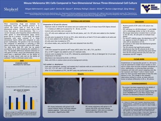
Data are averages of 5 replicates; corrections for background
absorbance were made by subtracting media and ECM control
data for each selected point in time. p value for one tailed t-
test is 0.0005.
DISCUSSION
RESULTS
Preparation of 2D and 3D cultures:
- Replicate wells of chilled 96-well plates were pre-coated with 10 mL of mouse tissue ECM (Sigma) diluted
1:1 in Dulbecco’s MEM and incubated for 15 - 30 min. at 37oC.
- Control wells and slides were uncoated.
- 4.5 x 10⁴ cells were added per well in the 96-well plates, and 1.8 x 10⁵ cells were added to the chamber
slides.
- All cells were incubated for 30 min at 37oC, after which 60 mL of Ham’s F12-K were added to all wells and
0.5 mL were added to the chamber slides.
- All cells were then incubated at 37oC.
- All culture media, sera, and the S91 cells were obtained from the ATCC.
MTT assay:
- Cells were assayed for growth by MTT assay (ATCC) after 24 h, 48 h, 72 h, and 96 h.
- 20 mL of MTT reagent was added to each of 5 replicate wells.
- Cultures were incubated for 3 h at 37oC, followed by solubilization in 100 mL of detergent for 2 h at room
temperature in the dark.
- Absorbance was measured by ELISA at 570 nm.
- Wells with ECM or medium alone served as background controls.
Cell number vs. absorbance:
- Cells were trypsinized, counted, and seeded into 5 replicate wells at concentrations of 1 x 10⁴, 2.5 x 10⁴,
5.0 x 10⁴, 7.5 x 10⁴, and 1 x 10⁵ cells per well.
- After 12 h of incubation at 37oC, the MTT assay was performed as above.
REFERENCES
Mouse Melanoma S91 Cells Compared in Two-Dimensional Versus Three-Dimensional Cell Culture
Megan Behrmann‡, Logan Lyda†, Denise M. Gipson*, Brittany Poling‡, David J. Klinke**, Burton Lidgerding‡, Qing Wang#.
‡Department of Biology, Shepherd University, Shepherdstown, WV; †Department of Chemistry, Shepherd University, Shepherdstown, WV; *Jefferson High School, Shenandoah Junction, WV;**Department of
Chemical Engineering, West Virginia University, Morgantown, WV; **Department of Microbiology, Immunology & Cell Biology, West Virginia University, Morgantown, WV; #Department of Computer
Sciences, Mathematics and Engineering, Shepherd University, Shepherdstown, WV
When researching drugs and methods for
eradicating cancer, the standard practice is to
culture the cancer cells in vitro on a two-
dimensional surface. However, cancer cells in the
living host grow in three-dimensions. This is a
concern because cells frequently behave differently
when grown in three-dimensional space versus
grown flat on a surface. In this study, S91 mouse
melanoma cells were cultured in a three
dimensional matrix (ECM) and compared to cells
grown traditionally. The growth in the two formats
was compared to determine if culturing in ECM
matrix culturing was successful using an MTT assay.
The data shows that S91 cells can be grown
successfully on an ECM simulating the conditions in
vivo. The possible differences between behaviors of
cells grown in three- versus two-dimensional culture
could have a significant effect on the ability of
drugs to treat cancer cells and on the ability of
cytotoxic T cells to eliminate cancer cells.
0
0.2
0.4
0.6
0.8
1
1.2
1.4
1.6
0 1 2 3 4 5 6 7 8 9 10 11
AverageAbsorbanceat570nm.
Cell Count (x 104)
Average absorbance at 570 nm. for 5 replicates vs. cell count.
MATERIALS AND METHODS
S91 mouse melanoma cells grown in 2D
culture without matrix; red areas indicate
melanin pigment. Cells were all on one
focal plane. Magnification 200X
S91 mouse melanoma cells grown in 3D
culture with ECM; red areas indicate
melanin pigment. Cells were on multiple
focal planes. Magnification 200x.
0
0.5
1
1.5
2
2.5
1 2 3 4
Absorbanceat570nm.
Time (days)
Growth of 3D vs. 2D Cells
3D Cells
2D Cells
FUTURE WORK
Establishment of reproducible growth in a 3D matrix
must be optimized to enable future studies in vitro
and to create more accurate computer simulations in
silico. Additionally, co-culturing these and other
melanoma cell lines with cancer-targeting cells like
the Cytotoxic and Helper T cells in matrix would
allow for more accurate observation of how the cells
behave In vivo. This change in the standard
observation method is crucial, as many cells are
known to behave differently in 2D vs. 3D culture.
D. J. Klinke II, N. Cheng, and E. Chambers, Quantifying crosstalk
among interferon – g, interleukin-12, and tumor necrosis factor
signaling pathways within a TH1 cell model. Sci. Signal. 5 (220), 1-
12 (2012).
G. Y. Lee, P. A. Kenny, E. H. Lee, and M. J. Bissell, Three-dimensional
culture models of normal and malignant breast epithelial cells.
Nat. Methods. 4 (4), 359-365 (2007). doi:10.1038/nmeth1015.
J. Odot, P. Albert, A. Carlier, M. Tarpin, J. Devy, and C. Madoulet, In
vitro and in vivo anti-tumoral effect of curcumin against
melanoma cells. Int. J. Cancer 111, 381-387 (2004).
ACKNOWLEDGMENTS
The project has been supported by the NIGMS of
the NIH grant as part of the WV-INBRE
(P20GM103434). It has also been Supported by NIH
Grant 5P20RR016477 to the West Virginia IDeA
Network for Biomedical Research Excellence.
- Routine growth of S91 cells in 2D culture was
achieved.
- S91 cells grew in a 3D ECM diluted 1:1 with DMEM
with a morphology similar to that in 2D culture.
- Microscope observation revealed that in contrast
to the 2D cells, 3D cells grew on multiple focal
planes.
- For the MTT viability assay, the absorbance at 570
nm. appears to level off slightly at 5 – 7.5 x 10⁴
cells with an absorbance of 0.8..
- All cultures in this study had absorbances greater
than this value.
- The 3D cultures had significantly greater
absorbances than the 2D cultures at all of the
time periods studied, indicating they grew better
than the 2D cultures because only live cells can
metabolize the MTT reagent.
DATA
STANDARD CURVE:
Absorbance Vs. Cell Count
MTT ASSAY OF GROWTH