2. indeed be a highly significant discovery, although previous work
in this area using MTH1(−/−) mice appeared somewhat
contradictory.4
Fortuitously, we had access to compounds that
were able to serve as leads from a previous internal project that
had sought agonists of Toll-like receptor 7 (TLR7) and
established cross-reactivity with MTH1 as described below.
Toll-like receptors (TLR) are pattern recognition receptors
that serve to recognize microbial pathogens and fragments of
bacterial DNA in order to elicit an immune response.5
Such
receptors are primed to recognize distinct pathogen-associated
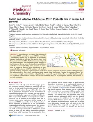
molecular patterns (PAMPs) within microbes. Imiquimod 4
(Figure 2), first developed by 3M Pharmaceuticals, is the only
approved small molecule agonist of toll-like receptor 7 (TLR7)
and is used in the topical treatment of basal cell carcinoma
(BCC), genital warts, and actinic keratosis. Agonism of TLR7
results in stimulation of the innate immune system via
induction of cytokines such as interferon alpha (IFNα) and
IL-12 and can result in significant antitumor activity. Clearance
rates in BCC following imiquimod treatment have been
reported in the range 73−87%, and for many patients the
therapy is effectively curative.6
During the course of historical
research into agonists of TLR7,7
a photoactivatable, cross-
linkable TLR7 agonist probe based on an imiquimod template,
5 (pEC50 of 1 μM for the induction of IL-12 in THP-1 cells),
was synthesized to identify which cellular proteins the probe
interacted with. At the time of this research, such TLR7
agonists had not been shown to mediate their effects via a direct
interaction with TLR7. Initial investigations were carried out on
THP-1 cells as these could be differentiated to be TLR7
responsive. No displaceable binding was observed in a protein
of molecular mass consistent with membrane associated TLR7,
but displaceable binding was observed to a cytosolic 20 kDa
protein. The interaction was observed in both differentiated
and undifferentiated cells. This protein was determined to be
MTH1. At this stage, a high-throughput screen was carried out
against MTH1 with the intention of identifying additional novel
equity against this target and several series of interest were
identified.
A working hypothesis that MTH1 may be the true effector of
the efficacy of TLR7 agonists was ultimately discounted based
on a number of key experiments, including the observation that
TLR7 agonist activity was unaltered in splenocytes from MTH1
knockout mice and that an 85% reduction in protein expression
of MTH1 via siRNA knock-down had no observable effect on
TLR7 signaling. Intriguingly, further testing of TLR7 agonists
from different chemical series also indicated cross-reactivity of
these ligands with MTH1 and we speculate this may be due to
recognition of the similar molecular architecture of the oxidized
nucleotides that act as MTH1 substrates and the nucleotide
bases present in the bacterial PAMPs.8
With the link to TLR7
disproven, internal research on MTH1 inhibitors was halted in
2003. When, more than a decade later, a role in oncology re-
emerged for MTH1, this information proved crucial in enabling
us to rapidly develop our own potent and selective cell probes
with which to investigate this target as described below.
■ CHEMISTRY
The synthesis of macrocyclic MTH1 inhibitors followed closely
the routes described for a related series of TLR7 agonists.9
Treatment of dichloropyrimidine 6 with ammonia gave
aminopyrimidine 7, which was protected as its ethylcarbamate
derivative 8 in good yield (Scheme 1). Alkylation of the
carbamate nitrogen with a protected phenolic benzyl group to
give 9 was followed by displacement of the second chlorine
atom to install the key methylamino motif in 10. Introduction
of the ether side chain to give 12 was achieved following
displacement of the sulfone 11, formed by oxidation of
thioether 10, with 2,2′-oxidiethanol. Removal of the tert-
butyldimethylsilyl protecting group gave phenol 13, which was
subject to a Mitsunobu induced macrocyclization to give 14.
Finally, formation of the imidazolone ring was achieved by iron-
mediated reduction of the nitro group with concomitant ring-
closure onto the carbamate to give macrocycle 15. An
analogous strategy was adopted for lactams 30−33 (see
Table 2), although in these cases the key macrocyclization
step involved macrolactamization in place of Mitsunobu
reaction (see Supporting Information).
Quinoline amides were constructed as shown in Scheme 2
from the 4-hydroxyquinoline ester 16 by conversion to the
chloride 17 using phosphoryl trichloride then subsequent
displacement with the requisite aniline, affording intermediates
18 and 20. The ester functionality was then reacted with
methylamine to give amide inhibitors 19 and 21. Procedures for
the synthesis of other members of this aniline series are
described in the Supporting Information. The ether linked
example was made by displacement of 17 with phenol under
basic conditions and then hydrolysis of the ester 22, conversion
Figure 1. Literature inhibitors of MTH1, 1−3.
Figure 2. TLR7 agonist Imiquimod (4) and a photoaffinity probe
TLR7 agonist (5).
Journal of Medicinal Chemistry Article
DOI: 10.1021/acs.jmedchem.5b01760
J. Med. Chem. XXXX, XXX, XXX−XXX
B
3. to the acid chloride, and reaction with methylamine to give 23.
Aminoquinazoline MTH1 inhibitor 24 was made using a
Suzuki reaction between 4-chloroquinazolin-2-amine and
phenyldimethylacetamide 4-boronic ester as shown in Scheme
3.
■ RESULTS AND DISCUSSION
Table 1 shows the MTH1 inhibitory potencies for a range of
TLR7 modulators known in the literature. Imiquimod 4 itself
has reasonable submicromolar potency, but Resiquimod, 25,10
known to be a much more potent TLR7 agonist, shows slightly
Scheme 1. Synthesis of Macrocycle MTH1 Inhibitor 15a
a
Reagents and conditions: (i) NH3, i-PrOH, Et3N, 0 °C, 5 h; (ii) EtOCOCl, THF, Et3N, 0 °C, 5 h, 76% (2 steps); (iii) m-OTBDMS benzyl chloride,
NaI, K2CO3, acetone, 20 °C, 16 h; (iv) MeNH2, THF, 20 °C, 16 h, 100% (2 steps); (v) mCPBA, DCM, 20 °C, 2 h; (vi) 2,2′-oxidiethanol, NaH,
THF, 20 °C, 17 h, 71% (2 steps); (vii) TBAF, THF, 20 °C, 16 h, 86%; (vii) DTAD, Ph3P, THF, 20 °C, 20 h, (ix) Fe, AcOH, water, 60 °C, 4 h, 7%
(2 steps).
Scheme 2. Synthesis of Quinoline Amide MTH1 Inhibitorsa
a
Reagents and conditions: (i) POCl3, 100 °C, 1 h, 90%; (ii) 4-chloro-2-fluoroaniline, AcOH, i-PrOH, 100 °C, 16 h, 99%; (iii) MeNH2, Me3Al, THF,
60 °C, 2 h, 79%; (iv) aniline, AcOH, DMF, 100 °C, 1 h, 76%; (v) MeNH2, EtOH, 120 °C, 3 h, 81%; (vi) phenol, K2CO3, MeCN, 80 °C, 16 h, 100%;
(vii) (a) NaOH, THF, 20 °C, 17 h, (b) SOCl2, 80 °C, 1 h, (c) MeNH2, DMF/THF, 20 °C, 1 h, 74%.
Journal of Medicinal Chemistry Article
DOI: 10.1021/acs.jmedchem.5b01760
J. Med. Chem. XXXX, XXX, XXX−XXX
C
4. reduced affinity for MTH1. Compound 26 is from a distinct
series of orally bioavailable purinone TLR7 agonists, first
reported by Dainippon Sumitomo Pharmaceuticals,11
that
elicits a TLR7-mediated cellular response with similar potency
to these clinical agents. Critical for this work, N-methylation of
26, to give 27, is observed to completely ablate TLR7 activity in
this series and also results in a modest increase in MTH1
affinity. This observation has previously been reported for a
closely related series of TLR7 agonist purinones and indicates
that the exocyclic NH2 motif is critical for TLR7 activity.12
In a
similar fashion, it has been reported that N-methylation of 4
also results in complete loss of TLR7 agonist activity.13
These
data show that MTH1 is able to bind agonists of TLR7 as well
as inactive close exemplars.
A crystal structure of compound 25 solved in complex with
full length p18 isoform MTH1 (M1−156) with an N-terminal
6-His TEV protease cleavable tag is shown in Figure 3.
Compound 25 (Figure 3a) makes hydrogen bonds between the
quinoline nitrogen and residue D119 and the amino group and
D120. These hydrogen bonds with D119 and D120 are
common features of MTH1 ligands as demonstrated in the
overlay with product 8-oxo-dGMP (Figure 3c) and also feature
Scheme 3. Synthesis of Aminoquinazoline MTH1 Inhibitor
24a
a
Reagents and conditions: (i) Pd-118, K2CO3, 1,4-dioxane/H2O, 100
°C, 2 h, 28%.
Table 1. MTH1 Potencies of Assorted TLR7 Ligandsa
a1
Inhibition of MTH1 enzyme was assessed by detecting the inorganic pyrophosphate generated when the nucleoside triphosphate substrate, 8-oxo-
dGTP, is hydrolyzed. 2
Induction of IL-12 in human PBMC cells. All data are in μM and are the mean of at least n = 2 determinations and have SEM
within 0.2 log units.
Figure 3. X-ray crystal structures of human MTH1 in complex with
(a) 25 (pink, resiquimod), (b) complex with 25 rotated to show the
hydrophobic residues (white), and (c) complexes with 25 and 8-oxo-
dGMP (orange) superimposed. Selected carbon atoms, protein
backbone cartoon, and close polar contacts are represented in the
complex color. Compounds are shown as sticks, and protein residue
side chains are shown as thinner sticks. Close polar contacts are
represented as dotted lines.
Journal of Medicinal Chemistry Article
DOI: 10.1021/acs.jmedchem.5b01760
J. Med. Chem. XXXX, XXX, XXX−XXX
D
5. in protein−ligand structures of 1 and 3. Compound 25 displays
an additional hydrogen bonding contact with the backbone
carbonyl of T8, another interaction it has in common with 8-
oxo-dGMP (Figure 3c). The interaction with T8 is
hypothesized to contribute to selectivity for the deoxyribose
group of 8-oxo-dGMP.14
The dimethylhydroxy group of 25
pushes “down” into the pocket where the ribose of the
substrate/product sits (Figure 3b and 3c). Two polar
interactions made by the product 8-oxo-dGMP are not seen
with inhibitors 25; an interaction between the guanosine base
and N33 and between the α-phosphate group and K23 terminal
amino group. N33 is a mobile residue which can move to
accommodate different chemotypes binding to MTH1. The
quinoline ring of 25 occupies the same space as the nucleobase
of 8-oxo-dGMP, nestling among a number of residues
highlighted in Figure 3b. These residues form a pocket around
the ligands making various hydrophobic and π-stacking
interactions. The hydrophobic residues include W117 (above
the purine binding site), L9, I70, F72, M81, and V83 (below
the base binding site) and F74 and F139 (on the right-hand
side of the nucleobase binding site). Finally, the side-chain of
F27 is at the tip of a flexible loop (residues 23−31) that folds
over the nucleobase binding site (not shown in Figure 1 for
clarity). The mobility of this loop, along with minor shifts in
other hydrophobic residues, readily accommodates different
sizes and shapes of ligands. In the complex with 25, rather
unusually, the loop is open and the position that F27 normally
adopts is occupied by a second molecule of 25, which may be
an artifact of the crystallographic system and high compound
concentration used.
The X-ray crystal structure of compound 27 (Figure 4a)
shows that it binds at the same site as compound 25 and the
base of 8-oxo-dGTP, making the usual hydrogen bonding
interactions with side chains of D119 and D120. The phenyl
group binds in the hydrophobic site. The asparagine side chain
of N33 moves in toward the binding site to occupy a similar
conformation to that seen in the 8-oxo-dGMP complex.
Examination of the crystal structure of 27 bound in MTH1
highlighted the relatively close proximity of the pendant
methoxyethyl side chain to the N1-benzyl group, with the
distance to the meta-position of the order of 3.5 Å (Figure 4a).
We hypothesized that this close proximity of groups was
unlikely to be a dominant conformation in solution,
contributing to the moderate affinity observed. A macro-
cyclization strategy that connects the ether side chain to the
meta-position of the benzyl group was expected to lock the
molecule in its active binding conformation and result in
significant potency gains through reduced entropic penalty of
association and increased energy barrier of dissociation.
Intriguingly, synthetic precedent for such a macrocyclization
strategy had been reported in the patent literature for a series of
purinone TLR7 agonists related to 26.9
It is unclear given the
lack of structural information for TLR7 how such a design
strategy was arrived at, although homology models of TLR7
receptors have been reported.15
Gratifyingly, macrocyclization of 27 utilizing an n-pentyl bis-
ether linker gave a compound with sub-nM potency against
MTH1, 28, representing over a 1700-fold increase in potency,
validating our hypothesis around the relatively high energy
binding conformation of the former (Table 2). This compound
was relatively lipophilic, and so a contribution to this increase in
potency from additional lipophilicity could not be ruled out
(because compound 27 has a log D = 2.6, 28 is nearly a log unit
more lipophilic). One consequence of this increased lip-
ophilicity is an increase in in vitro clearance as measured in
human microsomes, although in vitro permeability is high and
the compound demonstrates acceptable free binding levels in
human plasma. Shortening the linker to an n-butyl bis-ether
linker, compound 29, gave a profile that was largely similar, but
the reduced lipophilicity of this compound was not enough to
improve in vitro clearance. A breakthrough came when
introducing a polar ether linker into the all carbon side chain
of 28 to give 15. This compound maintained its exceptional
potency against MTH1 but did so at lipophilicity less than that
of the uncyclized analogue 27, representing a 1000-fold
increase in potency for no increase in log D. With a reduction
in log D came a significant improvement in in vitro clearance,
with a profile comparable to 27. Encouraged by this result, we
sought to further improve properties by increasing the polarity
of the linking group. Both secondary and tertiary amides 30 and
31 have substantially lowered log D (= 1.3), and this confers
increased metabolic stability in vitro, together with high free
levels. In each case, however, potency against MTH1 was
compromised, as was permeability. The regioisomeric amides
32 and 33 were also synthesized, and the former at least
demonstrated potent activity. Despite a log D of 1.8 for this
compound, clearance remained very high, and indeed this
compound proved to be unstable in human plasma. The
tertiary amide analogue 33 had a similar profile but was
substantially less active, more lipophilic, and similarly unstable
in human plasma.
Figure 4. (a) X-ray crystal structures of human MTH1 in complex
with 27 (green). (b) Complexes with 27 (green) and 15 (yellow)
overlaid. Selected carbon atoms, protein backbone cartoon, and close
polar contacts are represented in the complex color. Compounds are
shown as sticks, and protein residue side chains are shown as thinner
sticks. Close polar contacts are represented as dotted lines. Some
atoms of the loop covering the active site (residues 23−31) have been
removed for clarity. (c) Example conformational ensemble for 15.
Lowest-energy conformation (green) has a ring orientation similar to
that seen for 27 (cyan) in the protein−ligand structure. (d) Modeled
conformation of amide-containing macrocycle 30 (yellow) overlaid on
X-ray structure conformation of 27 (cyan) shows differential
orientation of phenyl ring. (e) Conformational ensemble of 29
derived using NMR suggests that the dominant conformations in
solution (gray) are very similar to the conformation seen in the
protein−ligand structure (maroon).
Journal of Medicinal Chemistry Article
DOI: 10.1021/acs.jmedchem.5b01760
J. Med. Chem. XXXX, XXX, XXX−XXX
E
6. The SAR seen for the macrocycles is likely to be closely
linked to their ability to adopt the correct conformation.
Modeling suggests that macrocycles with linkers of 6 and 7
atoms are able to mimic the conformation of uncyclized
analogue 27 in the binding pocket (Figure 4c) to varying
degrees. Linkers shorter than six atoms were considered, but
their effect on conformation appeared more pronounced than
with the longer linkers, and additionally, they proved less
tractable chemically. Taking into account the difference in
lipophilicity, the drop in potency between triether macrocycle
15 and amide-containing macrocycle 30 is approximately 30-
fold. Conformational analysis of 30 showed that the preferred
Table 2. MTH1 Potencies and in Vitro DMPK Profiles of Purinone Macrocyclesa
a1
Inhibition of MTH1 enzyme was assessed by detecting the inorganic pyrophosphate generated when the nucleoside triphosphate substrate, 8-oxo-
dGTP, is hydrolyzed. 2
log D at pH7.4. 3
In vitro clearance in human microsomes (μL/min/mg). 4
Percent free in human plasma. 5
A to B permeability
in Caco2 cells at pH7.4 (1 × 10⁻⁶
·cm/s). 6
LLE = pIC50 − log D. All activity data are reported in μM and are the mean of at least n = 2 determinations
and have SEM within 0.2 log units.
Journal of Medicinal Chemistry Article
DOI: 10.1021/acs.jmedchem.5b01760
J. Med. Chem. XXXX, XXX, XXX−XXX
F
7. orientation of the phenyl of the amide macrocycle (Figure 4d)
was different from the phenyl orientation seen for 27 and 15.
However, a protein crystal structure of 30 (not shown) exhibits
a phenyl orientation that is very similar to that of 27 and 15,
and we hypothesize that the energetic cost of inducing this
optimal orientation of the phenyl negatively affects the potency
of 30. Such an effect is likely to play a role in the lower potency
seen for 31 as well. The ligand-lipophilicity efficiency of
alternative amide-containing macrocycle 32 and triether
macrocycle 15 are similar, and comparison of their protein−
ligand structures suggests that both are very similar in shape
and conformation. The amide NH of 32 picks up an additional
hydrogen bonding contact (not shown) to the backbone
carbonyl of T8, but it is likely that the difference in potency
between these compounds is largely caused by their difference
in lipophilicity. Amide 33, an N-methylated analogue of 32,
exhibits a markedly lower potency, which may be explained by
abrogation of the amide hydrogen bond to T8 seen for 32 as
well as conformational effects resulting from the N-methylation.
The conformational hypothesis was corroborated by NMR
experiments that showed unambiguously that conformations of
the 6-linker compound 29 in solution closely resemble its
modeled and protein-bound conformers (Figure 4c−e). NMR
experiments on the nonmacrocyclic precursor 27 did not yield
a conformational ensemble, suggesting that the unlinked chain
is very flexible.
In addition to the observations of cross-reactivity of TLR7
ligands with MTH1, the historical project also generated high-
throughput screening data with the aim of identifying further
chemical equity. Among the hits identified, a series of 3-
amidoquinolines, exemplified by 19 and 34, stood out as
worthy of further profiling (Table 3). Both primary amide 34
and secondary amide 19 had high potency against MTH1 (12.5
and 0.9 nM, respectively), but the dimethyl analogue 35
showed a significant loss of potency, providing a closely related
compound with significantly less activity (>130-fold lower
activity of 35 vs 19). The permeabilities of 19 and 35 as
measured in a Caco-2 assay were high (Papp 24 × 10−6
and 33 ×
10−6
cm/s, respectively) with no evidence of efflux (efflux ratios
of 0.5 and 0.6, respectively), giving confidence that the
molecules penetrate into cells. Further exploration of secondary
amide substitution (data not shown) showed no benefits in
terms of potency or LLE, with methyl judged as being optimal.
Deletion of both methoxy groups on the core gave 36 and led
to a small potency decrease but with LLE and properties
remaining similar. Further truncation of the quinoline to the
pyridyl 37 somewhat surprisingly retained potency and showed
some benefits in terms of solubility, although this remained a
challenge for the series. The high potency of this fragment
shows the tolerance of MTH1 for binding a diverse range of
structural motifs, provided key pharmacophoric elements are
met, and was suggestive of a high degree of substrate
promiscuity.
The X-ray crystal structure of 19 in complex with MTH1 is
shown in Figure 5. The compound occupies the same site as
the base of 8-oxo-dGMP. Compound 19 makes the typical
hydrogen bonds with D119 and D120, and the methoxy groups
Table 3. MTH1 Potencies and in Vitro DMPK Profiles of 3-Amidoquinolines
ID R1 R2 R3 X MTH1 IC50
a
log Db
Clint Hu Micsc
Hu PPB (% free)d
Sol (μM)e
LLEf
34 NH2 2-F, 4-Cl Me NH 0.0125 3.5 23 3.4 2 4.4
19 NHMe 2-F, 4-Cl Me NH 0.0009 3.3 13 3.9 15 5.7
35 NMe2 2-F, 4-Cl Me NH 1.22 2.5 13 6.6 6 3.4
36 0.002 3.3 6 6.2 8 5.5
37 0.009 3.1 10 8.9 39 4.9
21 NHMe H Me NH 0.0005 2.7 14 9.1 108 6.6
38 NHMe 2-(CH2)2SO2Me Me NH 0.0005 1.7 8 22 30 7.6
23 NHMe H Me O 5.75 2.1 13 14.7 35 3.1
39 NHMe H CH2-4-N-Me-piperidine NH 0.00008 1.6 8 51 >1000 8.4
a
Inhibition of MTH1 enzyme was assessed by detecting the inorganic pyrophosphate generated when the nucleoside triphosphate substrate, 8-oxo-
dGTP, is hydrolyzed. b
log D at pH7.4. c
In vitro clearance in human microsomes (μL/min/mg). d
Percent free in human plasma. e
Aqueous solubility
at pH7.4. f
LLE = pIC50 − log D. All activity data are reported in μM and are the mean of at least n = 2 determinations and have SEM within 0.2 log
units.
Figure 5. X-ray crystal structures of human MTH1 in complex with 19
(green). Selected carbon atoms, protein backbone cartoon, and close
polar contacts are represented in the complex color. Compounds are
shown as sticks, and protein residue side chains are shown as thinner
sticks. Close polar contacts are represented as dotted lines. The 2-
chloro-4-fluoro-phenyl group adopts a dual conformation.
Journal of Medicinal Chemistry Article
DOI: 10.1021/acs.jmedchem.5b01760
J. Med. Chem. XXXX, XXX, XXX−XXX
G
8. sit among the hydrophobic residues. The side chain of N33
makes a hydrogen bond with the aniline nitrogen of 19. The
amide group makes an additional hydrogen bond with the
backbone nitrogen of G34. A hit expansion library was carried
out on lead 19 based on aniline substitution. Deletion of both
halogens gave unsubstituted phenyl 21, which was both more
potent and less lipophilic, with improved LLE and
physicochemical properties as a consequence. The permeability
of 21 as measured in a Caco-2 assay was high (Papp 27 × 10−6
cm/s) with no evidence of efflux (efflux ratio 0.4) that could
limit any cellular activity. The SAR showed a clear and
consistent preference for ortho > meta > para substitution
(based on six matched triplets, F, Cl, Me, CN, SO2Me, and
P(O)Me2, data not shown). Introduction of polar substituents
in the ortho position, exemplified by ethyl sulphone 38, allowed
further lowering of log D while increasing LLE. Importantly,
switching the aniline to an aryl ether gave 23, which reduced
activity against MTH1 by >1000-fold relative to 19, consistent
with removal of a key interaction seen in the X-ray crystal
structure. In an attempt to improve solubility in this series, we
investigated addition of basic functionality at the 6-position as
this looked most likely to be tolerated from inspection of the X-
ray crystal structure. Bases such as 39 further enhanced potency
(80 pM) and improved LLE (8.4), giving highly soluble, sub-
nM inhibitors of MTH1.
A comparison of the rat pharmacokinetics of macrocycle 15,
in comparison to its uncyclized analogue 27, is shown in Table
4. These data show a largely similar profile for the two
compounds, with increased bioavailability seen for the
macrocycle suggestive of better permeability. Clearances are
moderate, with a low volume of distribution typical of
uncharged molecules. The moderate clearance and low volume
combine to give a relatively short half-life of around 1 h in both
cases. It is clear that macrocyclization itself has had an overall
neutral impact on the pharmacokinetic properties of this
scaffold and a rather significant impact on potency at equivalent
log D. The rat pharmacokinetics of quinoline 19 shows that the
compound has low clearance and low volume, leading to a
moderate half-life. Bioavailability of 37% is consistent with the
reasonable solubility and high permeability of this compound as
described above and is indicative of the potential of this series
to develop tool compounds suitable for in vivo studies.
A third series of inhibitors based on a 2-aminoquinazoline
template was also identified from the HTS, with hit 40 showing
modest potency against MTH1 (Table 5). Dimethylation of the
amine to give 41 resulted in reduced activity, while removal of
the methyl (42) significantly increased potency against MTH1.
A hit expansion library around 40 based on Suzuki couplings
showed that meta and para substitution was favored over ortho.
Transfering this SAR into the des-methyl series led to the
identification of compounds such as dimethylamide 24 with
enhanced MTH1 potency and acceptable physicochememical
properties for further profiling. Permeability of 24 measured in
a Caco-2 assay was high (Papp 34 × 10−6
cm/s) with no
significant efflux (efflux ratio 0.7) that would limit cellular
penetration. The X-ray crystal structure of 24 in complex with
MTH1 is shown in Figure 6. The compound occupies the same
site as the base of 8-oxo-dGMP. An overlay of the complex of
24 with 1 (Figure 6a) shows that the aminopyrimidine rings of
the two compounds make identical typical hydrogen bonds
Table 4. Rat Pharmacokinetics of Macrocycle 15, Its
Uncyclized Analogue 27, and Quinoline Amide 19a
ID bioavailability (%) clearance (mL/min/kg) Vdss (L/kg) t1/2 (h)
27 31 29 1.1 1.2
15 64 26 0.8 1.3
19 37 9 0.9 3.0
a
Rat pharmacokinetics in Wistar Han rats followed a dose of 3 μMols/
kg (po) and 1.5 μMols/kg (iv).
Table 5. MTH1 Potencies and in Vitro DMPK Profiles of 2-Aminoquinazolines
ID R1 R2 MTH1 IC50
a
log Db
Clint Hu Micsc
Hu PPB (% free)d
Sol (μM)e
LLEf
40 NHMe H 3.32 3.6 215 0.9 7 1.9
41 NMe2 H 12.5 4.0 120 0.1 1 0.9
42 NH2 H 0.021 2.9 71 5.1 214 4.8
24 NH2 CH2C(O)NMe2 0.009 2.0 5 4.1 118 6.1
a
Inhibition of MTH1 enzyme was assessed by detecting the inorganic pyrophosphate generated when the nucleoside triphosphate substrate, 8-oxo-
dGTP, is hydrolyzed. b
log D at pH7.4. c
In vitro clearance in human microsomes (μL/min/mg). d
Percent free in human plasma. e
Aqueous solubility
at pH7.4. f
LLE = pIC50 − log D. All activity data are in μM and are the mean of at least n = 2 determinations and have SEM within 0.2 log units.
Figure 6. (a) X-ray crystal structures of human MTH1 in complex
with 24 (yellow). (b) Complexes with 24 and 1 (PDB code: 4N1U,
cyan) overlaid. Selected carbon atoms, protein backbone cartoon, and
close polar contacts are represented in the complex color. Compounds
are shown as sticks, and protein residue side chains are shown as
thinner sticks. Close polar contacts are represented as dotted lines.
Journal of Medicinal Chemistry Article
DOI: 10.1021/acs.jmedchem.5b01760
J. Med. Chem. XXXX, XXX, XXX−XXX
H
9. with D119 and D120. The phenyl ring of the quinoline group
sits in the same site as the cyclopropyl group among the
hydrophobic residues of the nucleotide base binding pocket.
In addition to potent activity in an MTH1 enzyme assay,
binding affinity for MTH1 protein was assessed independently
by surface plasmon resonance (SPR) (Table 6). This data
confirmed the high affinities seen and correlated well with
potencies as measured in the biochemical assay, giving further
confidence in the data. Intriguingly, observed half-lives for
dissociation from MTH1 varied significantly according to
chemotype. S-Crizotinib 3 showed rapid dissociation (t1/2
measured in seconds), whereas 1 and the quinazoline/quinoline
scaffolds described here showed moderate half-lives (of the
order of minutes). The macrocyclic inhibitors, however,
showed significantly extended residence time with t1/2 in excess
of 1 h for both 15 and 28 in contrast with acyclic precursors
like 27, which dissociated rapidly (t1/2 in the range of seconds,
beyond the resolution of SPR). On the basis of their high
potencies and good permeability, examples from each series
were assessed for their ability to penetrate cells and bind to
MTH1 in a manner that was independent of a phenotypic end
point. This is important because any phenotype which
manifests itself predominantly as a cell killing effect as here
may be particularly susceptible to any number of off-target
effects that impact cell viability. For this, we utilized a whole cell
thermal stabilization assay (CETSA)16
to assess target engage-
ment in K562 cells, a technique that had also been employed
for profiling of 1. In this assay, cells are treated with inhibitor
and subject to small increases over a temperature gradient
before cells are lysed and protein quantified by Western blot.
Any compound binding to its target within the cell will have a
stabilizing effect on, and prevent the thermal denaturation of,
the protein relative to untreated cells. These primary melt
curves and ligand-induced shifts are established to aid selection
of testing conditions. The compound is then tested in
increasing concentration at a single temperature to establish
the CETSA EC50 of target engagement. As can be seen in Table
6, the high enzymatic inhibitory activity of the macrocycles
translated into very potent cellular target engagement with
EC50s of 9 and 3 nM for 28 and 15, respectively. These
compared favorably with 1 for which in this assay we measure
10 nM (target engagement in this assay format is reported for
this compound but potency is not disclosed). Nonmacrocyclic
analogue 27 has a much weaker in EC50 of 1.5 μM, in line with
its much reduced MTH1 enzyme activity. A similar observation
was made with the quinoline series, with 19 showing a potent
CETSA EC50 (7 nM) in marked contrast to dimethylated
quinoline amide 35, which was less active in both the enzyme
assay (IC50 1200 nM) and CETSA (EC50 950 nM) assays. The
aminoquinazoline 24 showed an intermediate CETSA EC50 (80
nM) in accordance with the lower MTH1 enzyme activity.
Figure 7 shows the excellent correlation between MTH1
inhibitory potency and measured target engagement in K562
cells, across each of these described series, together with 1 and
3. These data further support the conclusion that these diverse
agents are potent, cell-active inhibitors of MTH1 function and
highlight the value in pairing potent inhibitors with inactive or
less active structurally related counterparts to act as negative
controls in cell based experiments.
To further characterize the utility of these novel inhibitors,
and to ensure any phenotype would not be confounded by off-
target kinase inhibition, we profiled representative compounds
15, 19, 21, and 24 against an extended panel of kinases. Given
the heritage of anilinoquinolines as kinase inhibitors, it was
surprising to find that 15 did not show appreciable kinase
inhibition at all (<35% at 10 μM across 267 kinases). Profiling
of 19, 21, and 24 suggested no activity against any kinase at a
testing concentration of 1 μM. In addition, these compounds
Table 6. Primary Target Potencies, Lipophilicity, and Selected Tumor Cell Line Antiproliferative Effects of MTH1 Inhibitors
ID series MTH1 IC50
a
MTH1 Kd
b
MTH1 t1/2 (min)b
CETSA EC50
c
log Dd
U2OS GI50
e
A549 GI50
e
H358 GI50
e
MCF7 GI50
f
1 0.003 0.005 3.9 0.010 3.5 3.0 3.3 2.3 4.0
3 0.138 0.039 0.1 3.6 2.2 2.2 3.8 4.9 4.4
27 TLR7 0.536 0.440 1.5 2.6
28 macrocycle 0.0003 0.0002 151.7 0.009 3.5 12.1 29 8.2
15 macrocycle 0.0005 0.0004 81.2 0.003 2.4 >30 >30 >30 >30
19 quinoline 0.0009 0.0005 8.7 0.007 3.3 12.0 13.9 5.8 14.4
21 quinoline 0.0006 0.0002 3.5 NT 2.7 >30 >30 >30 >30
35 quinoline 1.22 0.95 2.5
24 aminoquinazoline 0.009 0.003 2.1 0.080 2.0
a
Inhibition of MTH1 enzyme was assessed by detecting the inorganic pyrophosphate generated when the nucleoside triphosphate substrate, 8-oxo-
dGTP, is hydrolyzed. b
MTH1 affinity and halflife for dissociation as measured by surface plasmon resonance. c
Whole cell thermal stability assay in
K562 cells heated at 52 °C for 3 min. d
log D at pH7.4. e
Growth inhibition in the indicated cell line following 5 day compound incubation. f
Growth
inhibition in the indicated cell line following 7 day compound incubation. All activity data are in μM and are the mean of at least n = 2
determinations and have SEM within 0.2 log units.
Figure 7. Correlation between MTH1 inhibitory potencies and target
engagement in K562 cells colored by series. Straight line is line of fit.
Journal of Medicinal Chemistry Article
DOI: 10.1021/acs.jmedchem.5b01760
J. Med. Chem. XXXX, XXX, XXX−XXX
I
10. were profiled in a diverse panel of 153 secondary pharmacology
targets and no notable off-target activities were observed that
might confound phenotypic studies (these data are available as
Supporting Information).
A selection of compounds was assessed for their ability to
impact growth on a small panel of tumor cell lines (Table 6).
Consistent with published data, 1 and 3 displayed modest but
consistent growth inhibition in the range 3−5 μM, even though
in our hands 3 exhibits relatively weak cellular target
engagement. Macrocycle 28 demonstrated much weaker
activity in this panel but did have an effect on viability at the
top concentrations tested. Critically, however, macrocycle 15,
which is significantly less lipophilic than 28, and has equivalent
MTH1 potency, had no impact on cell viability. In a similar
fashion, initial lipophilic quinoline screening hit 19 had modest
antiproliferative effects. However, lowering its lipophilicity, as in
example 21, resulted in a compound which had no such impact
on cell viability for equivalent potency. These assays used an
extended 5- and 7-day compound incubation, although no
further changes in potency were observed when compared to
more typical 3-day incubation. We conclude that the modest
effects observed with 19 and 28 are due to their increased
lipophilicity and represent off-target, nonspecific, and MTH1-
independent effects on cell growth. Analogue 15, in a more
acceptable lipophilicity range, shows potent engagement with
MTH1 in cells and yet has no effect on cell viability (analogue
21 similarly has no impact on viability). These observations
were also seen across much larger cell panels. Macrocycle 28,
despite showing some activity at high concentration in this
mini-panel, was largely inactive in a wider panel of 212 tumor
cell lines representing a diverse array of primary cancers. From
this panel, 182 of 212 lines showed no impact on growth at all,
and of those where a GI50 was determined, the highest potency
was 6.74 μM, nearly 750-fold weaker than its measured cellular
target engagement of MTH1. Quinoline 21 was tested in a
panel of 135 cell lines and was inactive in 134 of them (GI50 >
30 μM), showing weak activity (GI50 = 27.16 μM) in a single
cell line. However, consistent with the effects reported in the
original disclosures, in a panel of 210 cell lines, 1 displayed
broad antiproliferative activity consistent with a general
cytotoxicity: moderately potent effects on cell viability across
nearly all lines tested. Of the 211 cell lines assayed, only 10
showed no activity, with a mean GI50 of 6.5 μM across the
remaining 201 lines (range 1.5−26.4 μM). Testing of 3 gave a
largely similar profile.
In addition to limited effects on cell viability, cellular
responses were assessed for 19 (at a concentration of 10 μM)
through Western blotting of DNA damage response (DDR),
p53, and apoptotic markers after 24, 48, or 72 h continual
exposure. Compound 19 did not show increases in any of the
DDR signaling markers tested (p-Ser1981 ATM, p-Ser15 p53,
γH2AX, pSer4/8 RPA), nor did we observe induction of
caspase-3 mediated apoptosis (cleaved PARP1) in the manner
reported for 1 and 2 (Figure 8). Consistent with the literature
data, we did observe increased p-Ser15 p53, p-Ser1981 ATM
(abeit weakly), and caspase-mediated apoptosis induction (as
measured by appearance of cleaved PARP1) upon treatment of
U2OS cells with high concentrations of 1 (10 μM). However,
we did not detect increases in other typical DNA double-strand
break signaling markers such as γH2AX or pSer4/8 RPA and,
somewhat confusingly, we observed robust apoptotic induction
at time points (24 h) that preceded the appearance of maximal
p-Ser15 p53 or p-Ser1981 ATM induction (48 h). Although the
mechanisms of apoptotic induction/cell death with 1 are
unclear, the difference in DDR and apoptosis induction
between compounds suggest that inhibition of MTH1 may
not be sufficient to induce a meaningful DNA damage
response.
Having observed key differences in effects on cell viability
and DDR signaling for chemically diverse series of MTH1
inhibitors, we sought to revisit the genetic knockdown
validation experiments disclosed in the original papers. Using
a commercially available oligonucleotide targeting MTH1 that
had a distinct sequence from those reported, we were able to
induce complete silencing of MTH1, and yet this cell line was
entirely viable, in stark contrast to the reported validation data
(Figure 9a,b). Growth of this U2OS cell line was identical to
that observed from one using a nontargeted siRNA and
indicates that loss of MTH1 function is insufficient to affect cell
viability. The original work detailed genetic ablation of protein
using a sequence termed ‘oligo #3′. We obtained this siRNA
and repeated these experiments. In agreement with the data
reported, using oligo #3 resulted in complete knockdown of
MTH1 and a profound impact on cell viability. The observation
that different siRNA can lead to knockdown of MTH1 protein
expression but that only one leads to cell death is suggestive of
off-target, nonspecific effects of the published siRNA. In our
experience, such off-target liabilities of siRNA techniques,
although well established,17
are not widely appreciated, and
caution should be exercised when interpreting such results,
particularly for end-points focused on cell death. In addition to
siRNA knockdown experiments, we also generated CRISPR-
mediated MTH1 knockout SW480 cell lines (Figure 9c,d). We
were able to isolate multiple, stable cell clones that completely
lack MTH1 expression. One such clone is shown in Figure 9:
this clone grows as efficiently as its wild-type counterpart,
further demonstrating MTH1 expression is not essential for
cancer cell line viability.
Finally, in a further extension of the original validation work,
we treated cell lines in which MTH1 had been knocked down
with MTH1 inhibitors (Figure 10). Critically, 1 and 3 were
each observed to kill cells that were MTH1-null, with potencies
in line with those reported for endogenous tumor lines and
Figure 8. Induction of DDR signaling markers and apopotosis
following treatment of U2OS cells for 24, 48, or 72 h with 10 μM 1 or
19, compared to DMSO vehicle control (Con). Treatment with 1
leads to increased p-Ser1981 ATM, p-Ser15 p53 expression, and
cleaved PARP1 (apoptosis). 19 shows no impact on DDR signaling
activation or apoptosis. No changes in γH2AX or pSer4/8 RPA DDR
signalng were observed for either compound.
Journal of Medicinal Chemistry Article
DOI: 10.1021/acs.jmedchem.5b01760
J. Med. Chem. XXXX, XXX, XXX−XXX
J
11. with potencies that matched those lines treated with scrambled
siRNA. Compound 19 similarly showed no separation between
these different genetic backgrounds. These experiments, which
were not part of the original validation, indicate that the
compounds cause cell death in an MTH1-independent manner,
likely due to activity against unknown off-target mechanisms or
general cytotoxicity. Consistent with this, the original validation
experiments do highlight that MTH1 overexpression does not
alter response to 3 and also that expression of the bacterial
homologue of MTH1, MutT, could only partially rescue cells
from the cytotoxic effects of 1 and 2.
■ CONCLUSIONS
We have developed three distinct chemical series of MTH1
inhibitors to complement those recently described and to
further probe the claimed role of this interesting enzyme.
Critical to this was the knowledge this target had been tackled
previously by AstraZeneca over a decade earlier. Although it
may seem obvious, such continuity of information within a
large organization should not automatically be assumed, not
least because the original discoveries took place within a
different disease-focused department, on a different site now no
longer part of the AstraZeneca family, with data held on local
drives in a nonsearchable format. In this instance, we relied on a
small number of scientists who conducted the original
programs, still present, to highlight knowledge that might
easily have been lost or overlooked. In the work described
herein, probes were developed through the strategies of (i)
increasing potency of a weak hit through conformational
restriction without increasing lipophilicity (27 to 15), (ii)
reducing the lipophilicity of a potent inhibitor through atom
deletion (19 to 21), or (iii) increasing potency while reducing
lipophilicity through inclusion of polar functionality (40 to 24)
Figure 9. (a) Treatment of U2OS cells with siRNA to MTH1.
Incubation for 2 and 7 days with both AZ and literature (#3) siRNA
leads to significant knockdown of MTH1 protein relative to
nontargeted (NT) controls, but no impact on other markers of
DNA-damage response (DDR) activation as measured by p-Ser1981
ATM and p-Ser15 p53 expression. (b) MTH1 knockdown results in a
significant reduction in clonogenic cell survival for literature (#3)
siRNA but not a distinct AZ siRNA compared to control transfected
cells. (c) Isogenic gene knockouts of MTH1 using CRISPR
technology in SW480 cells. (d) Clones with complete knockout of
all MTH1 alleles had undetectable levels of protein expression and
were readily recoverable. Clones with complete knockout of MTH1
were not impaired in growth rate compared to SW480 parental cells.
Figure 10. MTH1 inhibitor cell viability is independent of MTH1. (a) Clonogenic survival of U2OS cells treated with 1, 3, or 19 following
nontargeting control siRNA (NT), AZ MTH1 siRNA, or Gad et al. MTH1 siRNA #3. Values represent mean % of colonies forming ± SD in
comparison to nontargeting control siRNA treated cells from at least three independent experiments unless otherwise stated. (b) Viability of SW480
cell growth in parental and SW480 MTH1−/−/−
clone three cells treated with 1, 3, or 19. Values represent mean % viability (CellTiter-Glo) relative
to DMSO control-treated cells from at least fi veindependent experiments ± SD. Table: calculated GI50 values in μM for 1, 3, and 19 for experiments
shown in (a) and (b).
Journal of Medicinal Chemistry Article
DOI: 10.1021/acs.jmedchem.5b01760
J. Med. Chem. XXXX, XXX, XXX−XXX
K
12. as highlighted in Figure 11. It is not clear what the true
target(s) of the published MTH1 inhibitors are that lead to the
observed broad cell-killing effects, although it is possible that
the mechanisms by which 1 and 3 achieve this are entirely
different. High lipophilicity leading to general promiscuity is
well documented18
and could be one such factor for 1. Being
the enantiomer of a marketed drug, 3 is in an acceptable
property range, but its weaker activity, combined with its
potential for additional off-target kinase activities, may
contribute to the observed antiproliferative activity. Recent
work highlights the value of tool compounds but also the
associated pitfalls in choosing poorly characterized or
promiscuous compounds with which to validate biology.19
The use of a photoaffinity probe based on 1, in much the same
way as is described here for TLR7 agonists, may be one way to
dissect out the targets responsible for the antiproliferative
activity in this series.
MTH1 inhibition has been proposed to “eradicate cancer”
through a novel nononcogene addiction by targeting its
function in hydrolyzing oxidized nucleotides, preventing their
incorporation into DNA during replication and suppressing
accumulation of otherwise lethal levels of DNA damage.
However, it is not known at what level these oxidized
nucleotides are expected to be lethal to cells (possibly due to
an absence of reliable measures to quantify cellular levels of free
oxidized nucleotides), therefore direct evidence linking MTH1
function and the consequences of their incorporation into the
DNA in cancer cells remains to be understood. Indeed, deleting
the bacterial MTH1 homologue or mouse MTH1 does not lead
to cellular lethality despite the accumulation of thousands of
mutations.20
As the Nudix family of enzymes to which MTH1
belongs comprises more than 20 genes and other DDR
pathways (e.g., base-excision repair) exist which repair oxidized
bases in DNA, it is possible that redundancy may allow for
cellular survival, a hypothesis that remains to be tested. In
summary, our data using selective small molecule inhibitors,
siRNA knock-down, and CRISPR-mediated knockout of
MTH1 does not support the claimed role of MTH1 being
essential for cancer-cell survival.
■ EXPERIMENTAL METHODS
Chemistry. Unless otherwise stated, commercially available
reagents were used as supplied. All reactions requiring anhydrous
conditions were conducted in dried apparatus under an atmosphere of
nitrogen. 1
H NMR spectra were recorded using a Bruker AV300 or
AV400 NMR. Chemical shifts δ are reported in ppm, and multiplicity
of signals are denoted s = singlet, d = doublet, t = triplet and m =
multiplet, respectively. HRMS were recorded using a Shimadzu
LCMS-2020 instrument (ESI+). Reactions and intermediates were
also characterized by mass spectroscopy following liquid chromatog-
raphy (LCMS or UPLC); purity of tested compounds was assessed to
be at least 95% by UV using LCMS or UPLC analysis unless indicated
otherwise. UPLC was carried out using a Waters UPLC fitted with
Waters QDa mass spectrometer (column temp 40, UV = 190−400
nm, MS = ESI with pos/neg switching) at a flow rate of 1 mL/min
using a solvent system of 95% A + 5% B to 5% A to 95% B over 8 min
(total runtime with equilibration back to starting conditions was 10
min), where A = 0.1% formic acid or 0.05% TFA in water (for acid
work) or 0.04% ammonia in water (for base work) B = acetonitrile.
For acid or base analysis, the column used was Waters Acquity BEH
1.7 μm, 2.1 mm × 100 mm. LCMS was carried out using a Shimadzu
UFLC fitted with a Shimadzu LCMS-2020 mass spectrometer and a
Waters BEH C18 (50 mm × 2.1 mm, 1.7 μm) or Shim-pack XR-ODS
(50 mm × 3.0 mm, 2.2 μm) or Phenomenex Gemini −NX 3μ C18
110A (50 mm × 3.0 mm, 3 μm) column at a flow rate of 1.2 mL/min
95% A to 95% B over 2.0 min with a 0.6 min hold. Ion exchange
purification was generally performed using a SCX-2 (Biotage,
propylsulfonic acid functionalized silica, manufactured using a
trifunctional silane, non-end-capped) cartridge. Column chromatog-
raphy was performed on a Biotage system using a SiO2 column with
UV detection. Individual purification methods referred to here are
detailed in the Supporting Information.
6-Chloro-2-(methylthio)-5-nitropyrimidin-4-amine (7). Ammonia
in 2-propanol (27.1 mL, 54.2 mmol) was added dropwise to 4,6-
dichloro-2-(methylthio)-5-nitropyrimidine 6 (10 g, 41.7 mmol) and
triethylamine (8.7 mL, 62.5 mmol) in THF (200 mL) at −20 °C
under nitrogen. The resulting mixture was stirred below 0 °C for 5 h.
The solvent was removed under reduced pressure and the reaction
mixture diluted with water (200 mL) and extracted with ethyl acetate
(3 × 200 mL). The organic layer was dried over Na2SO4, filtered, and
evaporated to afford 6-chloro-2-(methylthio)-5-nitropyrimidin-4-
amine (9.1 g, 99%) as a yellow solid. The product was used in the
next step directly without further purification. 1
H NMR (DMSO-d6,
300 MHz) δ 2.48 (3H, s), 8.11 (2H, s); m/z (ES+), [M + H]+
= 221;
acidic, HPLC tR = 1.023 min.
Ethyl (6-Chloro-2-(methylthio)-5-nitropyrimidin-4-yl)carbamate
(8). Ethyl carbonochloridate (4.7 g, 43.1 mmol) was added to 6-
chloro-2-(methylthio)-5-nitropyrimidin-4-amine 7 (9.1 g, 41 mmol)
and triethylamine (11.4 mL, 82 mmol) in THF (200 mL) at 0 °C
under nitrogen. The resulting mixture was stirred at room temperature
for 14 h. The reaction mixture was quenched with water (50 mL),
extracted with ethyl acetate (3 × 50 mL), and the organic layer dried
over Na2SO4, filtered, and evaporated to afford a yellow solid. The
crude product was purified by flash silica chromatography, elution
gradient 0−15% ethyl acetate in petroleum ether. Pure fractions were
evaporated to dryness to afford ethyl (6-chloro-2-(methylthio)-5-
nitropyrimidin-4-yl)carbamate (9.2 g, 77%) as a pale-yellow solid. 1
H
NMR (DMSO-d6, 300 MHz) δ 1.21 (3H, t), 2.56 (3H, s), 4.14 (2H,
q), 11.51 (1H, s); m/z (ES+), [M + H]+
= 293; acidic, HPLC tR =
1.129 min.
3-((tert-Butyldimethylsilyl)oxy)benzaldehyde. tert-Butylchlorodi-
methylsilane (9.3 g, 61.4 mmol) was added portionwise to 3-
hydroxybenzaldehyde (5 g, 40.9 mmol), triethylamine (11.4 mL, 81.9
mmol), and DMAP (0.25 g, 2.1 mmol) in DCM (100 mL) at room
temperature. The resulting mixture was stirred at room temperature
Figure 11. Plot of log D versus MTH1 enzyme potency for MTH1
inhibitors. Points are colored by chemical series, and arrows indicate
the direction of optimization from screening hit to cell probe
compound described in this study. The highlighted box is indicative of
a potency and lipophilicity range deemed desirable for utility in
probing target biology within cells in order to ensure potent and
specific target modulation.
Journal of Medicinal Chemistry Article
DOI: 10.1021/acs.jmedchem.5b01760
J. Med. Chem. XXXX, XXX, XXX−XXX
L
13. for 2 h. The reaction mixture was diluted with DCM (500 mL) and
washed sequentially with saturated aqueous ammonium chloride (150
mL × 2) and saturated brine (200 mL × 2). The organic layer was
dried over Na2SO4, filtered, and evaporated to afford 3-((tert-
butyldimethylsilyl)oxy)benzaldehyde (11 g) as a pale-yellow oil. The
product was used in the next step directly without further purification.
1
H NMR (chloroform-d, 300 MHz) δ 0.24 (6H, s), 1.01 (9H, s),
7.08−7.12 (1H, m), 7.30−7.54 (3H, m), 9.96 (1H, s); m/z (ES+), [M
+ MeCN]+
= 278; acidic, HPLC tR = 1.835 min.
(3-((tert-Butyldimethylsilyl)oxy)phenyl)methanol. Sodium borohy-
dride (0.77 g, 20.2 mmol) was added portionwise to 3-((tert-
butyldimethylsilyl)oxy)benzaldehyde (11 g, 40.5 mmol) in methanol
(100 mL) at 0 °C. The resulting mixture was stirred at 0 °C for 15
min. The solvent was removed under reduced pressure. The crude
product was purified by flash silica chromatography, elution gradient
0−20% ethyl acetate in petroleum ether. Pure fractions were
evaporated to dryness to afford (3-((tert-butyldimethylsilyl)oxy)-
phenyl)methanol (9.80 g) as a colorless oil which was used without
further purification. 1
H NMR (chloroform-d, 300 MHz) δ 0.21 (6H,
s), 0.96 (9H, s), 1.63−1.64 (1H, m) 4.65 (2H, s), 6.78 (1H, ddd), 6.87
(1H, s), 6.94 (1H, d), 7.22 (1H, dd); m/z (ES+), [M + H − H2O]+
=
221; acidic, HPLC tR = 1.297 min.
tert-Butyl(3-(chloromethyl)phenoxy)dimethylsilane. Thionyl
chloride (4.6 mL, 62.9 mmol) was added to (3-((tert-
butyldimethylsilyl)oxy)phenyl)methanol (10 g, 42 mmol) in DCM
(150 mL) at 0 °C under nitrogen. The resulting mixture was stirred at
room temperature for 5 h. The reaction mixture was quenched with ice
water (100 mL) and extracted with DCM (2 × 100 mL). The organic
layer was washed sequentially with saturated NaHCO3 (100 mL × 3)
and saturated brine (100 mL × 2), and the organic layer dried over
Na2SO4, filtered, and evaporated to afford tert-butyl(3-(chloromethyl)-
phenoxy)dimethylsilane (10 g) as a colorless liquid. 1
H NMR
(chloroform-d, 300 MHz) δ 0.21 (6H, s), 0.99 (9H, s), 4.53 (2H,
s), 6.78 (1H, dd), 6.80 (1H, t), 6.97 (1H, dt), 7.21 (1H, t); m/z (ES
+), [M+] = 256, GCMS tR = 8.350 min.
Ethyl 3-((tert-Butyldimethylsilyl)oxy)benzyl(6-chloro-2-(methyl-
thio)-5-nitropyrimidin-4-yl)carbamate (9). tert-Butyl(3-
(chloromethyl)phenoxy)dimethylsilane (1.93 g, 7.5 mmol) was
added to ethyl (6-chloro-2-(methylthio)-5-nitropyrimidin-4-yl)-
carbamate 8 (2 g, 6.8 mmol), sodium iodide (1 g, 6.8 mmol), and
K2CO3 (1.42 g, 10.3 mmol) in acetone (40 mL) at 20 °C. The
resulting mixture was stirred at room temperature for 16 h. The solid
was filtered, and the solvent was removed under reduced pressure to
afford crude ethyl 3-((tert-butyldimethylsilyl)oxy)benzyl(6-chloro-2-
(methylthio)-5-nitropyrimidin-4-yl)carbamate (4 g, >100%) as a pale-
yellow gum. The product was used in the next step directly without
further purification; m/z (ES+), [M + H]+
= 513; acidic, HPLC tR =
1.616 min.
Ethyl 3-((tert-Butyldimethylsilyl)oxy)benzyl(6-(methylamino)-2-
(methylthio)-5-nitropyrimidin-4-yl)carbamate (10). Methylamine
(6.6 mL, 13.3 mmol) was added to ethyl 3-((tert-butyldimethylsilyl)-
oxy)benzyl(6-chloro-2-(methylthio)-5-nitropyrimidin-4-yl)carbamate
9 (4 g, 6.6 mmol) in THF (50 mL) at room temperature under
nitrogen. The resulting mixture was stirred at room temperature for 16
h. The solvent was removed under reduced pressure, and the crude
product was purified by flash silica chromatography, elution gradient
0−5% ethyl acetate in petroleum ether. Pure fractions were evaporated
to dryness to afford ethyl 3-((tert-butyldimethylsilyl)oxy)benzyl(6-
(methylamino)-2-(methylthio)-5-nitropyrimidin-4-yl)carbamate (3.1
g, 92%) as a yellow gum. 1
H NMR (chloroform-d, 300 MHz) δ
0.17 (6H, s), 0.96 (9H, s), 1.23 (3H, t), 2.47 (3H, s), 3.12 (3H, d),
4.15 (2H, q), 5.17 (2H, s), 6.71 (1H, dd), 6.91 (1H, d), 6.99 (1H, d),
7.15 (1H, t), 8.10 (1H, s); m/z (ES+), [M + H]+ = 508; acidic, HPLC
tR = 1.595 min.
Ethyl 3-((tert-Butyldimethylsilyl)oxy)benzyl(6-(methylamino)-2-
(methylsulfonyl)-5-nitropyrimidin-4-yl)carbamate (11). mCPBA
(2.55 g, 14.8 mmol) was added to ethyl 3-((tert-butyldimethylsilyl)-
oxy)benzyl(6-(methylamino)-2-(methylthio)-5-nitropyrimidin-4-yl)-
carbamate 10 (3 g, 5.9 mmol) in DCM (40 mL) at room temperature.
The resulting mixture was stirred at room temperature for 2 h. The
reaction mixture was diluted with DCM (100 mL) and washed
sequentially with saturated aqueous NaHCO3 (50 mL × 3) and
saturated brine (50 mL × 2). The organic layer was dried over
Na2SO4, filtered, and evaporated to afford ethyl 3-((tert-
butyldimethylsilyl)oxy)benzyl(6-(methylamino)-2-(methylsulfonyl)-5-
nitropyrimidin-4-yl)carbamate (3.3 g, >100%) as a yellow gum. The
product was used in the next step directly without further purification.
1
H NMR (chloroform-d, 300 MHz) δ 0.16 (6H, s), 0.95 (9H, s), 1.22
(3H, t), 3.14 (3H, s), 3.22 (3H, d), 4.21 (2H, q), 5.22 (2H, s), 6.71
(1H, ddd), 6.88 (1H, t), 6.96 (1H, d), 7.15 (1H, t), 7.93−8.10 (1H,
m); m/z (ES+), [M + H]+
= 540; acidic, HPLC tR = 1.398 min.
Ethyl 3-((tert-Butyldimethylsilyl)oxy)benzyl(2-(2-(2-
hydroxyethoxy)ethoxy)-6-(methylamino)-5-nitropyrimidin-4-yl)-
carbamate (12). Sodium hydride (0.73 g, 18.3 mmol) was added to
2,2′-oxidiethanol (3.89 g, 36.7 mmol) in THF (80 mL) at room
temperature under nitrogen. The resulting mixture was stirred at room
temperature for 1 h. Ethyl 3-((tert-butyldimethylsilyl)oxy)benzyl(6-
(methylamino)-2-(methylsulfonyl)-5-nitropyrimidin-4-yl)carbamate
11 (3.3 g, 6.1 mmol) was added to the mixture, and the mixture was
stirred at room temperature for 16 h. The reaction was quenched with
saturated aqueous ammonium chloride (2 mL). The solvent was
removed under reduced pressure and the crude product purified by
flash silica chromatography, elution gradient 0−5% methanol in DCM.
Pure fractions were evaporated to dryness to afford ethyl 3-((tert-
butyldimethylsilyl)oxy)benzyl(2-(2-(2-hydroxyethoxy)ethoxy)-6-
(methylamino)-5-nitropyrimidin-4-yl)carbamate (2.4 g, 69%) as a
yellow gum. 1
H NMR (chloroform-d, 300 MHz) δ 0.16 (6H, s), 0.96
(9H, s), 1.19 (3H, t), 3.05−3.23 (3H, m), 3.56−3.68 (2H, m), 3.73−
3.79 (4H, m), 4.17 (2H, q), 4.39−4.51 (2H, m), 5.17 (2H, s), 6.66−
6.76 (1H, m), 6.87 (1H, m), 7.00 (1H, m), 7.14 (1H, td), 8.19 (1H, s),
1× OH not observed; m/z (ES+), [M + H]+
= 566; acidic, HPLC tR =
1.365 min.
Ethyl 3-Hydroxybenzyl(2-(2-(2-hydroxyethoxy)ethoxy)-6-(methyl-
amino)-5-nitropyrimidin-4-yl)carbamate (13). TBAF in THF (5.1
mL, 5.1 mmol) was added to ethyl 3-((tert-butyldimethylsilyl)oxy)-
benzyl(2-(2-(2-hydroxyethoxy)ethoxy)-6-(methylamino)-5-nitropyri-
midin-4-yl)carbamate 12 (2.4 g, 4.2 mmol) in THF (40 mL) at room
temperature under nitrogen. The resulting solution was stirred at room
temperature for 16 h. The reaction mixture was diluted with ethyl
acetate (200 mL) and washed with saturated brine (50 mL × 4). The
organic layer was dried over Na2SO4, filtered, and evaporated to afford
crude product which was purified by flash C18-flash chromatography,
elution gradient 0−60% methanol in water. Pure fractions were
evaporated to dryness to afford ethyl 3-hydroxybenzyl(2-(2-(2-
hydroxyethoxy)ethoxy)-6-(methylamino)-5-nitropyrimidin-4-yl)-
carbamate (1.64 g, 86%) as a yellow gum. 1
H NMR (DMSO-d6, 400
MHz) δ 1.08 (3H, t), 2.97 (3H, d), 3.18 (1H, s), 3.43−3.50 (4H, m),
3.68 (2H, t), 4.04 (2H, q), 4.39 (2H, t), 5.01 (2H, s), 6.55−6.66 (1H,
m), 6.72−6.81 (2H, m), 7.09 (1H, t), 8.62 (1H, broad d), 9.34 (1H,
s); m/z (ES+), [M + H]+
= 452; acidic, HPLC tR = 1.299 min.
Ethyl 18-(Methylamino)-19-nitro-9,12,15-trioxa-2,17,20-
triazatricyclo[14.3.1.1−4,8∼]henicosa-1(20),4(21),5,7,16,18-hex-
aene-2-carboxylate (14). A solution of (E)-di-tert-butyl diazene-1,2-
dicarboxylate (1.63 g, 7.1 mmol) in THF (20 mL) was added dropwise
to a stirred solution of ethyl 3-hydroxybenzyl(2-(2-(2-hydroxyethoxy)-
ethoxy)-6-(methylamino)-5-nitropyrimidin-4-yl)carbamate 13 (1.6 g,
3.5 mmol) and triphenylphosphine (1.86 g, 7.1 mmol) in THF (120
mL) at room temperature under nitrogen. The resulting solution was
stirred at room temperature for 20 h. The reaction mixture was
quenched with water (2 mL). The solvent was removed under reduced
pressure and the crude product purified by flash silica chromatography,
elution gradient 0−100% DCM in petroleum ether. Pure fractions
were evaporated to dryness to afford 14 (1.6 g, > 100%) as a yellow oil
which solidified on standing and was used in the next step without
further purification; m/z (ES+), [M + H]+
= 434; acidic, HPLC tR =
1.136 min.
20-(Methylamino)-4,5,7,8-tetrahydro-15H-2,19-(azenometheno)-
1 4 , 1 0 - ( m e t h e n o ) i m i d a z o [ 5 , 1 - d ] [ 1 , 1 2 , 1 5 , 3 , 5 ] -
trioxadiazacycloheptadecin-17(18H)-one (15). Iron (2.22 g, 39.7
mmol) was added to 14 (1.6 g, 4 mmol) in acetic acid (16 mL) and
Journal of Medicinal Chemistry Article
DOI: 10.1021/acs.jmedchem.5b01760
J. Med. Chem. XXXX, XXX, XXX−XXX
M
14. water (2 mL) at room temperature. The resulting mixture was stirred
at 60 °C for 4 h. The reaction mixture was filtered through Celite and
the solvent removed under reduced pressure. The crude product was
purified by flash C18-flash chromatography, elution gradient 0−50%
acetonitrile in water (containing 0.1% ammonia). Pure fractions were
evaporated to dryness to afford 15 as a pale-yellow solid. This solid
was purified by preparative HPLC (XSelect CSH Prep C18 OBD
column, 5 μ silica, 19 mm diameter, 150 mm length), using
decreasingly polar mixtures of water (containing 0.1% NH4HCO3)
and acetonitrile as eluents. Fractions containing the desired compound
were evaporated to dryness to afford 15 (0.1 g, 7%) as a white solid.
1
H NMR (700 MHz, DMSO) 2.88 (3H, d, J = 4.7 Hz), 3.58−3.60
(2H, m), 3.61−3.64 (2H, m), 4.01−4.05 (2H, m), 4.65−4.69 (2H, m),
4.82 (2H, s), 6.47−6.50 (1H, m), 6.77 (1H, dd, J = 2.1, 8.0 Hz), 6.95
(1H, d, J = 7.5 Hz), 7.16 (1H, t, J = 7.8 Hz), 7.74 (1H, 1H), 9.87 (1H,
s); m/z (ES+), [M + H]+
= 358; base, HPLC tR = 3.20 min. HRMS
ESI+ m/z observed 358.1499, C17H20N5O4 requires 358.1515
Ethyl 4-Chloro-6,7-dimethoxyquinoline-3-carboxylate (17). Phos-
phoryl trichloride (150 mL, 59.9 mmol) was added to ethyl 4-hydroxy-
6,7-dimethoxyquinoline-3-carboxylate 16 (16.6 g, 59.9 mmol). The
resulting mixture was stirred at 100 °C for 1 h. The solvent was
removed under reduced pressure, and the reaction mixture was
adjusted to pH = 8 with saturated NaHCO3 with ice bath cooling. The
reaction mixture was filtered through Celite and the solid filtered then
dried in an oven under reduced pressure to afford 17 (16 g, 90%) as a
gray solid. 1
H NMR (DMSO-d6, 300 MHz) δ 1.38 (3H, t), 3.97 (6H,
s), 4.40 (2H, q), 7.50 (1H, s), 7.53 (1H, s), 8.97 (1H, s); m/z (ES+),
[M + H]+
= 296; acid, HPLC tR = 1.115 min.
Ethyl 4-((4-Chloro-2-fluorophenyl)amino)-6,7-dimethoxyquino-
line-3-carboxylate (18). Acetic acid (3.1 mL, 54.1 mmol) was
added to ethyl 4-chloro-6,7-dimethoxyquinoline-3-carboxylate 17 (4 g,
13.5 mmol) and 4-chloro-2-fluoroaniline (2.363 g, 16.23 mmol) in 2-
propanol (60 mL) at room temperature under nitrogen. The resulting
mixture was stirred at 100 °C for 16 h. The solvent was removed
under reduced pressure, and the crude residue was triturated with
ethyl acetate/diethyl ether to give a solid which was collected by
filtration and dried under vacuum to give 18 (5.4 g, 99%) as a gray
solid. 1
H NMR (DMSO-d6, 300 MHz) δ 1.23 (3H, t), 3.63 (3H, s),
3.94 (3H, s), 4.11 (2H, q), 7.00 (1H, t), 7.13−7.24 (2H, m), 7.37 (1H,
d), 7.42−7.60 (1H, m), 8.84 (1H, s), 9.42 (1H, s); m/z (ES+), [M +
H]+
= 296; acidic, HPLC tR = 1.0 min.
4-((4-Chloro-2-fluorophenyl)amino)-6,7-dimethoxy-N-methylqui-
noline-3-carboxamide (19). Trimethylaluminum (2 M in hexane)
(3.7 mL, 7.4 mmol) was added to ethyl 4-((4-chloro-2-fluorophenyl)-
amino)-6,7-dimethoxyquinoline-3-carboxylate 18 (1.5 g, 3.7 mmol)
and methylamine (2 M in THF, 5.56 mL, 11.1 mmol) in THF (20
mL) at room temperature. The resulting mixture was stirred at 60 °C
for 2 h. The reaction mixture was poured into ice water (100 mL),
diluted with DCM:methanol (10:1) (100 mL), and the mixture was
stirred at room temperature for 15 min. The mixture was filtered
through a Celite pad and washed with DCM:methanol (10:1) (100
mL), extracted with DCM:methanol (10:1) (3 × 100 mL), and then
the organic layer was dried over Na2SO4, filtered, and evaporated to
afford a pale-yellow solid. The crude product was purified by flash
C18-flash chromatography, elution gradient 0−70% acetonitrile in
water (containing 0.05% ammonia). Pure fractions were evaporated to
dryness to afford 4-((4-chloro-2-fluorophenyl)amino)-6,7-dimethoxy-
N-methylquinoline-3-carboxamide 19 (1.14 g, 79%) as an off-white
solid. 1
H NMR (400 MHz, DMSO) 2.66 (3H, d, J = 4.5 Hz), 3.61
(3H, s), 3.93 (3H, s), 6.82 (1H, t, J = 8.9 Hz), 7.06 (1H, s), 7.08−7.15
(1H, m), 7.34 (s, 1H), 7.46 (1H, dd, J = 2.2, 10.9 Hz), 8.61 (1H, d, J =
4.5 Hz), 8.73 (1H, s), 9.92 (1H, s); m/z (ES+), [M + H]+
= 390;
acidic, HPLC tR = 1.247 min. HRMS ESI+ m/z observed 390.1014,
C19H18N3O3ClF requires 390.1021.
Ethyl 6,7-Dimethoxy-4-(phenylamino)quinoline-3-carboxylate
(20). Acetic acid (0.097 mL, 1.7 mmol) was added to ethyl 4-
chloro-6,7-dimethoxyquinoline-3-carboxylate 17 (1 g, 3.4 mmol) and
aniline (0.37 mL, 4.1 mmol) in DMF (20 mL) at 25 °C. The resulting
mixture was stirred at 100 °C for 1 h. The reaction was evaporated to
dryness, and the crude product was purified by flash silica
chromatography, elution gradient 10−100% ethyl acetate (containing
2% triethylamine) in heptane. Pure fractions were evaporated to
dryness to afford 20 (0.9 g, 76%) as a white solid. 1
H NMR
(chloroform-d, 400 MHz) δ 1.45 (3H, t), 3.39 (3H, s), 4.00 (3H, s),
4.43 (2H, q), 6.86 (1H, s), 7.00−7.17 (3H, m), 7.22−7.41 (3H, m),
9.13 (1H, s), 10.31 (1H, s); m/z (ES+), [M + H]+
= 353.
6,7-Dimethoxy-N-methyl-4-(phenylamino)quinoline-3-carboxa-
mide (21). Methylamine (33% in ethanol, 7.0 mL, 0.96 mmol) was
added to ethyl 6,7-dimethoxy-4-(phenylamino)quinoline-3-carboxylate
20 (340 mg, 0.96 mmol) at 25 °C. The resulting mixture was stirred at
120 °C for 3 h. The reaction was evaporated and the crude product
was purified by flash silica chromatography, elution gradient 0−15%
methanol (containing 1% NH4OH/methanol) in DCM. Pure fractions
were evaporated to dryness, triturated with diethyl ether, and filtered
to afford 21 (265 mg, 81%) as a cream solid. 1
H NMR (400 MHz,
CDCl3) 3.04 (3H, d, J = 4.8 Hz), 3.45 (3H, s), 4.00 (3H, s), 6.36 (1H,
s), 6.90 (1H, s), 6.94−7.00 (2H, m), 7.03 (1H, t, J = 7.4 Hz), 7.25
(3H, d, J = 8.6 Hz), 8.70 (1H, s), 10.26 (1H, s); m/z (ES+), [M + H]+
= 338. HRMS ESI+ m/z observed 338.1492, C19H20N3O3 requires
338.1505
Ethyl 6,7-Dimethoxy-4-phenoxyquinoline-3-carboxylate (22).
Potassium carbonate (140 mg, 1 mmol) was added to ethyl 4-
chloro-6,7-dimethoxyquinoline-3-carboxylate 17 (150 mg, 0.5 mmol)
and phenol (95 mg, 1 mmol) in acetonitrile (3 mL) at room
temperature under nitrogen. The resulting mixture was stirred at 80
°C for 16 h. The solvent was removed under reduced pressure and the
crude product was purified by flash silica chromatography, elution
gradient 0−1% methanol in DCM. Pure fractions were evaporated to
dryness to afford 22 (180 mg, 100%) as a pale-yellow gum. 1
H NMR
(methanol-d4, 300 MHz) δ 1.11 (3H, t), 3.82 (3H, s), 4.06 (3H, s),
4.14 (2H, q), 6.83−6.94 (2H, m), 7.07 (1H, t), 7.29−7.36 (3H, m),
7.47 (1H, s), 9.03 (1H, s); m/z (ES+), [M + H]+
= 354; acidic, HPLC
tR = 1.017 min.
6,7-Dimethoxy-N-methyl-4-phenoxyquinoline-3-carboxamide
(23). NaOH (1.7 mL, 1.7 mmol) was added to ethyl 6,7-dimethoxy-4-
phenoxyquinoline-3-carboxylate 22 (150 mg, 0.42 mmol) in THF (5
mL), and the resulting mixture was stirred at room temperature for 17
h. The reaction mixture was adjusted to pH = 6 with 2 M HCl, and
then the solvent was removed under reduced pressure. The solid was
collected by filtration then sulfurous dichloride (5 mL) was added and
the mixture was stirred at 80 °C for 1 h. The solvent was removed
under reduced pressure, and then the residue was dissolved in DMF (5
mL), methylamine (2 M in THF, 2.0 mL, 4.0 mmol) was added, and
the mixture was stirred at room temperature for 1 h. The solvent was
removed under reduced pressure and the crude product was purified
by preparative HPLC (XSelect CSH Prep C18 OBD column, 5 μ
silica, 19 mm diameter, 150 mm length), using decreasingly polar
mixtures of water (containing 0.1% NH4HCO3) and acetonitrile as
eluents. Fractions containing the desired compound were evaporated
to dryness to afford 23 (92 mg, 74%) as a white solid. 1
H NMR (300
MHz, DMSO) 2.58 (3H, s), 3.74 (3H, s), 3.96 (3H, s), 6.90 (2H, d, J
= 7.8 Hz), 7.07 (1H, t, J = 7.4 Hz), 7.16 (1H, s), 7.26−7.38 (2H, m),
7.48 (1H, s), 8.30 (1H, d, J = 4.6 Hz), 8.77 (1H, s); m/z (ES+), [M +
H]+
= 339; acidic, HPLC tR = 1.110 min. HRMS ESI+ m/z observed
339.1363, C19H19N2O4 requires 339.1345
2-(4-(2-Aminoquinazolin-4-yl)phenyl)-N,N-dimethylacetamide
(24). Pd 118 (35 mg, 0.05 mmol) was added to N,N-dimethyl-2-(4-
(4,4,5,5-tetramethyl-1,3,2-dioxaborolan-2-yl)phenyl)acetamide (435
mg, 1.5 mmol), potassium carbonate (139 mg, 1 mmol), and 4-
chloroquinazolin-2-amine (180 mg, 1 mmol) in 1,4-dioxane (5 mL)
and water at 25 °C under nitrogen. The resulting mixture was stirred
at 100 °C for 2 h. The reaction was evaporated and the crude product
was purified by flash silica chromatography, elution gradient 0−10%
methanol in DCM. Pure fractions were evaporated to dryness and
triturated with ether, filtered, and dried to afford 24 (85 mg, 28%) as a
light-brown solid. 1
H NMR (400 MHz, CDCl3) 3.00 (3H, s), 3.06
(3H, s), 3.83 (2H, s), 5.22 (2H, s), 7.22 (1H, ddd, J = 1.4, 6.7, 8.2 Hz),
7.45 (2H, d, J = 8.3 Hz), 7.62−7.66 (1H, m), 7.66−7.74 (3H, m),
7.83−7.9 (1H, m); m/z (ES+), [M + H]+ = 307. HRMS ESI+ m/z
observed 307.1552, C18H19N4O requires 307.1559.
Journal of Medicinal Chemistry Article
DOI: 10.1021/acs.jmedchem.5b01760
J. Med. Chem. XXXX, XXX, XXX−XXX
N
15. ■ ASSOCIATED CONTENT
*S Supporting Information
The Supporting Information is available free of charge on the
ACS Publications website at DOI: 10.1021/acs.jmed-
chem.5b01760.
Protocols for the enzyme, SPR, NMR, and cell assays,
synthetic methods for the remaining examples, together
with crystallographic information, and kinase panel and
secondary pharmacology selectivity data for compounds
(PDF)
Molecular formula strings (CSV)
■ AUTHOR INFORMATION
Corresponding Author
*Phone: +44 (0)1625 517920. Fax: +44(0)1625519749. E-
mail: jason.kettle@astrazeneca.com.
Notes
The authors declare no competing financial interest.
■ ACKNOWLEDGMENTS
We thank all past members of the original MTH1 program
from AstraZeneca Charnwood without whom this work would
not have been possible. We also thank David Whittaker, Calum
Cook, David Perkins, and Scott Boyd for aspects of synthesis.
We thank Pelago Bioscience for performing cellular thermal
stability assays.
■ ABBREVIATIONS USED
BCC, basal cell carcinoma; CETSA, cellular thermal shift assay;
CRISPR, clustered regularly interspaced short palindromic
repeats; DDR, DNA-damage response; LLE, ligand-lipophilic
efficiency; PAMPs, pathogen-associated molecular patterns;
PBMC, peripheral blood mononuclear cells; SEM, standard
error of means; SPR, surface plasmon resonance
■ REFERENCES
(1) Freudenthal, B. D.; Beard, W. A.; Perera, L.; Shock, D. D.; Kim,
T.; Schlick, T.; Wilson, S. H. Uncovering the polymerase-induced
cytotoxicity of an oxidized nucleotide. Nature 2015, 517, 635−639.
(2) Gad, H.; Koolmeister, T.; Jemth, A.-S.; Eshtad, S.; Jacques, S. A.;
Ström, C. E.; Svensson, L. M.; Schultz, N.; Lundbäck, T.; Einarsdottir,
B. O.; Saleh, A.; Göktürk, C.; Baranczewski, P.; Svensson, R.;
Berntsson, R. P.-A.; Gustafsson, R.; Strömberg, K.; Sanjiv, K.; Jacques-
Cordonnier, M.-C.; Desroses, M.; Gustavsson, A.-L.; Olofsson, R.;
Johansson, F.; Homan, E.-J.; Loseva, O.; Bräutigam, L.; Johansson, L.;
Höglund, A.; Hagenkort, A.; Pham, T.; Altun, M.; Gaugaz, F. Z.;
Vikingsson, S.; Evers, B.; Henriksson, M.; Vallin, K. S. A.; Wallner, O.
A.; Hammarström, L. G. J.; Wiita, E.; Almlöf, I.; Kalderén, C.;
Axelsson, H.; Djureinovic, T.; Puigvert, J. C.; Häggblad, M.; Jeppsson,
F.; Martens, U.; Lundin, C.; Lundgren, B.; Granelli, I.; Jensen, A. J.;
Artursson, P.; Nilsson, J. A.; Stenmark, P.; Scobie, M.; Berglund, U.
W.; Helleday, T. MTH1 inhibition eradicates cancer by preventing
sanitation of the dNTP pool. Nature 2014, 508, 215−221.
(3) Huber, K. V. M; Salah, E.; Radic, B.; Gridling, M.; Elkins, J. M.;
Stukalov, A.; Jemth, A.-S.; Göktürk, C.; Sanjiv, K.; Strömberg, K.;
Pham, T.; Berglund, U. W.; Colinge, J.; Bennett, K. L.; Loizou, J. I.;
Helleday, T.; Knapp, S.; Superti-Furga, G. Stereospecific targeting of
MTH1 by (S)-crizotinib as an anticancer strategy. Nature 2014, 508,
222−227.
(4) Egashira, A.; Yamauchi, K.; Yoshiyama, K.; Kawate, H.; Katsuki,
M.; Sekiguchi, M.; Sugimachi, K.; Maki, H.; Tsuzuki, T. Mutational
specificity of mice defective in the MTH1 and/or the MSH2 genes.
DNA Repair 2002, 1, 881−893.
(5) Kobold, S.; Wiedemann, G.; Rothenfußer, S.; Endres, S. Modes of
action of TLR7 agonists in cancer therapy immunotherapy.
Immunotherapy 2014, 6, 1085−1095.
(6) Raasch, B. Management of superficial basal cell carcinoma: focus
on imiquimod. Clin., Cosmet. Invest. Dermatol. 2009, 2, 65−67.
(7) Greiff, L.; Cervin, A.; Ahlstroem-Emanuelsson, C.; Almqvist, G.;
Andersson, M.; Dolata, J.; Eriksson, L.; Hoegestaett, E.; Kaellen, A.;
Norlen, P.; Sjölin, I.-L.; Widegren, H. Repeated intranasal TLR7
stimulation reduces allergen responsiveness in allergic rhinitis. Respir.
Res. 2012, 13, 53.
(8) Diebold, S. S. Recognition of viral single-stranded RNA by toll-
like receptors. Adv. Drug Delivery Rev. 2008, 60, 813−823.
(9) Bonfanti, J.-F.; Fortin, J. M. C.; Muller, P.; Doublet, F. M. M.;
Raboisson, P. J-M. B.; Arnoult, E. P. A. Preparation of macrocyclic
purines for the treatment of viral infections. PCT Int. Appl.
WO2014009509, 2014.
(10) Meyer, T.; Surber, C.; French, L. E.; Stockfleth, E. Resiquimod,
a topical drug for viral skin lesions and skin cancer. Expert Opin. Invest.
Drugs 2013, 22, 149−159.
(11) Kurimoto, A.; Ogino, T.; Ichii, S.; Isobe, Y.; Tobe, M.; Ogita,
H.; Takaku, H.; Sajiki, H.; Hirota, K.; Kawakami, H. Synthesis and
evaluation of 2-substituted 8-hydroxyadenines as potent interferon
inducers with improved oral bioavailabilities. Bioorg. Med. Chem. 2004,
12, 1091−1099.
(12) Kazaoka, K.; Sajiki, H.; Hirota, K. Synthesis of 6-substituted 9-
benzyl-8-hydroxypurines with potential interferon-inducing activity.
Chem. Pharm. Bull. 2003, 51, 608−611.
(13) Gerster, J. F.; Lindstrom, K. J.; Miller, R. L.; Tomai, M. A.;
Birmachu, W.; Bomersine, S. N.; Gibson, S. J.; Imbertson, L. M.;
Jacobson, J. R.; Knafla, R. T.; Maye, P. V.; Nikolaides, N.; Oneyemi, F.
Y.; Parkhurst, G. W.; Pecore, S. E.; Reiter, M. J.; Scribner, L. S.;
Testerman, T. L.; Thompson, N. J.; Wagner, T. L.; Weeks, C. E.;
Andre, J.-D.; Lagain, D.; Bastard, Y.; Lupu, M. Synthesis and structure-
activity-relationships of 1H-imidazo[4,5-c]quinolines that induce
interferon production. J. Med. Chem. 2005, 48, 3481−3491.
(14) Svensson, L. M.; Jemth, A. S.; Desroses, M.; Loseva, O.;
Helleday, T.; Högbom, M.; Stenmark, P. Crystal structure of human
MTH1 and the 8-oxo-dGMP product complex. FEBS Lett. 2011, 585,
2617−2621.
(15) Wei, T.; Gong, J.; Jamitzky, F.; Heckl, W. M.; Stark, R. W.;
Rössle, S. C. Homology modeling of human toll-like receptors TLR7,
8, and 9 ligand-binding domains. Protein Sci. 2009, 18, 1684−1691.
(16) Molina, D. M.; Jafari, R.; Ignatushchenko, M.; Seki, T.; Larsson,
E. A.; Dan, C.; Sreekumar, L.; Cao, Y.; Nordlund, P. Monitoring drug
target engagement in cells and tissues using the cellular thermal shift
assay. Science 2013, 341, 84−87.
(17) Persengiev, S. P.; Zhu, X.; Green, M. R. Nonspecific,
concentration-dependent stimulation and repression of mammalian
gene expression by small interfering RNAs (siRNAs). RNA 2004, 10,
12−18.
(18) Tarcsay, Á.; Keserű, G. M. Contributions of molecular
properties to drug promiscuity. J. Med. Chem. 2013, 56, 1789−1795.
(19) Arrowsmith, C. H.; Audia, J. E.; Austin, C.; Baell, J.; Bennett, J.;
Blagg, J.; Bountra, C.; Brennan, P. E.; Brown, P. J.; Bunnage, M. E.;
Buser-Doepner, C.; Campbell, R. M.; Carter, A. J.; Cohen, P.;
Copeland, R. A.; Cravatt, B.; Dahlin, J. L.; Dhanak, D.; Edwards, A. M.;
Frederiksen, M.; Frye, S. V.; Gray, N.; Grimshaw, C. E.; Hepworth, D.;
Howe, T.; Huber, K. V. M.; Jin, J.; Knapp, S.; Kotz, J. D.; Kruger, R.
G.; Lowe, D.; Mader, M. M.; Marsden, B.; Mueller-Fahrnow, A.;
Müller, S.; O’Hagan, R. C.; Overington, J. P.; Owen, D. R.; Rosenberg,
S. H.; Ross, R.; Roth, B.; Schapira, M.; Schreiber, S. L.; Shoichet, B.;
Sundström, M.; Superti-Furga, G.; Taunton, J.; Toledo-Sherman, L.;
Walpole, C.; Walters, M. A.; Willson, T. M.; Workman, P.; Young, R.
N.; Zuercher, W. J. The promise and peril of chemical probes. Nat.
Chem. Biol. 2015, 11, 536−541.
(20) (a) Tajiri, T.; Maki, H.; Sekiguchi, M. Functional cooperation of
MutT, MutM and MutY proteins in preventing mutations caused by
spontaneous oxidation of guanine nucleotide in escherichia coli. Mutat.
Res., DNA Repair 1995, 336, 257−267. (b) Rotman, E.; Kuzminov, A.
Journal of Medicinal Chemistry Article
DOI: 10.1021/acs.jmedchem.5b01760
J. Med. Chem. XXXX, XXX, XXX−XXX
O
16. The mutT defect does not elevate chromosomal fragmentation in
escherichia coli because of the surprisingly low levels of MutM/MutY-
recognized DNA modifications. J. Bacteriol. 2007, 189, 6976−6988.
(c) Tsuzuki, T.; Egashira, A.; Kura, S. Analysis of MTH1 gene function
in mice with targeted mutagenesis. Mutat. Res., Fundam. Mol. Mech.
Mutagen. 2001, 477, 71−78.
■ NOTE ADDED AFTER ASAP PUBLICATION
This paper was published ASAP on February 16, 2016. The
Supporting Information PDF was updated with additional data.
The paper was reposted on February 25, 2016.
Journal of Medicinal Chemistry Article
DOI: 10.1021/acs.jmedchem.5b01760
J. Med. Chem. XXXX, XXX, XXX−XXX
P