Artifacts in Nuclear Medicine with Identifying and resolving artifacts.
OCT Angiography - Sustaining Innovation
1. OCT Angiography : High Resolution B Scans Show Persistent and Recurrent Exudation
Every diagnostic test should be evaluated with the following question:
What information was obtained, was it clinically useful and did it change the treatment plan for
the patient?
OCT Angiography introduces an innovative method to visualize vessels without the costly and
potentially dangerous use of intravenous imaging agents. Will OCT Angiography become the standard of
care and perhaps replace the iconic use of IVFA in clinical trials? Does it add any value to the diagnosis
and treatment of Exudative ARMD? What is the value to the stakeholders in this patient experience
(patients, physicians, payers and pharmaceutical industry)? What are the different types of innovation
(sustaining versus disruptive)?
Innovation comes in two forms: sustaining and disruptive. Let’s look at the former. Simply, OCT
Angiography is a sustaining innovation as the capture device has the improved capability to obtain
higher resolution ( 6 microns spacing between the adjacent B scans). The material question is how do
we interpret and use this information? Can we use this information to treat patients earlier? What is
the proper degree of exudation before we “pull the trigger” and start anti-VEGF therapy? During the
course of anti-VEGF therapy, do we really want to know that leakage resolution is incomplete? CATT
already shows the prevalence of such leakage – do we really want to know that a higher resolution OCT
device further shows the limits of monotherapy? Is any recurrent or persistent leakage an indication for
retreatment?
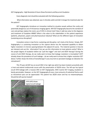
This 79 year old WF has an occult CNV in her right eye which has been treated successfully with
a treat and extend ranibizumab course with the preservation of 20/20 vision. The left eye has typical
occult IVFA leakage with a massive CNV on OCT Angiography. With routine OCT imaging, exudation is
barely perceivable. However, on the OCT Angiography B scans, trace amounts of subretinal fluid as well
as intraretinal cysts can be appreciated. This patient has 20/20 vision and has no metamorphopsia.
Should the left eye be treated?
IVFA (left) shows
typical occult leakage
2. The following questions must be answered:
Is it a mandatory to have all exudation (intraretinal and subretinal) resolved?
Do we know the natural history of persistent subretinal or intraretinal exudation?
OCT Angiography
OCT 200 micron space
between B scans
OCT Angiography – 6 microns
between B scans