SlideShare a Scribd company logo
1 of 44
Download to read offline
Special Series/Guidelines
ISPD peritonitis guideline recommendations:
2022 update on prevention and treatment
Philip Kam-Tao Li1,2
, Kai Ming Chow1,2
, Yeoungjee Cho3,4
,
Stanley Fan5
, Ana E Figueiredo6
, Tess Harris7
, Talerngsak Kanjanabuch8,9
,
Yong-Lim Kim10
, Magdalena Madero11
, Jolanta Malyszko12
,
Rajnish Mehrotra13
, Ikechi G Okpechi14
, Jeff Perl15
, Beth Piraino16
,
Naomi Runnegar17
, Isaac Teitelbaum18
, Jennifer Ka-Wah Wong19
,
Xueqing Yu20,21
and David W Johnson3,4
Abstract
Peritoneal dialysis (PD)-associated peritonitis is a serious complication of PD and prevention and treatment of such is important
in reducing patient morbidity and mortality. The ISPD 2022 updated recommendations have revised and clarified definitions for
refractory peritonitis, relapsing peritonitis, peritonitis-associated catheter removal, PD-associated haemodialysis transfer,
peritonitis-associated death and peritonitis-associated hospitalisation. New peritonitis categories and outcomes including pre-
PD peritonitis, enteric peritonitis, catheter-related peritonitis and medical cure are defined. The new targets recommended for
overall peritonitis rate should be no more than 0.40 episodes per year at risk and the percentage of patients free of peritonitis
per unit time should be targeted at >80% per year. Revised recommendations regarding management of contamination of PD
systems, antibiotic prophylaxis for invasive procedures and PD training and reassessment are included. New recommendations
regarding management of modifiable peritonitis risk factors like domestic pets, hypokalaemia and histamine-2 receptor
antagonists are highlighted. Updated recommendations regarding empirical antibiotic selection and dosage of antibiotics and
also treatment of peritonitis due to specific microorganisms are made with new recommendation regarding adjunctive oral
1
Department of Medicine and Therapeutics, Prince of Wales Hospital,
The Chinese University of Hong Kong, Hong Kong, China
2
Carol andRichardYuPeritoneal DialysisResearch Centre, Departmentof
Medicine and Therapeutics, The Chinese University of Hong Kong, Hong
Kong, China
3
Australasian Kidney Trials Network, University of Queensland,
Brisbane, Australia
4
Department of Nephrology, Princess Alexandra Hospital, Brisbane,
Australia
5
Translational Medicine and Therapeutic, William Harvey Research
Institute, Queen Mary University, London, UK
6
Nursing School Escola de Ciências da Saúde e da Vida
Pontificia Universidade Catolica do Rio Grande do Sul,
Porto Alegre, Brazil
7
Polycystic Kidney Disease Charity, London, UK
8
Division of Nephrology, Department of Medicine, Chulalongkorn
University, Bangkok, Thailand
9
Center of Excellence in Kidney Metabolic Disorders, Faculty of
Medicine, Chulalongkorn University, Bangkok, Thailand
10
Department of Internal Medicine, School of Medicine, Kyungpook
National University, Daegu, South Korea
11
Division of Nephrology, Department of Medicine, National Heart
Institute, Mexico City, Mexico
12
Department of Nephrology, Dialysis and Internal Diseases, The Medical
University of Warsaw, Poland
13
Division of Nephrology, Department of Medicine, Harborview
Medical Center, University of Washington, Seattle, Washington, DC, USA
14
Department of Medicine, Faculty of Health Sciences, University of Cape
Town and Groote Schuur Hospital, South Africa
15
St Michael’s Hospital, University of Toronto, ON, Canada
16
Department of Medicine, Renal Electrolyte Division, University of
Pittsburgh, PA, USA
17
Infectious Management Services, Princess Alexandra Hospital,
University of Queensland, Brisbane, Australia
18
Division of Nephrology, Department of Medicine, University of
Colorado, Aurora, CO, USA
19
Pharmacy Department, Prince of Wales Hospital, Shatin, Hong Kong,
China
20
Department of Nephrology, Guangdong Provincial People’s Hospital,
Guangzhou, China
21
Guangdong Academy of Medical Sciences, Guangzhou, China
Correction (March 2023): Article updated online to correct Figure 8.
Corresponding authors:
Philip Kam-Tao Li, Carol and Richard Yu Peritoneal Dialysis Research
Centre, Department of Medicine and Therapeutics, The Chinese
University of Hong Kong, Hong Kong, China.
Email: philipli@cuhk.edu.hk
David Johnson, Department of Nephrology, Level 2, ARTS Building,
Princess Alexandra Hospital, 199 Ipswich Road, Woolloongabba,
Brisbane, QLD 4102, Australia.
Email: david.johnson2@health.qld.gov.au
Peritoneal Dialysis International
2022, Vol. 42(2) 110–153
ª The Author(s) 2022
Article reuse guidelines:
sagepub.com/journals-permissions
DOI: 10.1177/08968608221080586
journals.sagepub.com/home/ptd
N-acetylcysteine therapy for mitigating aminoglycoside ototoxicity. Areas for future research in prevention and treatment of
PD-related peritonitis are suggested.
Keywords
Guideline, ISPD, peritonitis, prevention, treatment
What’s new with the 2022 update
of the ISPD peritonitis guidelines?
 Revised, clarified definitions for refractory peritoni-
tis, relapsing peritonitis, peritonitis-associated cathe-
ter removal, peritonitis-associated haemodialysis
transfer, peritonitis-associated death and peritonitis-
associated hospitalization (page 5).
 Definitions for new peritonitis categories and outcomes:
pre-PD peritonitis, enteric peritonitis, catheter-related
peritonitis and medical cure (page 3-4).
 Revised, updated recommendations for calculating
and reporting peritonitis rates before and after PD
commencement (page 4-6).
 New targets recommended for overall peritonitis
rate, proportion of patients free of peritonitis and
culture-negative peritonitis (page 5).
 Revised recommendations regarding management
of contamination of PD systems (page 7).
 Revised recommendations regarding antibiotic pro-
phylaxis for invasive procedures (page 7).
 Revised recommendations regarding PD training
and reassessment (page 8).
 New recommendations regarding PD patients with
pets (page 9).
 New recommendations regarding management of
modifiable peritonitis risk factors (hypokalaemia,
histamine-2 receptor antagonists) (page 10)
 Update on novel diagnostic techniques for peritoni-
tis (page 13).
 Updated recommendations regarding empirical anti-
biotic selection (page 13) and dosage of antibiotics
(page 14).
 New recommendation regarding adjunctive oral N-
acetylcysteine therapy for mitigating aminoglyco-
side ototoxicity (page 14).
 Revised recommendations regarding treatment of
peritonitis in patients receiving APD (page 18).
 Revised recommendation regarding consideration of
expectant management in patients longer than 5 days
if PD effluent white cell count is decreasing towards
normal, instead of mandatory PD catheter removal if
effluent does not clear up by day 5 (page 19).
 Updated recommendations for treatment of peritoni-
tis due to coagulase-negative staphylococci (page
22, Corynebacteria (page 23), enterococcus (page
23), Pseudomonas (page 24, Acinetobacter (page
25), Stenotrophomonas (page 25) and non-
tuberculous mycobacteria (page 30).
Introduction
Peritoneal dialysis (PD)-associated peritonitis is a seri-
ous complication of PD,1,2
which is a critically impor-
tant outcome to all key stakeholders including patients,
caregivers, clinicians, researchers and policymakers.3
It
is the most common type of PD-related infection result-
ing in increased healthcare utilisation and is associated
with significant harms including pain, treatment costs,
transfer to haemodialysis and death, as well as altera-
tions of the peritoneal membrane and peritoneal adhe-
sions which can make long-term treatment with PD
challenging.4–7
Recommendations on the prevention and treatment of
peritonitis have been published previously under the aus-
pices of the International Society for Peritoneal Dialysis
(ISPD) in 1983, 1993, 1996, 2000, 2005, 2010 and
2016.8–13
The present recommendations are organised into
five broad sections focusing on:
1. definitions and measurement of peritonitis;
2. prevention of peritonitis;
3. treatment of peritonitis: initial and subsequent;
4. monitoring response to peritonitis treatment includ-
ing indications for catheter removal and
5. return to PD after cessation of PD due to peritonitis-
related catheter removal.
These recommendations are evidence-based where evi-
dence is available, and if multiple reports are available,
findings from the more recent publications have been
incorporated by the committee. In general, these recom-
mendations follow the Grades of Recommendation
Assessment, Development and Evaluation system for
classification of the level of evidence and grade of recom-
mendations in clinical guideline reports.14
Within each
recommendation, the strength of recommendation is indi-
cated as Level 1 (We recommend), Level 2 (We suggest)
or Not Graded, and the quality of the supporting evidence
if shown as A (high quality), B (moderate quality), C (low
quality) or D (very low quality).14
The recommendations
related to treatment are not intended to be implemented
indiscriminately and may require adaptation according to
local conditions, such as pattern of infection, causative
organisms and microbial resistance. Clinicians caring for
paediatric PD patients should refer to the latest consensus
guidelines for the prevention and treatment of catheter-
related infections and peritonitis in paediatric patients
receiving PD.15
Li et al. 111
Definition and measurement of peritonitis
Definition
Standardisation of the definition of outcomes and its mea-
sures is pivotal to enabling assessment of the comparative
effects of interventions for peritonitis. It also facilitates
benchmarking of performance to improve and address
practice variations. A systematic review of 77 studies (three
randomised controlled trials) demonstrated large variability
in definitions of peritonitis (29% of studies did not describe
peritonitis definition used and 42% of studies modified
ISPD recommended diagnostic criteria for peritonitis) and
reporting of outcome measures (e.g. peritonitis rate,
peritonitis-related death).16
In another systematic review,
59 clinical trials of PD-related infections included 383 dif-
ferent outcome measures.3
The definitions related to peri-
tonitis can be further classified according to cause,
association with exit-site/tunnel infections, timing in rela-
tion to previous episodes and outcomes.
Peritonitis
 We recommend that peritonitis should be diagnosed
when at least two of the following are present:
1) clinical features consistent with peritonitis, that is,
abdominal pain and/or cloudy dialysis effluent;
2) dialysis effluent white cell count  100/mL or
 0.1  109
/L (after a dwell time of at least 2 h),
with  50% polymorphonuclear leukocytes (PMN);
3) positive dialysis effluent culture (1C).
Cause-specific peritonitis
 We recommend a diagnosis of peritonitis according
to organisms identified on culture (e.g., Staphylo-
coccus aureus peritonitis; 1C).
 We suggest culture-negative peritonitis is defined
when peritonitis is diagnosed using the criteria above
(criteria one and two), but no organism is identified
on culture of dialysis effluent (Not Graded).
 We suggest catheter-related peritonitis is defined as
peritonitis that occurs in temporal conjunction
(within 3 months) with a catheter infection (either
exit-site or tunnel17
) with the same organism at the
exit-site or from a tunnel collection and in the efflu-
ent or one site sterile in the context of antibiotic
exposure (Not Graded).
 We suggest enteric peritonitis be defined as perito-
nitis arising from an intestinal source involving pro-
cesses such as inflammation, perforation or ischemia
of intraabdominal organs. If a peritonitis episode in
this context is culture negative, we suggest that it be
classified/recorded as enteric peritonitis rather than
as culture-negative peritonitis (Not Graded).
The cause of peritonitis can be broadly divided accord-
ing to organism or concomitant event (e.g. tunnel infection)
to inform treatment. When no organism is identified after
the culture of dialysis effluent, culture-negative peritonitis
is diagnosed.11
All cases of culture-negative peritonitis that
meet the ISPD diagnostic criteria for peritonitis should be
counted in the peritonitis statistics. Culture-negative peri-
tonitis can be due to infectious or non-infectious causes.
For example, infectious causes may occur in the context of
recent antibiotic exposure, suboptimal sample collection or
culture methods or misclassification from slowly growing
atypical organisms (e.g. mycobacteria, fungus). Non-
infectious causes may include eosinophilic or chemical
(e.g. icodextrin) peritonitis but neutrophil predominance
of the elevated white blood cells (WBC) count may not
be present.11
Hemoperitoneum, characterised by the predo-
minant presence of red blood cells in the dialysis effluent,
should not be confused with peritonitis.
The association between catheter-related infections,
such as exit-site and tunnel infections, and peritonitis is
well established.18,19
Catheter-related peritonitis can be
diagnosed with a high degree of certainty when it occurs
concomitantly with an exit-site and/or tunnel infection.
Alternatively, one site (e.g. exit-site or PD effluent) may
be culture negative in catheter-related peritonitis in the
context of recent antibiotic exposure for treatment of the
initial infection. However, at present, there are no data
available to inform the precise temporal criterion for diag-
nosing catheter-related peritonitis.20
Interestingly, a case–
control study of 962 incident PD patients demonstrated that
the odds of peritonitis after an exit-site infection by organ-
ism class at 3, 6 and 9 months were significantly more
likely to be from the same class of organism at 3 months
(odds ratio (OR) at 3 months: 2.00, 95% confidence inter-
val (CI) 1.15–3.47, p ¼ 0.01), especially for gram-positive
organisms (OR at 3 months: 2.27, 95% CI 1.19–4.31,
p ¼ 0.01 compared to at 9 months: OR 1.91, 95% CI
1.29–2.83, p ¼ 0.001).18
Peritonitis from enteric causes (e.g. strangulated bowel,
ischemic colitis, appendicitis) can pose a diagnostic chal-
lenge with attendant delays in appropriate treatment and
resultant increased morbidity and a mortality rate of
approximately 50%.21,22
Identification of multiple organ-
isms (particularly both gram-positive and gram-negative) is
highly suggestive of an enteric cause for peritonitis; how-
ever, this has been reported to occur in less than 20%
of cases of enteric (sometimes known as ‘surgical’)
peritonitis.21,23
Enteric peritonitis can present as culture
negative if the process involves the peritoneal membrane
through a contiguous, non-infective, inflammatory reaction
(e.g. pancreatitis).24
Time-specific peritonitis
 Pre-PD peritonitis (before PD commencement)
 We suggest pre-PD peritonitis be defined as a
peritonitis episode occurring after PD catheter
insertion and prior to commencement of PD
112 Peritoneal Dialysis International 42(2)
treatment. The date of PD initiation is defined as
the day when the first PD exchange is performed
with the intention of continuing long-term PD
treatment from that day (i.e. first day of PD train-
ing or PD treatment in a hospital or at home with
the intention of continuing PD long-term, which-
ever occurs first). The intermittent flushing of a
PD catheter for the purpose of maintaining
catheter patency does not qualify as PD initiation
(Not Graded).
 For the purpose of pre-PD peritonitis rate repor-
ting,time at risk starts from the day of PD cathe-
ter insertion and ends with PD commencement,
PD catheter removal or death, whichever comes
first (Not Graded).
 PD-related peritonitis (after PD commencement)
 We suggest that, for the purpose of standard
peritonitis rate reporting for PD-related peritoni-
tis, time at risk starts from the day of PD com-
mencement (i.e. first day of PD training or PD
treatment in hospital or at home with the inten-
tion of continuing PD long-term, whichever
occurs first) and continues while a patient
remains on PD regardless of the setting (home,
hospital, residential aged care facility, etc.) or
who is performing the PD exchanges (Not
Graded).
 PD catheter insertion-related peritonitis
 We suggest that PD catheter insertion-related
peritonitis be defined as an episode of peritonitis
that occurs within 30 days of PD catheter inser-
tion (Not Graded).
Peritonitis occurring prior to PD training is an under-
recognised problem. Most units, including clinical regis-
tries, only capture peritonitis after patients commence
PD. One observational study in Hong Kong reported the
incidence of pre-training peritonitis to be 4.2% in 1252
patients newly started on PD.25
Another long-term study
in Germany confirmed that peritonitis incidence would be
underestimated by 0.03 per patient-year at risk if peritonitis
episodes occurring before completion of PD training were
not counted.26
In line with the ISPD Guidelines on Creating and Main-
taining Optimal PD Access in the Adult Patient,27
PD
catheter insertion-related peritonitis is defined as an epi-
sode of peritonitis that occurs within 30 days of PD catheter
insertion and should be 5% of PD catheter insertions
(Table 1).
Outcome-specific definitions of peritonitis.
 We recommend using the definitions outlined in
Table 2 to describe outcomes following peritonitis
(Not Graded). All outcomes associated with the peri-
tonitis episode should be captured.
Measuring, monitoring and reporting peritonitis
 We recommend that every programme should mon-
itor, at least on a yearly basis, the incidence and
outcomes of peritonitis (1C).
 We recommend that the parameters monitored
should include the PD-related peritonitis rate,
organism-specific peritonitis rates, antimicrobial
susceptibilities of the infecting organisms, culture-
negative peritonitis and peritonitis outcomes (1C).
 We suggest PD units also measure and report other
peritonitis parameters, including mean time to first
peritonitis episode (where time counts from the first
day of PD commencement), percentage of patients
free of peritonitis per unit time (target 80% per
year) and pre-PD peritonitis (2C).
 We suggest that the rate of peritonitis be reported as
number of episodes per patient-year (Not Graded).
 We suggest that organism-specific peritonitis rates
should be reported as absolute rates, that is, as num-
ber of episodes per year (Not Graded).
 We recommend that the overall peritonitis rate
should be no more than 0.40 episodes per year at
risk (1C).
 In addition to reporting peritonitis rate measured as
number of episodes per patient-year, we suggest the
culture-negative peritonitis be reported as a percent-
age of all peritonitis episodes per unit time (Not
Graded).
 We recommend the proportion of culture-negative
peritonitis should be less than 15% of all peritonitis
episodes (1C).
At regular intervals, all PD programmes should monitor
the incidence of peritonitis as part of a continuous quality
improvement (CQI) programme.28
Application of a stan-
dardised metric to measure outcomes is critical to bench-
mark performance and monitor progress. Peritonitis rate
should be measured as number of peritonitis episodes
divided by number of patient years at risk (i.e. number of
years on PD starting from the time of PD commencement),
reported as episodes per patient years. For the purpose of
calculating peritonitis rates, PD commencement is defined
as the first day on which the first PD exchange was per-
formed with the intention of continuing ongoing PD treat-
ment (i.e. the first day of PD training or PD treatment in a
hospital or at home with the intention of continuing PD
long-term, whichever occurs first); this does not include
intermittent flushing post-surgery to maintain catheter
patency. Number of patient years at risk should be fully
inclusive counting circumstances such as hospitalisation
episodes where patients may not be performing their own
PD. In terms of episodes, all subsequent relapsing episodes
should be considered as an extension of the original episode
and only the original episode captured as part of the
Li et al. 113
peritonitis rate determination. Peritonitis episodes that
occur during hospitalisations where nurses, patients or
caregivers perform PD should also be counted as events
for the purpose of calculating peritonitis rates. For quality
improvement purposes, they should preferably be identi-
fied and characterised separately.
A recent study has proposed an alternative simplified
formula for calculating peritonitis rates in which the denomi-
nator of patient years at risk is replaced by the average of the
numbers of patients at the start and beginning of a year.29
While this demonstrated reasonable overall agreement
against the gold standard method when analysing Australian,
New Zealand and French registry data, we suggest that peri-
tonitis rates only be calculated using the gold standard
method (i.e. number of episodes per patient year at risk) for
the purpose of benchmarking using a standardised approach,
and because the accuracy of the simplified method is sensi-
tive to centre characteristics (i.e. less accurate in smaller
centres or when centres are rapidly or unevenly losing or
gaining patients over a year). The simplified method has also
not been validated over shorter time periods than a year or
outside of Australia, New Zealand and France.
Globally, there is a substantial (up to 20-fold) variation
in peritonitis rates between PD units in different
Table 1. Outcome specific definition following peritonitis.
Outcome Definition
Medical cure Complete resolution of peritonitis together with NONE of the following complications: relapse/
recurrent peritonitis, catheter removal, transfer to haemodialysis for 30 days or death
Refractory Peritonitis episode with persistently cloudy bags or persistent dialysis effluent leukocyte count 100
 109
/L after 5 days of appropriate antibiotic therapy
Recurrent Peritonitis episode that occurs within 4 weeks of completion of therapy of a prior episode but with a
different organism
Relapsing Peritonitis episode that occurs within 4 weeks of completion of therapya
of a prior episode with the
same organism or one sterile (culture negative) episode (i.e. specific organism followed by the
same organism, culture negative followed by a specific organism or specific organism followed by
culture negative).
Repeat Peritonitis episode that occurs more than 4 weeks after completion of therapya
of a prior episode
with the same organism
Peritonitis-associated catheter
removal
Removal of PD catheter as part of the treatment of an active peritonitis episode
Peritonitis-associated
haemodialysis transfer
Transfer from PD to haemodialysis for any period of time as part of the treatment for a peritonitis
episode
Peritonitis-associated death Death occurring within 30 days of peritonitis onset or death during hospitalisation due to peritonitis
Peritonitis-associated
hospitalisation
Hospitalisation precipitated by the occurrence of peritonitis for the purpose of peritonitis treatment
delivery
PD: peritoneal dialysis.
a
Completion of therapy is defined as the last day of antibiotic administration.
Table 2. Measurement and reporting of peritonitis.
Unit of measure Minimum frequency Target
Peritonitis rates (overall and organism-
specific)
Episodes per patient year Yearly 0.4 episodes per patient-
year
Culture-negative peritonitis % of all peritonitis episodes Yearly 15% of all peritonitis
episodes
Time to first peritonitis episode Mean unit time to first episode
peritonitis
Quarterly (local report) –
Proportion of patients free of peritonitis % per unit time Quarterly (local report) 80% per year
Pre-PD peritonitis % of all peritonitis episodes Quarterly (local report) –
PD catheter insertion-related peritonitis % of all PD catheter insertions Quarterly (local report) 5%
Medical cure % of all peritonitis episodes Quarterly (local report) –
Recurrent peritonitis % of all peritonitis episodes Quarterly (local report) –
Relapsing peritonitis % of all peritonitis episodes Quarterly (local report) –
Peritonitis-associated catheter removal % of all peritonitis episodes Quarterly (local report) –
Peritonitis-associated haemodialysis
transfer
% of all peritonitis episodes Quarterly (local report) –
Peritonitis-associated death % of all peritonitis episodes Quarterly (local report) –
PD: peritoneal dialysis.
114 Peritoneal Dialysis International 42(2)
countries.30
The PD Outcomes and Practice Patterns Study
(PDOPPS) similarly reported variation in overall peritonitis
rates from participating PD units (7051 adult PD patients in
209 facilities from seven countries) ranging from 0.26
(95% CI 0.24–0.27) episodes/patient-year in the United
States to 0.40 (95% CI 0.36–0.46) episodes/patient-year
in Thailand.31
In separate studies, peritonitis rates have
been reported to be as low as 0.16–0.20 episodes/patient-
year in some PD units in China.32–34
In a systematic review
based on a random-effects Poisson model of registries from
33 countries, the peritonitis rate has been steadily decreas-
ing from 0.60 to 0.30 episodes/patient-year from 1992 to
2019.35
We recommend that the overall peritonitis rate
should be no more than 0.40 episodes per year at risk.13
This is an improvement of standard of 0.5 episodes per year
at risk as endorsed in the 2016 guideline.13
From global
review of data of reports from registries and studies, this is
an achievable standard and should be used as an initiative
to reduce peritonitis rates worldwide.
In addition to overall peritonitis rates, regular monitor-
ing of organism-specific peritonitis and associated antimi-
crobial sensitivities can be helpful in informing appropriate
empirical antibiotic regimens at a local level. Culture-
negative peritonitis has been reported to affect between
13.4% and 40% of all episodes of peritonitis.36–38
The large
variability in incidence of culture-negative peritonitis has
been attributed to differences in the definition and tech-
nique of microbiological isolation.39
Direct inoculation of
sediment from centrifuged PD effluent into culture bottles
has been shown to be most effective in identifying organ-
isms responsible for peritonitis where appropriate resources
are accessible.39
The culture-negative peritonitis rate
should be reported as percentage of all peritonitis episodes.
We recommend the proportion of culture-negative perito-
nitis should be less than 15% of all peritonitis episodes.
We also suggest PD units measure and report other peri-
tonitis parameters including, time to first peritonitis episode
(where time counts from the first day of PD training/com-
mencement), percentage of patients free of peritonitis per
unit time (target 80% per year) and pre-PD peritonitis (epi-
sodes per year). Death associated with peritonitis may also
be collected at a unit level, which can be defined as
described in Table 1.5
These additional outcomes may be
captured and reported at a unit level on a monthly basis or at
least quarterly to inform local practice (Table 2).
Prevention of peritonitis
Catheter placement
 We recommend that systemic prophylactic antibio-
tics be administered immediately prior to catheter
placement (1A).
Detailed description of the recommended practice of PD
catheter insertion has been covered in the 2019 ISPD
position paper.27
There are four randomised, controlled
trials on the use of perioperative intravenous cefuroxime,40
gentamicin,41
vancomycin42
and cefazolin41,42
as compared
to no treatment. The overall benefit of prophylactic perio-
perative intravenous antibiotics was confirmed by a sys-
tematic review of these four trials, but its effect on the
risk of exit-site/tunnel infection is uncertain.43
Although
first-generation cephalosporin may be slightly less effec-
tive than vancomycin,42
the former is still commonly used
because of the concern regarding vancomycin resistance.
Each PD programme should determine its own choice of
antibiotic for prophylaxis after considering the local spec-
trum of antibiotic resistance. No data exist on the effective-
ness of routine screening and eradication of S. aureus nasal
carriage before catheter insertion (such as intranasal
mupirocin).
Exit-site care
Detailed description of exit-site care to prevent peritonitis
should be referred to another guideline from ISPD.17
At
present, topical application of antibiotic cream or oint-
ment to the PD catheter exit site is recommended although
such practice varied among centres internationally.44
Proper PD catheter immobilisation and avoidance of
mechanical stress on the exit site may be useful to lower
exit-site infection rate.45
Prompt treatment of exit-site or
catheter tunnel infection is mandatory to reduce subse-
quent peritonitis risk.17–19
Contamination of PD system
 We suggest advice be sought immediately from the
treatment team if contamination during PD
exchange is noted (Not Graded).
 We suggest prophylactic antibiotics after wet
contamination of the PD system to prevent perito-
nitis (2D).
PD patients should be instructed to immediately seek
advice from their dialysis centre if the sterility of PD
exchange has been breached. When patients report contam-
ination during an exchange procedure, the need for treat-
ment is driven by distinguishing ‘dry contamination’
(contamination outside a closed PD system, such as dis-
connection distal to a closed clamp) from ‘wet contamina-
tion’ (referring to contamination with an open system,
when either dialysis fluid is infused after contamination
or if the catheter administration set has been left open for
an extended period). Examples of wet contamination
include leaks from dialysate bags, leaks or breaks in tubing
proximal to the tubing clamp, breach of aseptic technique
or touch contamination of the connection during a PD
exchange. Prophylactic antibiotics is only recommended
after wet contamination.46,47
If it is unclear whether the
tubing clamp was closed or open during contamination, wet
Li et al. 115
contamination should be considered, for benefit of doubt.
The common practice is thus change of a sterile transfer set.
A PD effluent should preferably be obtained for cell count
and culture after wet contamination.47
A wet contamination
should be monitored closely for an extended period, and a
broader spectrum of organisms might lead to peritonitis,
particularly in tropical centers.48
One retrospective study involving 548 episodes of
touch contamination revealed a relatively low rate of
peritonitis (3.1%), and peritonitis occurred only after wet
contamination (5.6%). Most episodes were coagulase-
negative staphylococcal or culture-negative episodes, and
the risk was significantly reduced by prophylactic anti-
biotics.46
There is no standard regimen of prophylactic
antibiotic.
Although short course of oral fluoroquinolones has been
used previously,46
the drug has now been discouraged by
Food and Drug Administration (FDA)49
unless there is no
alternative options. One dose of intraperitoneal (IP) cefa-
zolin is a reasonable option.
Invasive gastrointestinal and gynaecological
procedures
 We suggest antibiotic prophylaxis prior to colono-
scopy (2C) and invasive gynaecological procedure
(2D).
 We suggest drainage of PD fluid to keep the abdomen
empty before endoscopic gastrointestinal and inva-
sive or instrumental gynaecological procedures (2D).
Peritonitis commonly follows endoscopic gastrointest-
inal and invasive or instrumental gynaecological proce-
dures (e.g. gastroscopy, colonoscopy, hysteroscopy) in
PD patients.50–57
The highest peritonitis complication rate
after endoscopic or instrumental procedures is reported
after invasive gynaecological procedures, ranging from
26.9%57
to 38.5%.58
Reported rates of peritonitis after
colonoscopy without antibiotic prophylaxis ranged
between 3.4% and 8.5%.55,56
The rates of peritonitis after
gastroscopy in PD patients range from 1.2%58
to 3.9%.59
Concerns about invasive or instrumental gynaecologi-
cal procedures and peritonitis in PD patients come from
the proximity of the pelvis to the peritoneal cavity. The
most commonly reported bacterial pathogens in reported
cases are Streptococcus, followed by Escherichia coli,
Enterococcus, Staphylococcus and infrequently Can-
dida.57
Data supporting antibiotic prophylaxis come from
two small retrospective studies.57,58
In a retrospective
study of 26 gynaecological procedures on 18 PD patients,
none of the 11 procedures with antibiotic prophylaxis was
followed by peritonitis, as opposed to a peritonitis occur-
rence of 47% among those procedures performed without
antibiotic prophylaxis.57
An earlier study reported a sim-
ilar finding of less common peritonitis occurrence after
antibiotic prophylaxis, but the difference did not reach
statistical significance: none of four patients with prophy-
lactic antibiotic administration developed peritonitis
whereas 55.6% without antibiotic prophylaxis developed
peritonitis.58
Because of limited data, there is no standar-
dised recommendation of antibiotic choice and adminis-
tration route. However, reasonable regimens should cover
gram-positive and gram-negative (aerobic and anaerobic)
bacterial isolates from the upper tract of female reproduc-
tive tracts. Examples include intravenous cefazolin or cef-
triaxone before the procedure or oral cefadroxil 500 mg
once daily for 3 days.57
More than half of reported peritonitis episodes occurr-
ing after colonoscopy are caused by E. coli.55,60
In a
single-centre study of 97 colonoscopies performed in 77
continuous ambulatory peritoneal dialysis (CAPD) patients,
none of the 18 patients having a colonoscopy procedure
with antibiotic prophylaxis developed peritonitis, as
opposed to a 6.3% peritonitis occurrence among those
undergoing colonoscopy without antibiotic prophylaxis.50
This is consistent with a more extensive multicentre study
of 236 colonoscopy procedures, in which none of the 65
patients who received antibiotic prophylaxis developed
peritonitis, compared to a peritonitis rate of 3.8% for those
without prophylactic antibiotics.55
Furthermore, thera-
peutic procedures, such as polypectomy and endoscopic
mucosal resection, are predictive of peritonitis.55,60
The
optimal antibiotic regimen for preventing peritonitis after
colonoscopy has not been determined by clinical study.
The only randomised controlled trial of prophylactic anti-
biotics used IP ceftazidime (1 g IP 1 h before the proce-
dure) and recruited 93 patients receiving APD without a
history of peritonitis in the last 12 months from a single
centre in Saudi Arabia. The peritonitis rate did not differ
with (6.5%) and without (8.5%) IP ceftazidime prophy-
laxis (p ¼ 0.27).56
For intravenous antibiotic prophylaxis,
potential choices include cephalosporins (such as ceftriax-
one or ceftazidime), amoxicillin–clavulanate, ampicillin–
sulbactam, ampicillin plus aminoglycoside,50,58
with an
aim to target most of the organisms described above that
cause peritonitis after colonoscopy. Interestingly, the
alternative option of oral antibiotic prophylaxis was sug-
gested by a recent case series of 49 PD patients who
received oral ampicillin 1000 mg, ciprofloxacin 500 mg
and/or metronidazole 250 mg 1 to 2 h before colonoscopy
and did not experience any post-procedure episodes of
peritonitis.61
Finally, PD effluent should be drained to
keep patient’s abdomen empty before colonoscopy (and
gynaecological) procedure.62
The argument for emptying
the abdomen before colonoscopy is to enhance host
defence,63
because the peritoneal macrophage phagocytic
function and polymorphonuclear cell function are sup-
pressed by the presence of dialysate.64
Furthermore, high
fluid volumes can compromise efficiency of bacterial kill-
ing by disrupting the volume-to-surface-area ratio.65
The risk of PD patients developing peritonitis after gas-
troscopy is more uncertain. Other than case reports66,67
and
116 Peritoneal Dialysis International 42(2)
a small case series,58
a single-centre observational study of
408 gastroscopy procedures in 216 PD patients showed a
3.9% incidence of peritonitis within 1 week of endo-
scopy.59
Patient’s age and the number of endoscopic biop-
sies predicted peritonitis risk. One-quarter of the 16
peritonitis episodes were polymicrobial, commonly caused
by organisms either enteric in origin or arising from the oral
cavity, such as Streptococcus.59
Although there is insuffi-
cient evidence to recommend antibiotic prophylaxis prior
to gastroscopy in PD patients, the study confirmed a lower
odds of peritonitis after gastroscopy, after adjustment for
confounding factors, when antibiotics were used within
7 days of gastroscopy.59
Training programme
 We suggest that the characteristics of an optimal PD
training programme (how, how long, where, when
and by whom) remain uncertain (2C).
 We recommend that PD exchange technique and
knowledge be regularly reassessed and updated,
with an emphasis on direct inspection of practice
of PD technique (1C).
Detailed description on the recommended practice of
PD training has been covered in another ISPD guideline,68,69
which each PD programme should consult while prepar-
ing the trainer and developing a specific curriculum for
PD training. Unfortunately, limited data are available to
guide when, how or how long PD training is optimal. The
PDOPPS noted marked variation in training practices
across 120 facilities across seven countries; timing of
commencement, duration of training, location or use of
competency assessments were not predictive of peritonitis
risk.70
Taken together, flexibility should be allowed to
deliver training according to local resources and indivi-
dualised to patients’ needs. Furthermore, distance learn-
ing and remote monitoring have been increasingly
utilised. Previously, hybrid PD education programme with
online video material has been developed and shown to be
associated with lower peritonitis rate.71
On the other hand,
a single-centre study reported that face-to-face patient–
doctor contact intervals less frequent than every 2 months
was associated with higher peritonitis rate.72
In essence, all PD trainers should receive adequate edu-
cation to perform training and further education to update
and hone their teaching skills. Each programme should
have an established curriculum that is followed in teaching
the patient the procedure, theory of PD and self-care, taking
into account the individual’s learning style. Testing the
patient’s practical skills at the end of training is essential.
After PD training is completed and patients are started
on home PD, a home visit by the PD nurse is often helpful
in detecting problems with exchange technique, adherence
to protocols and other environmental and behaviour issues
which increase the risk of peritonitis. Observational studies
reported a non-significantly lower peritonitis rates associ-
ated with home visit programmes in paediatric73
and
adult74
PD patients. Another registry data set showed an
independent association of nurse visits before starting PD
with a lower likelihood of peritonitis.75
In addition to the initial training, refresher course or
retraining plays an important role in reducing mistakes
according to learning specialists.76
Previous studies
showed that adherence with exchange protocols was signif-
icantly associated with peritonitis rate,76
which applied
even during the coronavirus pandemic when behaviour for
personal hygiene is anticipated to be enhanced.77
The pur-
pose of retraining is to target patients who have begun to
take shortcuts or simply deviate from the standard steps
which they were taught previously. An observational study
found that 6 months after the initiation of PD, around half
of the patients took shortcuts, modified the standard
exchange method, failed to follow appropriate hand
hygiene protocols properly or follow the aseptic tech-
nique.78
Despite common usage of the term ‘retraining’
in literature, the healthcare providers should be mindful
of the implicit negative connotation of this word. Emphasis
for updating of knowledge and technique should be used to
address the benefit. Home visits by PD nurses or trained
personnel may be a good way to determine which patients
require retraining.76
Other indications of retraining are
listed in Table 3.68,79
Certainly, all patients must be
retrained whenever the equipment to perform PD is chan-
ged. Evidence for retraining PD is evolving as an increas-
ing number of randomised controlled trials have been
completed.80–82
The optimal timing and frequency of
retraining remain uncertain but a randomised controlled
trial lends strong support for more frequent retraining at
home. As compared to 53 PD patients receiving conven-
tional retraining (two home visits within two months after
starting dialysis), 51 incident PD patients randomised to
frequent retraining (regular repeated home visits every 1–
3 months over 2 years) showed a significantly lower rates
of exit site infection and peritonitis.80
Moreover, subgroup
analysis demonstrated a significant beneficial effect on the
first episode of peritonitis in patients older than 60 years.80
Their results were not able to be replicated in another ran-
domised controlled trial, with a larger sample size, of
retraining intervention targeting incident PD patients who
failed regular testing of PD knowledge and practical PD
skill assessment.81
Furthermore, it has been proposed that
practical assessment of PD technique is more important
than testing of theory. Patients might not be aware of their
making mistakes in PD procedures until the visiting nurse
discovers them. The best support for emphasis on practical
assessment of patients’ techniques comes from a controlled
trial randomising incident PD patients to retraining via
technique inspection, oral education or usual care.82
The
oral education group (retraining every 2 months using a
checklist and focus on knowledge) did not reduce the risk
of peritonitis, whereas the technique inspection group
Li et al. 117
(retraining every 2 months and focus on behaviour by
nurses’ inspection of PD technique) demonstrated a lower
risk of first non-enteric peritonitis.82
In other words, the
most effective learning is through direct feedback immedi-
ately after return demonstration of PD steps.
Domestic pet and zoonotic infection
 We recommend PD patients take extra precautions
to prevent peritonitis if domestic pets are kept (1C).
 We suggest pets not be allowed in the room where
PD exchange takes place, and where dialysis tubing,
equipment and machine are stored (2A).
PD patients should be asked about pets during training
and home visits or after a diagnosis of unusual organisms
suspicious of zoonoses because peritonitis due to zoonotic
organisms can occur in the context of close contact with
companion animals.83,84
With regard to cats, more than 40 cases of Pasteurella
multocida peritonitis have been reported in the literature.85
Despite the name ‘cat-bite peritonitis’,86
the aerobic gram-
negative coccobacillus P. multocida is found in the upper
respiratory tract and oral cavity of many domestic and wild
animals including dogs and hamsters. Direct contact with
animals, either through close contact with PD equipment or
patient, bites or scratches, can be implicated in PD-related
infections. The prevalence of colonisation with P. multo-
cida is higher in cats, including their claws.87
Other cat-
related organisms include Capnocytophaga canimorsus,
Capnocytophaga cynodegmi and Neisserria species.84,88
The frequency of cat-related peritonitis is higher in patients
on APD than CAPD, possibly secondary to the longer tub-
ing required or the prolonged environmental contact time
of equipment for APD. Cycler tubing moving with the
action of the cycler pump is another stimulus that may
entice a cat to play with the instrument. Furthermore, cats
enjoy the warmth of the cycler heat plate and may lay on
the dialysis machines.85
The hidden pet-related damage to PD tubing that
occurs when animals bite or scratch the tubing should
be emphasised as the damage may go unnoticed when
APD patients are sleeping. The small pinhole-shaped
damage, as opposed to a complete tear, can also be
challenging to detect until leakage of PD solution occurs.
Such minor but serious tubing defects have been reported
in PD patients who cohabitate with pets including cats,
hamsters and cockatoo.86,88–93
With the bonds between owners and domestic pets
being potentially very strong, and possible emotional and
quality-of-life benefits, it is not always possible to dis-
courage keeping pets. Around one-fifth of PD patients
surveyed in a single PD centre were keeping pets.94
To
minimise the risk of pet-related peritonitis, PD patients
should adhere to stringent hand washing before and after
PD exchanges and handling pets, as well as ensuring high
home environment hygiene standards. Domestic pets
should be strictly kept away from the dialysis equipment
and should not be allowed into the room during the dia-
lysis treatment procedure.
Other modifiable risk factors
 We suggest that avoidance and treatment of hypo-
kalaemia may reduce the risk of peritonitis (2C).
 We suggest that avoiding or limiting the use of
histamine-2 receptor antagonists may prevent
enteric peritonitis (2C).
A number of other modifiable risk factors for PD peri-
tonitis have been described. One of the investigation tools
is the undertaking of a large international cohort study,
such as the Peritoneal Dialysis and Outcomes Practice Pat-
terns Study (PDOPPS), to collect detailed information in a
uniform manner.31,70,95,96
The results obtained from
PDOPPS provide a high-level overview of peritonitis risk
factors and outcomes across countries and PD centres but
require further prospective interventional studies to estab-
lish causation.
Gastrointestinal problems, such as constipation and
enteritis, have been reported to be associated with perito-
nitis due to enteric organisms.97
PDOPPS also reported an
association of higher peritonitis risk with gastrointestinal
bleeding.31
A previous study reported an association of
hypokalaemia with a higher risk of enteric peritonitis.98
International data from seven countries, under PDOPPS,
showed that hypokalaemia persistent for 4 months was
associated with 80% higher subsequent peritonitis rates
after adjustment for confounders.95
The causative organ-
isms underpinning the excess of peritonitis were mostly
gram positive and culture negative. This concurs with
another Brazilian propensity-matched score study linking
hypokalaemia with higher infection-related mortality and
peritonitis risk.99
In addition to the degree of hypokalae-
mia, the duration of hypokalaemia was associated with the
risk of peritonitis in PD patients.100
Although there is no
compelling evidence that treatment of hypokalaemia, con-
stipation or gastroenteritis mitigates the risk of peritonitis,
such problems, which are common in the PD setting, merit
treatment in their own right. Based on previous
Table 3. Indications for PD Retraining.
 Following prolonged hospitalisation
 Following peritonitis and/or catheter infection
 Following change in dexterity, vision or mental acuity
 Following change to another supplier or a different type of
connection
 Following change in caregiver for PD exchange
 Following other interruption in PD (e.g. period of time on
haemodialysis)
PD: peritoneal dialysis.
118 Peritoneal Dialysis International 42(2)
observational and mechanistic studies of hypokalaemia in
PD studies, the main contributory factor of hypokalaemia is
low dietary potassium intake, rather than increased potas-
sium excretion or intracellular shift.101,102
Dietary inter-
vention is recommended to mitigate hypokalaemia.
Observational data from a single-centre study suggested
that regular lactulose use is associated with a lower rate
of peritonitis.103
However, the benefit of lactulose to
reduce peritonitis rate, compared with sennosides, has not
been confirmed in a single-centre randomised controlled
trial.104
There are emerging data to suggest that gastric acid
suppression, especially with histamine-2 receptor antago-
nists, is a modifiable risk factor for enteric peritonitis in PD
patients. The hazard ratio for enteric peritonitis, as demon-
strated in an observational cohort of 119 PD patients on
histamine-2 receptor antagonists, was 1.67 (95% confi-
dence interval 1.02–2.80). The increase in infectious mor-
tality among histamine-2 receptor antagonist users further
supported the burden of this risk.105
However, the risk of
peritonitis associated with proton pump inhibitors is less
consistently reported.106–108
A similar finding of heigh-
tened risk conferred by the use of histamine-2 receptor
antagonists, but not proton pump inhibitors, was found in
a case series of peritonitis after gastroscopy. Of note,
histamine-2 receptor antagonist users had a significantly
higher post gastroscopy peritonitis rate (9.4%) compared
to non-users (2.9%).59
A meta-analysis of six non-
randomised studies involving pooled data of 829 PD
patients showed that histamine-2 receptor antagonist use
was associated with an increased odds of enteric peritonitis
(OR 1.4, 95% CI 1.01–1.93).108
Notably, even though the
association between proton pump inhibitor use and perito-
nitis is less compelling, other concerns with proton pump
inhibitors (including but not limited to Clostridioides infec-
tion) do not justify a routine switching of histamine-2
receptor antagonist to proton pump inhibitor therapy.
Secondary prevention
 To prevent fungal peritonitis, we recommend that
anti-fungal prophylaxis be co-prescribed whenever
PD patients receive an antibiotic course, regardless
of the indication for that antibiotic course (1B).
The majority of fungal peritonitis episodes are preceded
by courses of antibiotics.109–112
A number of observational
studies113–120
and randomised trials121,122
have examined
the use of either oral nystatin (500,000 units qid) or fluco-
nazole (200 mg every 48 h) as prophylaxis during antibiotic
therapy. In essence, two randomised control trials121,122
and a systematic review43
showed a significant benefit.
Most of the other reports on the prophylactic use of
antifungals during antibiotic administration were non-
randomised studies and have yielded mixed results.
Unfortunately, nystatin is not available in some countries.
Observational data118–120
and one randomised controlled
trial122
showed that prophylactic fluconazole is effective.
The randomised controlled trial of oral fluconazole
included patients who received antibiotics for treating
exit-site and tunnel infection, in addition to the treatment
of peritonitis122
. However, there are potential problems
(including drug interactions, emergence of resistant strains)
with fluconazole prophylaxis. Overall, a Cochrane meta-
analysis of the two randomised controlled studies on anti-
fungal prophylaxis with oral nystatin or fluconazole
showed a risk ratio of 0.28 (95% CI 0.12–0.63) for fungal
peritonitis occurring after a patient has had an antibiotic
course.43
Furthermore, each episode of peritonitis should be con-
sidered a preventable event and hence evaluated.47
The
CQI programme provides a means in secondary prevention.
For each peritonitis episode, a root-cause analysis should
be performed to determine the aetiology and, whenever
possible, an intervention directed against any reversible
risk factor should be made to prevent another episode. For
example, Streptococcal viridans peritonitis could have
indicated dental problems although such link is based on
isolated case reports only.123,124
Peritonitis episodes caused
by coagulase-negative staphylococcal species are associ-
ated with touch contamination, while S. aureus infections
have been associated with touch contamination or catheter
infections. Identification of aetiology may involve review
of the exchange technique. Retraining is sometimes neces-
sary. Rarely, an outbreak of culture-negative peritonitis or
peritonitis secondary to unusual organisms should trigger
epidemiological investigation and field visit to look for
environment risk factors such as PD fluid, hospital air or
water contamination.125–127
Initial presentation and management
of peritonitis
The algorithm of initial management for PD patients pre-
senting with a clinical diagnosis is summarised in Figure 1.
 We recommend that peritonitis always be diagnosed
when at least two of the following are present:
(1) clinical features consistent with peritonitis, that
is, abdominal pain and/or cloudy dialysis effluent;
(2) dialysis effluent white cell count 100/mL or
0.1  109
/L (after a dwell time of at least 2 h),
with 50% PMN; and (3) positive dialysis effluent
culture (1C).
 We recommend that PD effluent be tested for cell
count, differential, gram stain and culture whenever
peritonitis is suspected (1B).
 We recommend that PD patients presenting with
cloudy effluent be presumed to have peritonitis and
treated as such until the diagnosis can be confirmed
or excluded (1C).
Li et al. 119
Patients with peritonitis usually present with cloudy PD
effluent and abdominal pain. Cloudy effluent almost
always represents infectious peritonitis, although there are
other differential diagnoses classified according to cellular
and non-cellular causes (Table 4).128
Some patients present
with cloudy effluent but no or minimal abdominal pain. On
the other hand, peritonitis should also be included in the
differential diagnosis of the PD patient presenting with
abdominal pain, even if the effluent is clear. In addition
to the presenting symptoms, the patient should be ques-
tioned about any recent contamination, accidental discon-
nection, endoscopic or gynaecological procedures, as well
as the presence of constipation or diarrhoea. In addition, the
patient should be questioned about past history of peritoni-
tis and exit-site infection.
On physical examination, abdominal tenderness is typi-
cally generalised and is less often associated with a
rebound. Localised pain or tenderness should raise the sus-
picion of underlying surgical pathology. Physical examina-
tion should also include a careful inspection of the catheter
tunnel and exit site. Any discharge from the exit site should
be cultured. Erythema, tenderness and the presence of fluid
collections (which may be confirmed with ultrasound)
along the PD catheter tunnel may be indicative of a tunnel
infection. The degrees of abdominal pain and tenderness
are important factors in deciding whether a patient requires
hospital admission. In general, patients with minimal pain
could be treated on an outpatient basis with IP antibiotic
therapy if this can be arranged. Follow-up within 3 days is
advisable to confirm resolution and appropriateness of the
antibiotic choice.
When peritonitis is suspected, dialysis effluent should
be drained, carefully inspected and sent for cell count with
differential, Gram stain and culture.129
An effluent cell
count with WBC  100/mL (after a dwell time of at least
2 h), with  50% PMN, is highly suggestive of peritoni-
tis.130
Abdominal X-ray is generally not necessary and may
be potentially misleading since pneumoperitoneum is com-
mon (around one-third of CAPD patients)131
secondary to
air entry into the peritoneal cavity via the PD catheter
during exchanges. Peripheral blood cultures are usually
negative132
and can be omitted unless the patient is clini-
cally septic133
or on immunosuppression.134
Bacteraemia
during peritonitis should raise the possibility of other
intra-abdominal events.135,136
To prevent delay in treat-
ment, antibiotic therapy (see below) should be initiated
once the appropriate dialysis effluent specimens have
been collected, without waiting for the results of laboratory
testing.
The WBC count in the effluent depends in part on the
length of the dwell. For patients on APD with rapid cycle
treatment, the clinician should use the percentage of PMN
rather than the absolute WBC count to diagnose peritonitis,
and a proportion above 50% PMN is strong evidence of
peritonitis, even if the absolute WBC count is less than
100/mL.130
On the other hand, APD patients without a day-
time exchange presenting with abdominal pain during the
daytime may have no effluent to drain. In this case, 1 L of
Figure 1. The algorithm of initial management for PD patients presenting with a clinical diagnosis of peritonitis. PD: peritoneal dialysis.
120 Peritoneal Dialysis International 42(2)
dialysis solution should be infused, dwelled for 2 h, and
then drained for inspection and laboratory testing.
Identification of causative organisms
 We recommend that the blood culture bottle(s) be
the preferred technique for bacterial culture of PD
effluent (1C).
 We suggest that sampling and culture methods be
reviewed and improved if more than 15% of perito-
nitis episodes are culture negative (2C).
Gram stain of the PD effluent should be performed even
though the result is often negative.137,138
An additional
benefit of Gram stain is its effectiveness in early detection
of fungal elements, facilitating timely diagnosis and man-
agement of fungal peritonitis.139
The diagnostic yield of the
Gram stain is increased if it is performed on a centrifuged
specimen. An appropriate method of culturing PD effluent
is the most important step in establishing the causative
organism. In some specialised centres, it has been possible
to achieve a culture-negative peritonitis rate of less than
10%. Identification of the organism and subsequent anti-
biotic sensitivities help to guide the choice of antibiotic,
and the type of organism often indicates the possible source
of infection. Bedside inoculation of 5–10 mL effluent in
two (aerobic and anaerobic) blood-culture bottles has a
reasonable sensitivity, and the culture-negative rate is typi-
cally around 10–20%.140-143
The yield of peritoneal fluid
culture is enhanced by inoculating the fluid directly into
rapid blood-culture bottle kits (e.g. BACTEC, Kent, UK;
Septi-Chek, Roche Diagnostics, Basel, Switzerland; BacT/
Alert, Biomerieux, Inc., Basingstoke, UK), centrifuging PD
fluid and culturing the pellet or the lysis centrifugation
technique as compared to inoculation into standard
blood-culture bottles. Specifically, centrifugation of 50
mL PD effluent at 3000 g for 15 min, followed by resus-
pension of the sediment in 3–5 mL supernatant and inocu-
lation on solid culture media or standard blood-culture
media, increases the yield by 5–10 times.39,142,143
The com-
bination of water lysis, Tween-80 blood agar and Triton-X
treatment of the PD effluent is also a sensitive culture
method.144,145
The specimens should arrive at the labora-
tory within 6 h. If immediate delivery to the laboratory is
not possible, the inoculated culture bottles should ideally be
incubated at 37
C. Inoculated bottles should not be refri-
gerated or frozen, since it may kill or retard the growth of
some microorganisms.146
The solid media should be incu-
bated in aerobic, microaerophilic and anaerobic environ-
ments. To fully assess yeast and filamentous fungal
pathogens, appropriate fungal media should be selected;
incubation of inoculated media under two temperature con-
ditions (room temperature and 35–37
C) can increase the
diagnostic yield.146
The speed with which bacteriological diagnosis can be
established is very important. Concentration methods not
only facilitate microbial identification, but also reduce the
time needed for a positive culture. In over 75% of cases,
microbiologic diagnosis can be established in less than 3
days. When the causative microorganism has been identi-
fied, subsequent cultures for monitoring may be performed
by only inoculating the effluent in blood-culture bottles.
In a prospective study using facility-level data over 22
PD centres, immediate transfer of specimens or inoculated
bottles to laboratories and the practice of PD effluent cen-
trifugation are associated with lower culture-negative peri-
tonitis rates.146
Notably, experience of the centre is
important because culture-negative peritonitis rates fre-
quently show an inverse relationship with the PD centre
size.38,146
When cultures remain negative after 3–5 days of incu-
bation, PD effluent should be sent for repeat cell count,
differential count, fungal and mycobacterial culture. In
addition, subculture on media with aerobic, anaerobic
and microaerophilic incubation conditions for a further
3–4 days may help to identify slow-growing fastidious
bacteria and yeasts that are undetectable in some automated
culture systems. Furthermore, culture of the PD catheter
can improve the diagnostic yield, especially for detection
of fungi and enterococci.147
Other novel diagnostic techniques
A number of novel diagnostic techniques have been
explored for the early diagnosis of peritonitis, including leu-
kocyte esterase reagent strips,148
biomarker assays (matrix
metalloproteinase-8 and -9,149
neutrophil gelatinase-
associated lipocalin150
and procalcitonin), polymerase chain
Table 4. Differential diagnosis of cloudy effluent.
Cellular causes
PMN leucocytes
Culture-positive infectious peritonitis
Infectious peritonitis with sterile cultures
Chemical peritonitis
Eosinophils
Dialysate eosinophilia
Chemical peritonitis
Monocyte/macrophages
Specimen taken from ‘dry’ abdomen (after prolonged
peritoneal rest)
Red blood cells
Hemoperitoneum
Malignant cells
Lymphoma
Peritoneal metastasis
Non-cellular causes
Fibrin
Triglycerides (milky white appearance of effluent)
Calcium channel blockers
Lymphatic obstruction
Acute pancreatitis
PMN: polymorphonuclear.
Li et al. 121
reaction (PCR) for bacterial-derived DNA fragments, PCR/
electrospray ionisation–mass spectrometry assay,151
16 S
rRNA gene sequencing,152
matrix-assisted laser desorption
ionisation-time of flight mass spectrometry153
and pathogen-
specific ‘immune fingerprints’.154,155
However, none of
them has been proved to be superior to conventional tech-
niques. Immune fingerprint, for instance, by multicolour
flow cytometry and multiplex enzyme-linked immunosor-
bent assay, has been shown to discriminate between
culture-negative, gram-positive, gram-negative episodes
of peritonitis but provides no information on antibiotic
resistance.155
Further refinement using mathematical
machine-learning algorithms can characterise specific
pathogens like streptococcal species and coagulase-
negative staphylococci in a point-of-care manner.154
Utility
of PD effluent phenotyping approach or immune fingerprint-
ing remains to be validated before application to clinical use.
In addition, a point-of-care device measuring levels of
matrix metalloproteinase-8 and interleukin-6 has been tested
to expedite diagnosis of peritonitis but is more useful
to exclude peritonitis with a high negative predictive value
over 98%.156
For rapid diagnosis of fungal peritonitis, PD effluent and
serum galactomannan index might offer a faster turnaround
time than the conventional culture method, but with a diag-
nostic accuracy of 65.2% sensitivity, 85.0% specificity
only.157,158
False-positive galactomannan results159
lead-
ing to unnecessary use of antifungals is a definite concern.
Empiric antibiotic selection
 We recommend that empirical antibiotic therapy be
initiated as soon as possible, using either IP or sys-
temic route, after appropriate microbiological speci-
mens have been obtained (1B).
 We recommend that empirical antibiotic regimens
be centre-specific and cover both gram-positive and
gram-negative organisms (1C).
 We recommend that gram-positive organisms be
covered by a first-generation cephalosporin or
vancomycin and gram-negative organisms by a
third-generation cephalosporin or an aminoglyco-
side (1B).
 We suggest that cefepime monotherapy may be an
acceptable alternative for empirical antibiotic regi-
mens (2B).
Once the diagnostic investigations have been com-
pleted, empirical antibiotics should be started to achieve
rapid resolution of inflammation, reduction of pain and
preservation of the peritoneal membrane. No single anti-
biotic regimen has been proven to be superior to others,160
and the choice should be centre-specific. There should be
adequate coverage for both gram-positive and gram-
negative organisms. A national registry confirmed that
centres with higher proportions of peritonitis episodes
receiving complete empirical coverage for both gram-
positive and gram-negative organisms at presentation had
higher odds of peritonitis cure by antibiotics.161
For the
coverage of gram-positive organisms, vancomycin or
first-generation cephalosporin is recommended. Cefazolin
might be preferred to vancomycin when there is concern
about emergence of organisms resistant to the latter. How-
ever, vancomycin should be considered in centres with a
high prevalence of methicillin-resistant organisms.162
The
threshold prevalence of methicillin resistance that justifies
empirical use of vancomycin remains controversial. No
discernible difference in peritonitis cure rate was found
between empirical cefazolin and vancomycin use for
gram-positive or culture-negative peritonitis, according to
observational data from PDOPPS.96
For the gram-negative
coverage, third-generation cephalosporin or aminoglyco-
side is suggested. Observational studies163,164
and one ran-
domised controlled trial165
showed that aminoglycoside
does not accelerate the loss of residual kidney function.
However, repeated or prolonged aminoglycoside treatment
was associated with a high incidence of vestibular toxicity
or ototoxicity.166
It is also important to mention that treat-
ment failure with ceftazidime is high with rising prevalence
of extended-spectrum beta-lactamases (ESBL)-producing
organisms. A recent analysis from PDOPPS reported that,
for treatment of gram-negative peritonitis, empirical ami-
noglycoside was associated with a higher likelihood of
medical cure than ceftazidime.96
Monotherapy for empiri-
cal treatment of peritonitis, instead of combination therapy,
has now been accepted as an effective strategy. Two ran-
domised controlled trials167,168
and one observational
prospective study169
testing the use of IP cefepime mono-
therapy have been published. Although there were differ-
ences in cefepime dosing (intermittent, continuous, with
and without adjustment for residual kidney function), all
three studies showed primary response rates exceeding
80% on day 10.167–169
In particular, the largest study used
a non-inferiority design and specified adjustment for resi-
dual kidney function by increasing the loading and main-
tenance doses of cefepime by 25% for urine volume more
than 100 mL daily. Cefepime monotherapy was shown to
be effective and non-inferior to standard dual therapy with
cefazolin plus ceftazidime.168
In contrast, monotherapy
with quinolones is not recommended because of the con-
cern with emergence of resistant organisms and declining
effectiveness.162,170
It is important to note that prompt administration of
antibiotics has been consistently shown to be associated
with better outcome of peritonitis treatment. In a prospec-
tive multicentre study of 159 peritonitis episodes in West-
ern Australia, the contact-to-treatment time was
independently associated with treatment failure, defined
as either catheter removal or death at 30 days. For each
hour of delay in administering antibiotic therapy from the
time of presentation to a hospital facility, the risk of PD
failure or death was higher by 5.5%.171
In another
122 Peritoneal Dialysis International 42(2)
retrospective study of 109 peritonitis episodes, a delay of
starting IP or intravenous antibiotics treatment from the
sign of peritonitis by 24 h conferred a threefold risk of
peritoneal catheter removal by multivariate analysis.172
For logistics consideration, immediate IP antibiotic
administration might not be feasible in the emergency
department or wards in which staff are not familiar with
PD. In order to avoid the adverse outcome of delayed
peritonitis treatment, the systemic route should be started
as a temporary measure when there is a foreseeable delay,
such as long wait for dialysis unit bed or presentation
outside the working hours of the ambulatory PD unit.
However, the route of administrating antibiotics should
still be switched to IP as soon as possible.
Dosage of antibiotics
 We recommend that IP antibiotics be the preferred
route of administration as long as the compatibility
and stability of the IP antibiotics allow, unless the
patient has features of systemic sepsis (1B).
 We suggest that IP aminoglycoside be administered
as daily intermittent dosing (2B).
 We recommend that prolonged courses of IP amino-
glycoside be avoided (1C).
 We suggest that adjunctive oral N-acetylcysteine
therapy may help to prevent aminoglycoside oto-
toxicity (2B).
 There is insufficient evidence to make a recommen-
dation as to whether patients on APD should be
temporarily switched to CAPD during treatment of
peritonitis (Not Graded).
The recommended dosage of antibiotics for the treatment
of PD-related peritonitis is summarised in Table 5 (IP anti-
biotics) and Table 6 (systemic antibiotics). However, the rec-
ommended dosages of many antibiotics are based on
published clinical experience rather than formal pharmacoki-
netic studies. Most studies of IP antibiotics have been con-
ducted in patients on CAPD rather than in patients on APD.
The importance of adequate dosing of antibiotics was
supported by an observational study of 339 episodes of
gram-positive, gram-negative and culture-negative PD-
related peritonitis, in which treatment failure was higher
for patients with greater residual kidney function defined
as urinary creatinine clearance more than 5 mL/min.247
The
observation suggests that better clearance of antibiotics
might lead to lower concentration of antibiotics, and hence
the reduced time above the minimum inhibitory concentra-
tion (MIC). Optimal dosing of antibiotics in patients with
significant residual kidney function remains unknown,
although fixed dosing irrespective of residual kidney func-
tion might not be the best solution for antibiotics (such as
cephalosporin) that exhibit time-dependent killing effects.
Limited data are available to guide the adjustment of anti-
biotics dosing, except a recent randomised controlled
study advocating a 25% increase in the loading and main-
tenance dose of cefepime, cefazolin and ceftazidime
when PD patients have residual urine volumes of more than
100 mL daily.168
Vancomycin is the drug of choice in centres with a high
prevalence of methicillin resistant gram-positive bacteria
or for directed therapy in patients with relevant pathogens.
IP administration is preferred because nearly 90% is
absorbed in the presence of peritonitis.248
The superiority
of treatment success rate with IP versus intravenous vanco-
mycin is supported by Cochrane systematic review.160
Optimal dosing of IP vancomycin is unknown, and guide-
line recommendations are variable regarding whether to
prefer fixed dosing or target-guided dosing according to
serum trough level. Although fixed dosing of IP vancomy-
cin had been reported in a randomised controlled trial,234
it
is unknown whether inter-individual variability of vanco-
mycin bioavailability warrants adjustment of maintenance
dose according to therapeutic drug monitoring of steady-
state serum vancomycin concentration. A retrospective
study reported that 60% of patients had subtherapeutic
trough level following the loading dose after a fixed dosing
of IP vancomycin 30 mg/kg every 5 days for CAPD and
every 3 days for CCPD, irrespective of the residual renal
function. However, all subsequent serum vancomycin lev-
els were above 15 mg/L.249
Several observational studies
did not show correlation between trough levels and cure
rates of peritonitis.250,141
On the other hand, one observa-
tional study reported a higher rate of peritonitis relapse with
intravenous vancomycin use when the cumulative 4-week
mean trough vancomycin levels were less than 12 mg/L.251
Another study of peritonitis due to methicillin-resistant
coagulase-negative staphylococci showed that higher
serum trough vancomycin levels achieved by IP vancomy-
cin were associated with a lower relapse rate.252
Regarding
the practice of trough-guided vancomycin dosing, there
was no consensus on the preferred timing of obtaining
trough vancomycin concentration. Based on a retrospective
analysis of 61 episodes of gram-positive or culture-
negative peritonitis, serum vancomycin levels lower than
10.1 mg/L on day 5, but not the level on day 3, were
associated with worse outcomes (including transfer to hae-
modialysis, death, persistent infection and relapse).208
Recently, trough-guided vancomycin dosing has been
increasingly replaced by the area under the 24-h time-
concentration curve (AUC)-guided dosing to optimise the
management of severe S. aureus infection. Although the
clinical significance of AUC pharmacokinetic parameters
for monitoring vancomycin dosing in peritonitis treatment
is incompletely understood, accumulating evidence sug-
gests that trough level might not be the best option. A
recent study of anuric patients on APD reported that peak
serum concentration level (30 min after IP administration),
but not trough vancomycin level, was associated with cure
of gram-positive peritonitis.212
Li et al. 123
Aminoglycosides remain useful for treating gram-
negative peritonitis. Since aminoglycosides exhibit
concentration-dependent activity, their maximal bacterial
killing occurs at high peak drug concentrations. In addition,
aminoglycosides continue to suppress bacterial growth
even after drug concentration falls below MIC of the bac-
teria, a characteristic known as the post-antibiotic effect.253
As a result of the post-antibiotic effect and concentration-
dependent bactericidal characteristics, we favour intermit-
tent daily dosing of IP aminoglycosides to minimise
toxicity and adaptive resistance while maintaining drug
efficacy. This has been confirmed in a randomised con-
trolled trial comparing once-daily gentamicin dose versus
continuous dosing; treatment success and relapse rate did
not differ between the two regimens. The once-daily dosing
strategy, nevertheless, was associated with lower trough
Table 5. IP antibiotic dosing recommendations for treatment of peritonitis.
Antibiotic Intermittent (1 exchange daily for at least 6 h) Continuous (all exchanges)
Aminoglycosides
Amikacin 2 mg/kg daily173
Not advised
Gentamicin 0.6 mg/kg daily174,175
Not advised
Netilmicin 0.6 mg/kg daily165
Not advised
Tobramycin 0.6 mg/kg daily Not advised
Cephalosporins
Cefazolin 15 mg/kg daily (for long dwell)176,177
20 mg/kg daily (for short dwell)178,176
LD 500 mg/L, MD 125 mg/Ld 168,179
Cefepime 1000 mg daily LD 500 mg/L, MD 125 mg/Ld 168
Cefoperazone No data LD 500 mg/L, MD 62.5–125 mg/L180
Cefotaxime 500–1000 mg daily181
no data
Ceftazidime 1000–1500 mg daily (for long dwell)
20 mg/kg daily (for short dwell)178
LD 500 mg/L, MD 125 mg/Ld 168,182
Ceftriaxone 1000 mg daily183
No data
Penicillins
Penicillin G No data LD 50,000 unit/L, MD 25,000 unit/L13
Amoxicillin No data MD 150 mg/L184
Ampicillina
4 gm daily185
MD 125 mg/L186
Ampicillin/
sulbactam
LD 1000 mg/500 mg, MD 133.3 mg/66.7
mg187,188
Piperacillin/
tazobactam
No data LD 4 gm/0.5 gm, MD 1 gm/0.125 gm189
Ticarcillin/clavulanic
acid
No data LD 3 gm/0.2 gm, MD 300 mg/20 mg/L190
Others
Aztreonam 2 gm daily191
LD 500 mg/L192
, MD 250 mg/L192,193
Ciprofloxacin No data MD 50 mg/L194
Clindamycin No data MD 600 mg/bag195
Daptomycin 300 mg daily196
LD 100 mg/L197,198,199
, MD 20 mg/L197,200
Fosfomycin 4 g daily201,202
No data
Imipenem/cilastatin 500 mg in alternate exchange203
LD 250 mg/L, MD 50 mg/L182
Ofloxacin No data LD 200 mg, MD 25 mg/L204
Polymyxin B No data MD 300,000 unit (30 mg)/bag188
Quinupristin/
dalfopristin
25 mg/L in alternate exchangesb205
No data
Meropenem 500 mg daily (for long dwell in APD)207
1000 mg daily (for short dwell in CAPD)208,209
MD 125 mg/L206
Teicoplanin 15 mg/kg every 5 days210
LD 400 mg/bag, MD 20 mg/L211,140
Vancomycin 15–30 mg/kg every 5–7 daysc141,212
for CAPD
15 mg/kg every 4 days213
for APD
LD 20–25 mg/kg, MD 25 mg/L214
Antifungal
Fluconazole IP 150–200 mg every 24 to 48 h215,216
(oral route is preferred:
see Table 6)
No data
Voriconazole IP 2.5 mg/kg daily217
(oral route is preferred: see Table 6) No data
LD: loading dose in mg; MD: maintenance dose in mg; IP: intraperitoneal; APD: automated peritoneal dialysis.
a
IP ampicillin is not recommended for treatment of enterococcal peritonitis.218
b
Given in conjunction with 500 mg intravenous twice daily.
c
Supplemental doses may be needed for APD patients and dwell time of at least 6 h is preferred.
d
Increase in doses by 25% may be needed for patients with significant residual kidney function.168
124 Peritoneal Dialysis International 42(2)
serum gentamicin level.174
After the initiation of IP ami-
noglycosides, a significant fraction of the drug can be
absorbed into the systemic circulation, especially when the
peritoneal solute transfer rate is increased during the
acutely inflamed phase. High mass active transfer coeffi-
cients for IP gentamicin and tobramycin were consistently
reported in pharmacokinetic studies of patients with active
peritonitis.175,254
In a case series of 24 PD patients with
peritonitis, 76% of the IP gentamicin dose was absorbed
into the systemic circulation and was higher among those
with high and high average membrane solute transfer
rates.175
Two studies in which outcomes were compared
between patients with different gentamicin levels have not
demonstrated any difference in gram-negative or culture-
negative peritonitis cure rates.141,255
A major concern with
aminoglycoside use in PD patients is ototoxicity. At the
currently recommended peritonitis treatment dosage of
aminoglycosides, ototoxicity could occur in PD patients,
resulting in either vestibular or cochlear damage. Such oto-
toxicity was reported even in the context of therapeutic
serum concentrations.256,257
Not unexpectedly, ototoxicity
occurs with IP aminoglycosides, similar to systemic admin-
istration, as confirmed in both animal models258
and
human.259,260
According to an observational study of PD
patients, risk factors for hearing loss include older age,
episodes of peritonitis and cumulative doses of amikacin
and vancomycin.166
The mechanism of aminoglycoside
ototoxicity is incompletely understood. Besides genetic
predisposition, reactive oxygen species damage to the inner
ear is the most accepted hypothesis. Based on three rando-
mised controlled trials of N-acetylcysteine, the preventive
approach with antioxidant protection of aminoglycoside-
induced ototoxicity appears promising. The largest study
involved 60 CAPD patients who received IP vancomycin
and amikacin. Compared with the control group, patients
randomly assigned to oral N-acetylcysteine 600 mg twice
daily had significantly better protection from ototoxicity as
measured by pure tone audiometry assessment of high-
frequency hearing function at the first and fourth weeks.261
Similar findings were reported in two other randomised
trials of N-acetylcysteine for PD patients receiving
amikacin.262,263
Only one of the three trials included a
control group with a placebo; the other two were open-
label. A protective benefit using the same dose strategy
of oral N-acetylcysteine on high tone frequency ototoxicity
had also been demonstrated in haemodialysis patients
receiving intravenous gentamicin for dialysis catheter-
related bloodstream infection.264
None of these randomised
Table 6. Systemic antibiotic dosing recommendations for treatment of peritonitis.
Drug Dosing
Antibacterial
Amoxicillin Oral 500 mg thrice daily219
Ciprofloxacin Oral 500–750 mg daily220
Oral 750 mg BD for CCPD221
Clarithromycin Oral 250 mg BD222,223
Colistin IV 300 mg loading (for critically ill patients), then 60–200 mg dailyb224-226
Dalbavancin IV 1500 mg over 30 min single dose227
Daptomycin IV 4–6 mg/kg every 48 h228
Ertapenema
IV 500 mg daily229
Levofloxacin Oral 250 mg daily230
or 500 mg every 48 h
Linezolid IV or oral 600 mg BD231,232
for 48 h, then 300 mg BD233
Moxifloxacin Oral 400 mg daily234,235
Rifampicin Oral or IV 450 mg daily for BW 50 kg; 600 mg daily for BW 50 kg
Ticarcillin/clavulanic acid IV 3 gm/0.2 gm every 12 h
Tigecycline IV 100 mg loading, then 50 mg every 12 h236,237
Trimethoprim/sulfamethoxazole Oral 160 mg/800 mg BD238,239
Anti-fungal
Amphotericin B desoxycholate IV 0.75–1.0 mg/kg/day over 4–6 h240
Amphotericin B (liposomal) IV 3–5 mg/kg/day241,242
Anidulafungin IV 200 mg loading, then 100 mg daily243,244
Caspofungin IV 70 mg loading, then 50 mg daily243
Fluconazole Oral 200 mg loading, then 100 mg daily240
Flucytosine Oral 1 gm daily240
Isavuconazole Oral or IV 200 mg every 8 h for 6 doses (48 h) loading, then 200 mg daily
Micafungin IV 100 mg daily243,245
Posaconazole Oral tablet 300 mg every 12 h loading for two doses, then 300 mg daily246
Voriconazole Oral 200 mg every 12 h
BD: twice a day; IV: intravenous; BW: body weight.
a
Ertapenem is not active against Pseudomonas or Acinetobacter species.
b
Expressed as colistin base activity in mg.
Li et al. 125
controlled trials assessed vestibular function. The pooled
relative risk for otoprotection at 4–6 weeks was 0.14 (95%
CI 0.05 to 0.45) according to meta-analysis.265
Notwith-
standing the potential risks of bias of these trials with rel-
atively small sample size, it is reasonable to consider
co-administration of N-acetylcysteine at 600 mg twice
daily for PD patients requiring aminoglycoside. In the
absence of high-quality evidence to ameliorate potentially
irreversible aminoglycoside ototoxicity, the best measure is
to minimise prolonged or repeated administration. When an
alternative drug of choice is available, early switch has
been shown to have comparable clinical outcomes com-
pared with continuing IP gentamicin.141
In other words,
avoiding prolonged aminoglycoside should be advocated
to prevent aminoglycoside ototoxicity.
Fluoroquinolones, including ciprofloxacin266
and
moxifloxacin,267
have been confirmed to be compatible
with PD solutions and shown to be highly active and
bactericidal in PD fluids with concentration-dependent
activity.268
A small randomised controlled study sup-
ported the safety and efficacy of IP vancomycin plus oral
moxifloxaicin but was not powered to be a non-inferiority
trial.234
Oral administration is an alternative and more
convenient choice for susceptible organisms, as both
ciprofloxacin and moxifloxacin can achieve adequate lev-
els within the peritoneum.235,221
Oral ciprofloxacin
should be administered in a once-daily dose of 500–750
mg instead of as a 250 mg twice daily dosing regimen,220
although higher dosing at 750 mg every 12 h has been
suggested in CCPD patients.221
Patients should be
instructed to avoid concomitant use of aluminium-
containing antacids and oral phosphate binders (including
calcium carbonate, lanthanum269
and sevelamer270
) to
avoid interference with absorption (and hence lower peak
concentration) of fluoroquinolones.271
Antibiotic delivery and stability
Stability and compatibility of antibiotics in PD solution
(Table 7), as reviewed recently,272
is one of the factors
which influences treatment success.
Gentamicin is stable for 14 days both at room tempera-
ture and under refrigeration in both dextrose-based and
icodextrin-based PD solutions, but the duration of stability
is reduced by admixture with heparin.13,273,274
Cefazolin is stable for 8 days at room temperature or for
14 days if refrigerated in dextrose-based PD solutions;
addition of heparin has no adverse effect.13,275
In
icodextrin-based PD solution, cefazolin is stable for 7 days
at room temperature or for 14 days if refrigerated.273
Cef-
tazidime is stable for 4 days at room temperature or 7 days
if refrigerated in dextrose-based PD solutions. It is stable in
icodextrin-based PD solution for 2 days at room tempera-
ture or 14 days at refrigerated temperature.273
Cefepime is
stable for 14 days in dextrose-based PD solutions when
refrigerated.13,276
Vancomycin is stable for 28 days in dextrose-based PD
solutions at room temperature, but the duration of stability
is reduced at higher ambient temperatures.274
Stability of
vancomycin in icodextrin-based PD solution has been con-
firmed for 14 days at 4
C and 25
C.273
For compatibility of combined antibiotics in PD solu-
tions, aminoglycosides and penicillins should not be added
to the same bag due to chemical incompatibility.275
There
are several antibiotics which can be mixed in the same PD
bag; gentamicin is compatible with cefazolin or vancomy-
cin, and ceftazidime is compatible with cefazolin or
vancomycin.272,273,277
Emerging data of piperacillin/tazobactam showed that,
when admixed with heparin in dextrose-based and
icodextrin-based PD solutions, both drugs are stable for
7 days when refrigerated.278
Data on the stability of newer antibiotics and PD solu-
tions are important to prepare the readiness for clinical use.
Potential candidates include ceftolozane-tazobactam for
gram-negative bacilli producing ESBL and Pseudomonas
aeruginosa; the drug’s stability in PD solution has been
confirmed.279
Special considerations for APD
Extrapolation of antibiotic dosing from CAPD to APD is
not recommended. First, patients on APD may have
greater peritoneal antibiotic clearance. The implication
of shorter antibiotic half-lives during the cycler exchanges
is inadequate serum and dialysate drug concentrations
throughout 24 h.
An important concern for treating APD patients with
peritonitis is the potential of underdosing, especially for
antibiotics that exhibit time-dependent killing. Under such
circumstances, it is important to use a dosing strategy that
allows antibiotic concentrations to exceed the MIC for at
least 50% of treatment time.
Sufficient dwell time should be allowed for drug absorp-
tion. Limited data are available to guide the optimal dwell
time of antibiotics. A close correlation between vancomy-
cin dwell time and bioavailability has been shown in phar-
macokinetic study of APD patients.213
Minimal dwell time
of 4 h should be used for vancomycin to achieve adequate
peritoneal concentration according to previous APD
experience,280
although dwelling for 6 h may be a more
reasonable strategy.212
While conversion to CAPD is not always feasible for
pragmatic reasons, this may be considered for antibiotics
requiring continuous dosing. When the conversion to
CAPD is difficult to implement, the treatment dose of IP
antibiotics administered to short dwells should ideally be
validated. For short-dwell automated cycling exchanges,
cefazolin and ceftazidime can still be used based on phar-
macokinetic studies on patients with281
and without
peritonitis.178
126 Peritoneal Dialysis International 42(2)
Adjunctive treatments
 We suggest that augmented peritoneal lavage should
not be performed for the purpose of improving peri-
tonitis cure (2B).
 We suggest that icodextrin be considered for volume
overload which occurs during acute peritonitis (2C).
Many patients with PD-related peritonitis could be man-
aged on an outpatient basis. According to a PDOPPS anal-
ysis of 1689 episodes of peritonitis internationally, only
half of them had a hospitalisation within 14 days of peri-
tonitis onset.31
The decision to hospitalise a patient
depends on many factors, including social support, hemo-
dynamic status of the patient, severity of signs and symp-
toms and, for APD patients, the type of treatment schedule
chosen as well as the ability to provide IP antibiotics as an
outpatient and the reliability of the patient. The rationale
for anti-fungal prophylaxis has been discussed in a previous
section (see Secondary prevention section).
Patients with cloudy effluent may benefit from the addi-
tion of heparin 500 units/L IP to prevent occlusion of the
catheter by fibrin. Depending on the severity of symptoms,
some patients require analgesics for pain control. At the
initial presentation and before IP antibiotics are initiated,
one or two rapid PD exchanges are often performed for
pain relief, although there are no data supporting this
approach. Two randomised controlled trials showed that
more extensive rapid-cycle peritoneal lavage, during the
first 24 h of peritonitis282
or from day 3 to 5,283
did not
improve the rate of complete cure or relapse.
IP urokinase has been advocated for the treatment of bio-
film, which may be the cause of refractory or relapsing
peritonitis. A retrospective study found that IP urokinase and
oral rifampicin, in addition to conventional antibiotics, could
facilitate catheter salvage among patients with persisting
asymptomatic infection following coagulase-negative staphy-
lococcus peritonitis.284
However, three randomised controlled
trials failed to show any benefit of IP urokinase for the treat-
ment of refractory peritonitis.285–287
The rates of complete
cure, catheter removal or relapsing episodes as well as overall
mortality were not affected by adjunctive treatment with IP
urokinase. In contrast, one randomised controlled study
showed that simultaneous catheter removal and replacement
was superior to IP urokinase in reducing relapsing peritonitis
episodes.288
Peritoneal permeability to water and solutes typically
increases during peritonitis. Reduced ultrafiltration is com-
monly observed and may result in the complication of fluid
overload. In addition to temporary use of hypertonic
exchanges, management of fluid overload might require short
dwell times, which can theoretically compromise local
defence mechanisms (owing to decreased macrophage pha-
gocytic capacity and immunoglobulin G concentration).289
Temporary use of icodextrin solution during acute peritonitis
has been shown to be a better therapeutic option in one
randomised controlled study.290
The primary cure rate of peri-
tonitis was similar between the icodextrin and original
glucose-based dialysis solution treatment groups in the
study,290
although PDOPPS reported that icodextrin use was
associated with a higher cure rate.96
Because of rapid glucose
absorption, glycemic control may worsen in diabetic patients.
Blood glucose monitoring with appropriate adjustments of
insulin dosage may be needed. Protein loss during peritonitis
is also increased. Screening for malnutrition should be
Table 7. Summary of IP antibiotics stability.
Antibiotics
PD solutions
Stability
Storage conditions Remarksa
Dextrose-based Icodextrin- based Room temperature Under refrigeration Tested for Stable for
Gentamicin P 14 days P P 14 days
P 14 days P P 14 days
Cefazolin P 8 days P 8 days
P 14 days P 14 days
P 7 days P 7 days
P 14 days P 14 days
Ceftazidime P 4 days P 4 days
P 7 days P 7 days
P 2 days P 2 days
P 14 days P 14 days
Cefepime P 14 days P 14 days
Vancomycin P 28 days P N/A
P 14 days P P 14 days
Piperacillin/
tazobactam
þ Heparin
P P 7 days P 7 days
PD: peritoneal dialysis.
a
’Stable for X days’ indicates that the antibiotic concentration retained at least 90% of its initial concentration up to day X. ‘Tested for X days’ indicates
the antibiotic concentration retained at least 90% of its initial concentration up to the study duration set for X days only.
Stability (Stable for X days) is interpreted according to the type of PD solutions and storage conditions specified.
Li et al. 127
undertaken in patients with prolonged peritoneal inflamma-
tion. There are currently no high-quality, randomised studies
that have examined the effects of dietary interventions or
nutrition supplements in patients with peritonitis.
Subsequent management of peritonitis
 We recommend that antibiotic therapy be adjusted
once results and sensitivities are known (1C).
The management algorithms for bacteria identified in
dialysis effluent are summarised in Figures 2 to 4. Within
48 h of initiating therapy, most patients with PD-related
peritonitis will show considerable clinical improvement.
The effluent should be visually inspected regularly to deter-
mine if clearing is occurring. Catheter lumen and exit site,
tunnel should be re-examined. If there is no improvement
after 48 h, cell counts and repeat cultures should be per-
formed. In addition, monitoring of WBC count in PD efflu-
ent may also predict treatment response. A retrospective
study with a validation cohort showed that dialysis effluent
WBC count 1090/mL on day 3 was an independent prog-
nostic marker for treatment failure.291
Another retrospec-
tive study further confirmed a prediction model
incorporating a dialysis effluent WBC count 1000/mL
on day 3–4 is associated with a substantially higher like-
lihood of treatment failure.292
Nonfermenting gram-negative bacilli are important
nosocomial pathogens contributing to serious peritonitis.
Notably, P. aeruginosa, Acinetobacter baumannii and Ste-
notrophomonas maltophilia are known to have a high
intrinsic resistance and deserve special attention in relation
to the choice of antimicrobial agents (see below).
Refractory peritonitis
 We recommend that PD catheter be removed in
refractory peritonitis episodes, defined as failure of
the PD effluent to clear after 5 days of appropriate
antibiotics (1D).
 We suggest that observation for antibiotic effect lon-
ger than 5 days is appropriate if PD effluent white
cell count is decreasing towards normal, instead of
mandatory PD catheter removal if effluent does not
clear up by day 5 (2C).
After initiation of antibiotic treatment, there is usually
clinical improvement in 72 h. Refractory peritonitis is
defined as failure of the PD effluent to clear up after 5 days
of appropriate antibiotics (Table 1). Catheter removal is
indicated in cases of refractory peritonitis, or earlier if the
patient’s clinical condition is deteriorating, in order to pre-
serve the peritoneum for future PD as well as prevent mor-
bidity and mortality. Prolonged attempts to treat refractory
peritonitis by antibiotics without catheter removal are asso-
ciated with extended hospital stay, peritoneal membrane
damage, increased risk of fungal peritonitis and excessive
mortality.293,294
The cut-off of 5 days in deciding PD catheter removal
should be considered an arbitrary reference tool. Data to
compare long-term outcomes between 5-day decision rule
and longer wait for antibiotic effect are lacking. In a single-
centre study including 190 consecutive peritonitis episodes,
substantial variation of PD effluent white cell count was
reported.295
The approach to less virulent organisms should
probably be less aggressive to minimise premature or unne-
cessary PD catheter removal. Instead of a ‘one-size-fits-all’
rule on day 5, the trajectory of effluent white cell count
should be taken into consideration. A large observational
study of 644 peritonitis episodes tracked the longitudinal
change of effluent white cell count. Three patterns of treat-
ment outcome were analysed: early response, delayed
response (defined by gradual decline in effluent white cell
count but still above 100/mL on day 5) and treatment failure
(defined as peritonitis not cured by antibiotics, change to
haemodialysis either temporarily or permanently or
peritonitis-associated death).296
This study highlighted the
varying rate or trajectory of effluent white cell count
decline. In one-fifth of the cases, patients showed delayed
response with 34% reduction of effluent white cell count by
day 5, without the need for PD catheter removal.296
Thus,
expectant of peritonitis episodes with longer antibiotic
treatment duration without immediate catheter removal can
be an option if the effluent white cell count is decreasing,
albeit not reaching the nadir 100/mL by day 5.
Relapsing, recurrent and repeat peritonitis
 We recommend timely PD catheter removal be con-
sidered for relapsing, recurrent or repeat peritonitis
episodes (1C).
 We suggest that simultaneous PD catheter removal
and reinsertion be considered after culture of the PD
effluent has become negative and the PD effluent
white cell count is below 100/mL, in the absence of
concomitant exit site or tunnel infection (2C).
The definitions of relapsing, recurrent and repeat peri-
tonitis are summarised in Table 1. Retrospective studies
showed that relapsing, recurrent and repeat peritonitis epi-
sodes are caused by different species of bacteria and prob-
ably represent distinct clinical entities.297–301
When
compared to non-relapsing episodes, relapsing ones are
associated with a lower rate of cure, more ultrafiltration
problems and higher rates of technique failure.297
Recur-
rent peritonitis episodes had a worse prognosis than relap-
sing ones.297,298
Centres with larger PD sizes are associated
with lower rates of relapsing and recurrent peritonitis.161
To manage or reduce the risk of relapsing, recurrent or
repeat peritonitis, simultaneous removal and reinsertion of
PD catheters have been proposed.302
This allows a
128 Peritoneal Dialysis International 42(2)
Figure 2. Management algorithm for Staphylococcus aureus peritonitis.
Figure 3. Management algorithm for Streptococci identified in dialysis effluent.
Li et al. 129
continuation of PD without transfer to HD. Such a strategy
should be considered only after culture of PD effluent has
been confirmed negative following appropriate treatment,
with a PD effluent white cell count lower than 100/mL
and in the absence of concomitant exit-site or tunnel
infection.27
Before the bacterial culture became negative,
it would be inappropriate to attempt simultaneous removal
and reinsertion of catheter because there could still be
planktonic bacteria. To optimise the eradication success
rate, we suggest deferring the procedure until the culture
is negative, indicating absence of planktonic bacteria
(when the bacteria are sequestered in biofilm). The simul-
taneous removal and reinsertion of catheter procedure
should be carried out under perioperative antibiotic
coverage.27,303
The long-term benefit of simultaneous
removal and reinsertion of PD catheters has been replicated
in several series, with reported 1-year technique survival
of 64%304
and median technique survival of more than
5 years.303
On the other hand, prolonged antibiotic treatment is not
recommended. A randomised controlled study showed that
extending antibiotic treatment duration for an additional
week beyond that recommended by the ISPD is not advi-
sable because such a strategy does not reduce the risk of
relapsing, recurrent or repeat peritonitis and may increase
the risk of repeat peritonitis. Another downside to pro-
longed antibiotic use is the risk of developing secondary
fungal peritonitis.305
A previous study suggested that bacterial DNA fragment
levels in PD effluent are significantly higher 5 days before
and on the date of completion of antibiotics amongst
patients who subsequently develop relapsing or recurrent
peritonitis.306
Despite the prognostic value of bacterial
DNA fragments, a subsequent study showed that bacterial
DNA levels do not decrease significantly with extended
antibiotic therapy.305
Coagulase-negative Staphylococcus
 We suggest that coagulase-negative staphylococci
be treated with IP cephalosporin or vancomycin,
according to susceptibility, for a period of 2 weeks
(2C).
 We suggest that retraining be considered for patients
with coagulase-negative staphylococcal peritonitis
(Not Graded).
The leading cause of pathogenic coagulase-negative sta-
phylococci peritonitis is Staphylococcus epidermidis, fol-
lowed by Staphylococcus haemolyticus.307
Despite lower virulence properties than S. aureus,
coagulase-negative staphylococci are more common, partly
because host fibrinogen antimicrobial defences can elimi-
nate the former but not coagulase-negative staphylococci.308
Coagulase-negative staphylococcal peritonitis is also
challenging due to the large proportion of methicillin-
resistant strains and biofilm formation. The methicillin resis-
tance rate of coagulase-negative Staphylococcus causing
peritonitis has been increasing to more than 50% in most
centres309–311
and up to 70%.162,307
Such a high prevalence
Figure 4. Management algorithm for other gram-positive organism including coagulase-negative Staphylococcus identified in dialysis
effluent.
130 Peritoneal Dialysis International 42(2)
ISPD peritonitis guideline recommendations 2022 update on prevention and treatment 2022.pdf

More Related Content

Similar to ISPD peritonitis guideline recommendations 2022 update on prevention and treatment 2022.pdf

Guias peritonitis
Guias peritonitisGuias peritonitis
Guias peritonitisEze Dzul
 
C13 nice type 2 diabetes 2008
C13 nice type 2 diabetes 2008C13 nice type 2 diabetes 2008
C13 nice type 2 diabetes 2008Diabetes for all
 
Acute kidney injury asa guidelines
Acute kidney injury   asa guidelinesAcute kidney injury   asa guidelines
Acute kidney injury asa guidelinesMohamedKhamis77
 
UK Diagnostics Summit 2019
UK Diagnostics Summit 2019UK Diagnostics Summit 2019
UK Diagnostics Summit 20194 All of Us
 
Impact of a clinical pharmacist stress ulcer prophylaxis management program o...
Impact of a clinical pharmacist stress ulcer prophylaxis management program o...Impact of a clinical pharmacist stress ulcer prophylaxis management program o...
Impact of a clinical pharmacist stress ulcer prophylaxis management program o...Eduardo Grisolia Luylla França
 
Surgical vs Conservative Management of Colonic Diverticulitis
Surgical vs Conservative Management of Colonic DiverticulitisSurgical vs Conservative Management of Colonic Diverticulitis
Surgical vs Conservative Management of Colonic DiverticulitisVedica Sethi
 
Surviving sepsis campaign guidelines 2012
Surviving sepsis campaign guidelines 2012Surviving sepsis campaign guidelines 2012
Surviving sepsis campaign guidelines 2012Yuri Liberato
 
Ssc guidelines
Ssc guidelinesSsc guidelines
Ssc guidelinesGordon1945
 
Ssc guidelines
Ssc guidelinesSsc guidelines
Ssc guidelineswarner
 
2015 ESC Guidelines on Infective Endocarditis ppt. by Dr Abhishek Rathore MD
2015 ESC Guidelines on Infective Endocarditis ppt. by Dr Abhishek Rathore MD2015 ESC Guidelines on Infective Endocarditis ppt. by Dr Abhishek Rathore MD
2015 ESC Guidelines on Infective Endocarditis ppt. by Dr Abhishek Rathore MDdrabhishekbabbu
 
Guidance isth _management_of_coagulopathy_in_covid-19 Dr. Freddy Flores Malpa...
Guidance isth _management_of_coagulopathy_in_covid-19 Dr. Freddy Flores Malpa...Guidance isth _management_of_coagulopathy_in_covid-19 Dr. Freddy Flores Malpa...
Guidance isth _management_of_coagulopathy_in_covid-19 Dr. Freddy Flores Malpa...Freddy Flores Malpartida
 
34 effect of-vit_d_supplematation_in_type_2_diabetes_patients_with_pulmonary_...
34 effect of-vit_d_supplematation_in_type_2_diabetes_patients_with_pulmonary_...34 effect of-vit_d_supplematation_in_type_2_diabetes_patients_with_pulmonary_...
34 effect of-vit_d_supplematation_in_type_2_diabetes_patients_with_pulmonary_...Khalil Alshawamri
 

Similar to ISPD peritonitis guideline recommendations 2022 update on prevention and treatment 2022.pdf (20)

Pancreatitis
PancreatitisPancreatitis
Pancreatitis
 
Guias peritonitis
Guias peritonitisGuias peritonitis
Guias peritonitis
 
C13 nice type 2 diabetes 2008
C13 nice type 2 diabetes 2008C13 nice type 2 diabetes 2008
C13 nice type 2 diabetes 2008
 
592128
592128592128
592128
 
Pneumonia Vaccination
Pneumonia VaccinationPneumonia Vaccination
Pneumonia Vaccination
 
Absceso hepatico1
Absceso hepatico1Absceso hepatico1
Absceso hepatico1
 
Acute kidney injury asa guidelines
Acute kidney injury   asa guidelinesAcute kidney injury   asa guidelines
Acute kidney injury asa guidelines
 
UK Diagnostics Summit 2019
UK Diagnostics Summit 2019UK Diagnostics Summit 2019
UK Diagnostics Summit 2019
 
Impact of a clinical pharmacist stress ulcer prophylaxis management program o...
Impact of a clinical pharmacist stress ulcer prophylaxis management program o...Impact of a clinical pharmacist stress ulcer prophylaxis management program o...
Impact of a clinical pharmacist stress ulcer prophylaxis management program o...
 
Surgical vs Conservative Management of Colonic Diverticulitis
Surgical vs Conservative Management of Colonic DiverticulitisSurgical vs Conservative Management of Colonic Diverticulitis
Surgical vs Conservative Management of Colonic Diverticulitis
 
Wjg 15-1613
Wjg 15-1613Wjg 15-1613
Wjg 15-1613
 
Ssc guidelines2013
Ssc guidelines2013Ssc guidelines2013
Ssc guidelines2013
 
Surviving sepsis campaign guidelines 2012
Surviving sepsis campaign guidelines 2012Surviving sepsis campaign guidelines 2012
Surviving sepsis campaign guidelines 2012
 
Ssc guidelines2013
Ssc guidelines2013Ssc guidelines2013
Ssc guidelines2013
 
Ssc guidelines
Ssc guidelinesSsc guidelines
Ssc guidelines
 
Ssc guidelines
Ssc guidelinesSsc guidelines
Ssc guidelines
 
EBP-NPWT-G8-Faisal.pptx
EBP-NPWT-G8-Faisal.pptxEBP-NPWT-G8-Faisal.pptx
EBP-NPWT-G8-Faisal.pptx
 
2015 ESC Guidelines on Infective Endocarditis ppt. by Dr Abhishek Rathore MD
2015 ESC Guidelines on Infective Endocarditis ppt. by Dr Abhishek Rathore MD2015 ESC Guidelines on Infective Endocarditis ppt. by Dr Abhishek Rathore MD
2015 ESC Guidelines on Infective Endocarditis ppt. by Dr Abhishek Rathore MD
 
Guidance isth _management_of_coagulopathy_in_covid-19 Dr. Freddy Flores Malpa...
Guidance isth _management_of_coagulopathy_in_covid-19 Dr. Freddy Flores Malpa...Guidance isth _management_of_coagulopathy_in_covid-19 Dr. Freddy Flores Malpa...
Guidance isth _management_of_coagulopathy_in_covid-19 Dr. Freddy Flores Malpa...
 
34 effect of-vit_d_supplematation_in_type_2_diabetes_patients_with_pulmonary_...
34 effect of-vit_d_supplematation_in_type_2_diabetes_patients_with_pulmonary_...34 effect of-vit_d_supplematation_in_type_2_diabetes_patients_with_pulmonary_...
34 effect of-vit_d_supplematation_in_type_2_diabetes_patients_with_pulmonary_...
 

Recently uploaded

💰Call Girl In Bangalore☎️63788-78445💰 Call Girl service in Bangalore☎️Bangalo...
💰Call Girl In Bangalore☎️63788-78445💰 Call Girl service in Bangalore☎️Bangalo...💰Call Girl In Bangalore☎️63788-78445💰 Call Girl service in Bangalore☎️Bangalo...
💰Call Girl In Bangalore☎️63788-78445💰 Call Girl service in Bangalore☎️Bangalo...gragneelam30
 
Circulatory Shock, types and stages, compensatory mechanisms
Circulatory Shock, types and stages, compensatory mechanismsCirculatory Shock, types and stages, compensatory mechanisms
Circulatory Shock, types and stages, compensatory mechanismsMedicoseAcademics
 
ANATOMY AND PHYSIOLOGY OF REPRODUCTIVE SYSTEM.pptx
ANATOMY AND PHYSIOLOGY OF REPRODUCTIVE SYSTEM.pptxANATOMY AND PHYSIOLOGY OF REPRODUCTIVE SYSTEM.pptx
ANATOMY AND PHYSIOLOGY OF REPRODUCTIVE SYSTEM.pptxSwetaba Besh
 
Call Girls Wayanad Just Call 8250077686 Top Class Call Girl Service Available
Call Girls Wayanad Just Call 8250077686 Top Class Call Girl Service AvailableCall Girls Wayanad Just Call 8250077686 Top Class Call Girl Service Available
Call Girls Wayanad Just Call 8250077686 Top Class Call Girl Service AvailableDipal Arora
 
7 steps How to prevent Thalassemia : Dr Sharda Jain & Vandana Gupta
7 steps How to prevent Thalassemia : Dr Sharda Jain & Vandana Gupta7 steps How to prevent Thalassemia : Dr Sharda Jain & Vandana Gupta
7 steps How to prevent Thalassemia : Dr Sharda Jain & Vandana GuptaLifecare Centre
 
Lucknow Call Girls Service { 9984666624 } ❤️VVIP ROCKY Call Girl in Lucknow U...
Lucknow Call Girls Service { 9984666624 } ❤️VVIP ROCKY Call Girl in Lucknow U...Lucknow Call Girls Service { 9984666624 } ❤️VVIP ROCKY Call Girl in Lucknow U...
Lucknow Call Girls Service { 9984666624 } ❤️VVIP ROCKY Call Girl in Lucknow U...Janvi Singh
 
ANATOMY AND PHYSIOLOGY OF RESPIRATORY SYSTEM.pptx
ANATOMY AND PHYSIOLOGY OF RESPIRATORY SYSTEM.pptxANATOMY AND PHYSIOLOGY OF RESPIRATORY SYSTEM.pptx
ANATOMY AND PHYSIOLOGY OF RESPIRATORY SYSTEM.pptxSwetaba Besh
 
❤️ Chandigarh Call Girls☎️98151-579OO☎️ Call Girl service in Chandigarh ☎️ Ch...
❤️ Chandigarh Call Girls☎️98151-579OO☎️ Call Girl service in Chandigarh ☎️ Ch...❤️ Chandigarh Call Girls☎️98151-579OO☎️ Call Girl service in Chandigarh ☎️ Ch...
❤️ Chandigarh Call Girls☎️98151-579OO☎️ Call Girl service in Chandigarh ☎️ Ch...Rashmi Entertainment
 
Lucknow Call Girls Just Call 👉👉8630512678 Top Class Call Girl Service Available
Lucknow Call Girls Just Call 👉👉8630512678 Top Class Call Girl Service AvailableLucknow Call Girls Just Call 👉👉8630512678 Top Class Call Girl Service Available
Lucknow Call Girls Just Call 👉👉8630512678 Top Class Call Girl Service Availablesoniyagrag336
 
Bhawanipatna Call Girls 📞9332606886 Call Girls in Bhawanipatna Escorts servic...
Bhawanipatna Call Girls 📞9332606886 Call Girls in Bhawanipatna Escorts servic...Bhawanipatna Call Girls 📞9332606886 Call Girls in Bhawanipatna Escorts servic...
Bhawanipatna Call Girls 📞9332606886 Call Girls in Bhawanipatna Escorts servic...Dipal Arora
 
Chennai ❣️ Call Girl 6378878445 Call Girls in Chennai Escort service book now
Chennai ❣️ Call Girl 6378878445 Call Girls in Chennai Escort service book nowChennai ❣️ Call Girl 6378878445 Call Girls in Chennai Escort service book now
Chennai ❣️ Call Girl 6378878445 Call Girls in Chennai Escort service book nowtanudubay92
 
Call Girls Kathua Just Call 8250077686 Top Class Call Girl Service Available
Call Girls Kathua Just Call 8250077686 Top Class Call Girl Service AvailableCall Girls Kathua Just Call 8250077686 Top Class Call Girl Service Available
Call Girls Kathua Just Call 8250077686 Top Class Call Girl Service AvailableDipal Arora
 
Cara Menggugurkan Kandungan Dengan Cepat Selesai Dalam 24 Jam Secara Alami Bu...
Cara Menggugurkan Kandungan Dengan Cepat Selesai Dalam 24 Jam Secara Alami Bu...Cara Menggugurkan Kandungan Dengan Cepat Selesai Dalam 24 Jam Secara Alami Bu...
Cara Menggugurkan Kandungan Dengan Cepat Selesai Dalam 24 Jam Secara Alami Bu...Cara Menggugurkan Kandungan 087776558899
 
Call Girls Rishikesh Just Call 9667172968 Top Class Call Girl Service Available
Call Girls Rishikesh Just Call 9667172968 Top Class Call Girl Service AvailableCall Girls Rishikesh Just Call 9667172968 Top Class Call Girl Service Available
Call Girls Rishikesh Just Call 9667172968 Top Class Call Girl Service AvailableSteve Davis
 
Call Girls in Lucknow Just Call 👉👉8630512678 Top Class Call Girl Service Avai...
Call Girls in Lucknow Just Call 👉👉8630512678 Top Class Call Girl Service Avai...Call Girls in Lucknow Just Call 👉👉8630512678 Top Class Call Girl Service Avai...
Call Girls in Lucknow Just Call 👉👉8630512678 Top Class Call Girl Service Avai...soniyagrag336
 
Call Girls Service Jaipur {9521753030 } ❤️VVIP BHAWNA Call Girl in Jaipur Raj...
Call Girls Service Jaipur {9521753030 } ❤️VVIP BHAWNA Call Girl in Jaipur Raj...Call Girls Service Jaipur {9521753030 } ❤️VVIP BHAWNA Call Girl in Jaipur Raj...
Call Girls Service Jaipur {9521753030 } ❤️VVIP BHAWNA Call Girl in Jaipur Raj...Janvi Singh
 
Call 8250092165 Patna Call Girls ₹4.5k Cash Payment With Room Delivery
Call 8250092165 Patna Call Girls ₹4.5k Cash Payment With Room DeliveryCall 8250092165 Patna Call Girls ₹4.5k Cash Payment With Room Delivery
Call 8250092165 Patna Call Girls ₹4.5k Cash Payment With Room DeliveryJyoti singh
 
Race Course Road } Book Call Girls in Bangalore | Whatsapp No 6378878445 VIP ...
Race Course Road } Book Call Girls in Bangalore | Whatsapp No 6378878445 VIP ...Race Course Road } Book Call Girls in Bangalore | Whatsapp No 6378878445 VIP ...
Race Course Road } Book Call Girls in Bangalore | Whatsapp No 6378878445 VIP ...dishamehta3332
 
👉 Chennai Sexy Aunty’s WhatsApp Number 👉📞 7427069034 👉📞 Just📲 Call Ruhi Colle...
👉 Chennai Sexy Aunty’s WhatsApp Number 👉📞 7427069034 👉📞 Just📲 Call Ruhi Colle...👉 Chennai Sexy Aunty’s WhatsApp Number 👉📞 7427069034 👉📞 Just📲 Call Ruhi Colle...
👉 Chennai Sexy Aunty’s WhatsApp Number 👉📞 7427069034 👉📞 Just📲 Call Ruhi Colle...rajnisinghkjn
 
Call girls Service Phullen / 9332606886 Genuine Call girls with real Photos a...
Call girls Service Phullen / 9332606886 Genuine Call girls with real Photos a...Call girls Service Phullen / 9332606886 Genuine Call girls with real Photos a...
Call girls Service Phullen / 9332606886 Genuine Call girls with real Photos a...call girls hydrabad
 

Recently uploaded (20)

💰Call Girl In Bangalore☎️63788-78445💰 Call Girl service in Bangalore☎️Bangalo...
💰Call Girl In Bangalore☎️63788-78445💰 Call Girl service in Bangalore☎️Bangalo...💰Call Girl In Bangalore☎️63788-78445💰 Call Girl service in Bangalore☎️Bangalo...
💰Call Girl In Bangalore☎️63788-78445💰 Call Girl service in Bangalore☎️Bangalo...
 
Circulatory Shock, types and stages, compensatory mechanisms
Circulatory Shock, types and stages, compensatory mechanismsCirculatory Shock, types and stages, compensatory mechanisms
Circulatory Shock, types and stages, compensatory mechanisms
 
ANATOMY AND PHYSIOLOGY OF REPRODUCTIVE SYSTEM.pptx
ANATOMY AND PHYSIOLOGY OF REPRODUCTIVE SYSTEM.pptxANATOMY AND PHYSIOLOGY OF REPRODUCTIVE SYSTEM.pptx
ANATOMY AND PHYSIOLOGY OF REPRODUCTIVE SYSTEM.pptx
 
Call Girls Wayanad Just Call 8250077686 Top Class Call Girl Service Available
Call Girls Wayanad Just Call 8250077686 Top Class Call Girl Service AvailableCall Girls Wayanad Just Call 8250077686 Top Class Call Girl Service Available
Call Girls Wayanad Just Call 8250077686 Top Class Call Girl Service Available
 
7 steps How to prevent Thalassemia : Dr Sharda Jain & Vandana Gupta
7 steps How to prevent Thalassemia : Dr Sharda Jain & Vandana Gupta7 steps How to prevent Thalassemia : Dr Sharda Jain & Vandana Gupta
7 steps How to prevent Thalassemia : Dr Sharda Jain & Vandana Gupta
 
Lucknow Call Girls Service { 9984666624 } ❤️VVIP ROCKY Call Girl in Lucknow U...
Lucknow Call Girls Service { 9984666624 } ❤️VVIP ROCKY Call Girl in Lucknow U...Lucknow Call Girls Service { 9984666624 } ❤️VVIP ROCKY Call Girl in Lucknow U...
Lucknow Call Girls Service { 9984666624 } ❤️VVIP ROCKY Call Girl in Lucknow U...
 
ANATOMY AND PHYSIOLOGY OF RESPIRATORY SYSTEM.pptx
ANATOMY AND PHYSIOLOGY OF RESPIRATORY SYSTEM.pptxANATOMY AND PHYSIOLOGY OF RESPIRATORY SYSTEM.pptx
ANATOMY AND PHYSIOLOGY OF RESPIRATORY SYSTEM.pptx
 
❤️ Chandigarh Call Girls☎️98151-579OO☎️ Call Girl service in Chandigarh ☎️ Ch...
❤️ Chandigarh Call Girls☎️98151-579OO☎️ Call Girl service in Chandigarh ☎️ Ch...❤️ Chandigarh Call Girls☎️98151-579OO☎️ Call Girl service in Chandigarh ☎️ Ch...
❤️ Chandigarh Call Girls☎️98151-579OO☎️ Call Girl service in Chandigarh ☎️ Ch...
 
Lucknow Call Girls Just Call 👉👉8630512678 Top Class Call Girl Service Available
Lucknow Call Girls Just Call 👉👉8630512678 Top Class Call Girl Service AvailableLucknow Call Girls Just Call 👉👉8630512678 Top Class Call Girl Service Available
Lucknow Call Girls Just Call 👉👉8630512678 Top Class Call Girl Service Available
 
Bhawanipatna Call Girls 📞9332606886 Call Girls in Bhawanipatna Escorts servic...
Bhawanipatna Call Girls 📞9332606886 Call Girls in Bhawanipatna Escorts servic...Bhawanipatna Call Girls 📞9332606886 Call Girls in Bhawanipatna Escorts servic...
Bhawanipatna Call Girls 📞9332606886 Call Girls in Bhawanipatna Escorts servic...
 
Chennai ❣️ Call Girl 6378878445 Call Girls in Chennai Escort service book now
Chennai ❣️ Call Girl 6378878445 Call Girls in Chennai Escort service book nowChennai ❣️ Call Girl 6378878445 Call Girls in Chennai Escort service book now
Chennai ❣️ Call Girl 6378878445 Call Girls in Chennai Escort service book now
 
Call Girls Kathua Just Call 8250077686 Top Class Call Girl Service Available
Call Girls Kathua Just Call 8250077686 Top Class Call Girl Service AvailableCall Girls Kathua Just Call 8250077686 Top Class Call Girl Service Available
Call Girls Kathua Just Call 8250077686 Top Class Call Girl Service Available
 
Cara Menggugurkan Kandungan Dengan Cepat Selesai Dalam 24 Jam Secara Alami Bu...
Cara Menggugurkan Kandungan Dengan Cepat Selesai Dalam 24 Jam Secara Alami Bu...Cara Menggugurkan Kandungan Dengan Cepat Selesai Dalam 24 Jam Secara Alami Bu...
Cara Menggugurkan Kandungan Dengan Cepat Selesai Dalam 24 Jam Secara Alami Bu...
 
Call Girls Rishikesh Just Call 9667172968 Top Class Call Girl Service Available
Call Girls Rishikesh Just Call 9667172968 Top Class Call Girl Service AvailableCall Girls Rishikesh Just Call 9667172968 Top Class Call Girl Service Available
Call Girls Rishikesh Just Call 9667172968 Top Class Call Girl Service Available
 
Call Girls in Lucknow Just Call 👉👉8630512678 Top Class Call Girl Service Avai...
Call Girls in Lucknow Just Call 👉👉8630512678 Top Class Call Girl Service Avai...Call Girls in Lucknow Just Call 👉👉8630512678 Top Class Call Girl Service Avai...
Call Girls in Lucknow Just Call 👉👉8630512678 Top Class Call Girl Service Avai...
 
Call Girls Service Jaipur {9521753030 } ❤️VVIP BHAWNA Call Girl in Jaipur Raj...
Call Girls Service Jaipur {9521753030 } ❤️VVIP BHAWNA Call Girl in Jaipur Raj...Call Girls Service Jaipur {9521753030 } ❤️VVIP BHAWNA Call Girl in Jaipur Raj...
Call Girls Service Jaipur {9521753030 } ❤️VVIP BHAWNA Call Girl in Jaipur Raj...
 
Call 8250092165 Patna Call Girls ₹4.5k Cash Payment With Room Delivery
Call 8250092165 Patna Call Girls ₹4.5k Cash Payment With Room DeliveryCall 8250092165 Patna Call Girls ₹4.5k Cash Payment With Room Delivery
Call 8250092165 Patna Call Girls ₹4.5k Cash Payment With Room Delivery
 
Race Course Road } Book Call Girls in Bangalore | Whatsapp No 6378878445 VIP ...
Race Course Road } Book Call Girls in Bangalore | Whatsapp No 6378878445 VIP ...Race Course Road } Book Call Girls in Bangalore | Whatsapp No 6378878445 VIP ...
Race Course Road } Book Call Girls in Bangalore | Whatsapp No 6378878445 VIP ...
 
👉 Chennai Sexy Aunty’s WhatsApp Number 👉📞 7427069034 👉📞 Just📲 Call Ruhi Colle...
👉 Chennai Sexy Aunty’s WhatsApp Number 👉📞 7427069034 👉📞 Just📲 Call Ruhi Colle...👉 Chennai Sexy Aunty’s WhatsApp Number 👉📞 7427069034 👉📞 Just📲 Call Ruhi Colle...
👉 Chennai Sexy Aunty’s WhatsApp Number 👉📞 7427069034 👉📞 Just📲 Call Ruhi Colle...
 
Call girls Service Phullen / 9332606886 Genuine Call girls with real Photos a...
Call girls Service Phullen / 9332606886 Genuine Call girls with real Photos a...Call girls Service Phullen / 9332606886 Genuine Call girls with real Photos a...
Call girls Service Phullen / 9332606886 Genuine Call girls with real Photos a...
 

ISPD peritonitis guideline recommendations 2022 update on prevention and treatment 2022.pdf

  • 1. Special Series/Guidelines ISPD peritonitis guideline recommendations: 2022 update on prevention and treatment Philip Kam-Tao Li1,2 , Kai Ming Chow1,2 , Yeoungjee Cho3,4 , Stanley Fan5 , Ana E Figueiredo6 , Tess Harris7 , Talerngsak Kanjanabuch8,9 , Yong-Lim Kim10 , Magdalena Madero11 , Jolanta Malyszko12 , Rajnish Mehrotra13 , Ikechi G Okpechi14 , Jeff Perl15 , Beth Piraino16 , Naomi Runnegar17 , Isaac Teitelbaum18 , Jennifer Ka-Wah Wong19 , Xueqing Yu20,21 and David W Johnson3,4 Abstract Peritoneal dialysis (PD)-associated peritonitis is a serious complication of PD and prevention and treatment of such is important in reducing patient morbidity and mortality. The ISPD 2022 updated recommendations have revised and clarified definitions for refractory peritonitis, relapsing peritonitis, peritonitis-associated catheter removal, PD-associated haemodialysis transfer, peritonitis-associated death and peritonitis-associated hospitalisation. New peritonitis categories and outcomes including pre- PD peritonitis, enteric peritonitis, catheter-related peritonitis and medical cure are defined. The new targets recommended for overall peritonitis rate should be no more than 0.40 episodes per year at risk and the percentage of patients free of peritonitis per unit time should be targeted at >80% per year. Revised recommendations regarding management of contamination of PD systems, antibiotic prophylaxis for invasive procedures and PD training and reassessment are included. New recommendations regarding management of modifiable peritonitis risk factors like domestic pets, hypokalaemia and histamine-2 receptor antagonists are highlighted. Updated recommendations regarding empirical antibiotic selection and dosage of antibiotics and also treatment of peritonitis due to specific microorganisms are made with new recommendation regarding adjunctive oral 1 Department of Medicine and Therapeutics, Prince of Wales Hospital, The Chinese University of Hong Kong, Hong Kong, China 2 Carol andRichardYuPeritoneal DialysisResearch Centre, Departmentof Medicine and Therapeutics, The Chinese University of Hong Kong, Hong Kong, China 3 Australasian Kidney Trials Network, University of Queensland, Brisbane, Australia 4 Department of Nephrology, Princess Alexandra Hospital, Brisbane, Australia 5 Translational Medicine and Therapeutic, William Harvey Research Institute, Queen Mary University, London, UK 6 Nursing School Escola de Ciências da Saúde e da Vida Pontificia Universidade Catolica do Rio Grande do Sul, Porto Alegre, Brazil 7 Polycystic Kidney Disease Charity, London, UK 8 Division of Nephrology, Department of Medicine, Chulalongkorn University, Bangkok, Thailand 9 Center of Excellence in Kidney Metabolic Disorders, Faculty of Medicine, Chulalongkorn University, Bangkok, Thailand 10 Department of Internal Medicine, School of Medicine, Kyungpook National University, Daegu, South Korea 11 Division of Nephrology, Department of Medicine, National Heart Institute, Mexico City, Mexico 12 Department of Nephrology, Dialysis and Internal Diseases, The Medical University of Warsaw, Poland 13 Division of Nephrology, Department of Medicine, Harborview Medical Center, University of Washington, Seattle, Washington, DC, USA 14 Department of Medicine, Faculty of Health Sciences, University of Cape Town and Groote Schuur Hospital, South Africa 15 St Michael’s Hospital, University of Toronto, ON, Canada 16 Department of Medicine, Renal Electrolyte Division, University of Pittsburgh, PA, USA 17 Infectious Management Services, Princess Alexandra Hospital, University of Queensland, Brisbane, Australia 18 Division of Nephrology, Department of Medicine, University of Colorado, Aurora, CO, USA 19 Pharmacy Department, Prince of Wales Hospital, Shatin, Hong Kong, China 20 Department of Nephrology, Guangdong Provincial People’s Hospital, Guangzhou, China 21 Guangdong Academy of Medical Sciences, Guangzhou, China Correction (March 2023): Article updated online to correct Figure 8. Corresponding authors: Philip Kam-Tao Li, Carol and Richard Yu Peritoneal Dialysis Research Centre, Department of Medicine and Therapeutics, The Chinese University of Hong Kong, Hong Kong, China. Email: philipli@cuhk.edu.hk David Johnson, Department of Nephrology, Level 2, ARTS Building, Princess Alexandra Hospital, 199 Ipswich Road, Woolloongabba, Brisbane, QLD 4102, Australia. Email: david.johnson2@health.qld.gov.au Peritoneal Dialysis International 2022, Vol. 42(2) 110–153 ª The Author(s) 2022 Article reuse guidelines: sagepub.com/journals-permissions DOI: 10.1177/08968608221080586 journals.sagepub.com/home/ptd
  • 2. N-acetylcysteine therapy for mitigating aminoglycoside ototoxicity. Areas for future research in prevention and treatment of PD-related peritonitis are suggested. Keywords Guideline, ISPD, peritonitis, prevention, treatment What’s new with the 2022 update of the ISPD peritonitis guidelines? Revised, clarified definitions for refractory peritoni- tis, relapsing peritonitis, peritonitis-associated cathe- ter removal, peritonitis-associated haemodialysis transfer, peritonitis-associated death and peritonitis- associated hospitalization (page 5). Definitions for new peritonitis categories and outcomes: pre-PD peritonitis, enteric peritonitis, catheter-related peritonitis and medical cure (page 3-4). Revised, updated recommendations for calculating and reporting peritonitis rates before and after PD commencement (page 4-6). New targets recommended for overall peritonitis rate, proportion of patients free of peritonitis and culture-negative peritonitis (page 5). Revised recommendations regarding management of contamination of PD systems (page 7). Revised recommendations regarding antibiotic pro- phylaxis for invasive procedures (page 7). Revised recommendations regarding PD training and reassessment (page 8). New recommendations regarding PD patients with pets (page 9). New recommendations regarding management of modifiable peritonitis risk factors (hypokalaemia, histamine-2 receptor antagonists) (page 10) Update on novel diagnostic techniques for peritoni- tis (page 13). Updated recommendations regarding empirical anti- biotic selection (page 13) and dosage of antibiotics (page 14). New recommendation regarding adjunctive oral N- acetylcysteine therapy for mitigating aminoglyco- side ototoxicity (page 14). Revised recommendations regarding treatment of peritonitis in patients receiving APD (page 18). Revised recommendation regarding consideration of expectant management in patients longer than 5 days if PD effluent white cell count is decreasing towards normal, instead of mandatory PD catheter removal if effluent does not clear up by day 5 (page 19). Updated recommendations for treatment of peritoni- tis due to coagulase-negative staphylococci (page 22, Corynebacteria (page 23), enterococcus (page 23), Pseudomonas (page 24, Acinetobacter (page 25), Stenotrophomonas (page 25) and non- tuberculous mycobacteria (page 30). Introduction Peritoneal dialysis (PD)-associated peritonitis is a seri- ous complication of PD,1,2 which is a critically impor- tant outcome to all key stakeholders including patients, caregivers, clinicians, researchers and policymakers.3 It is the most common type of PD-related infection result- ing in increased healthcare utilisation and is associated with significant harms including pain, treatment costs, transfer to haemodialysis and death, as well as altera- tions of the peritoneal membrane and peritoneal adhe- sions which can make long-term treatment with PD challenging.4–7 Recommendations on the prevention and treatment of peritonitis have been published previously under the aus- pices of the International Society for Peritoneal Dialysis (ISPD) in 1983, 1993, 1996, 2000, 2005, 2010 and 2016.8–13 The present recommendations are organised into five broad sections focusing on: 1. definitions and measurement of peritonitis; 2. prevention of peritonitis; 3. treatment of peritonitis: initial and subsequent; 4. monitoring response to peritonitis treatment includ- ing indications for catheter removal and 5. return to PD after cessation of PD due to peritonitis- related catheter removal. These recommendations are evidence-based where evi- dence is available, and if multiple reports are available, findings from the more recent publications have been incorporated by the committee. In general, these recom- mendations follow the Grades of Recommendation Assessment, Development and Evaluation system for classification of the level of evidence and grade of recom- mendations in clinical guideline reports.14 Within each recommendation, the strength of recommendation is indi- cated as Level 1 (We recommend), Level 2 (We suggest) or Not Graded, and the quality of the supporting evidence if shown as A (high quality), B (moderate quality), C (low quality) or D (very low quality).14 The recommendations related to treatment are not intended to be implemented indiscriminately and may require adaptation according to local conditions, such as pattern of infection, causative organisms and microbial resistance. Clinicians caring for paediatric PD patients should refer to the latest consensus guidelines for the prevention and treatment of catheter- related infections and peritonitis in paediatric patients receiving PD.15 Li et al. 111
  • 3. Definition and measurement of peritonitis Definition Standardisation of the definition of outcomes and its mea- sures is pivotal to enabling assessment of the comparative effects of interventions for peritonitis. It also facilitates benchmarking of performance to improve and address practice variations. A systematic review of 77 studies (three randomised controlled trials) demonstrated large variability in definitions of peritonitis (29% of studies did not describe peritonitis definition used and 42% of studies modified ISPD recommended diagnostic criteria for peritonitis) and reporting of outcome measures (e.g. peritonitis rate, peritonitis-related death).16 In another systematic review, 59 clinical trials of PD-related infections included 383 dif- ferent outcome measures.3 The definitions related to peri- tonitis can be further classified according to cause, association with exit-site/tunnel infections, timing in rela- tion to previous episodes and outcomes. Peritonitis We recommend that peritonitis should be diagnosed when at least two of the following are present: 1) clinical features consistent with peritonitis, that is, abdominal pain and/or cloudy dialysis effluent; 2) dialysis effluent white cell count 100/mL or 0.1 109 /L (after a dwell time of at least 2 h), with 50% polymorphonuclear leukocytes (PMN); 3) positive dialysis effluent culture (1C). Cause-specific peritonitis We recommend a diagnosis of peritonitis according to organisms identified on culture (e.g., Staphylo- coccus aureus peritonitis; 1C). We suggest culture-negative peritonitis is defined when peritonitis is diagnosed using the criteria above (criteria one and two), but no organism is identified on culture of dialysis effluent (Not Graded). We suggest catheter-related peritonitis is defined as peritonitis that occurs in temporal conjunction (within 3 months) with a catheter infection (either exit-site or tunnel17 ) with the same organism at the exit-site or from a tunnel collection and in the efflu- ent or one site sterile in the context of antibiotic exposure (Not Graded). We suggest enteric peritonitis be defined as perito- nitis arising from an intestinal source involving pro- cesses such as inflammation, perforation or ischemia of intraabdominal organs. If a peritonitis episode in this context is culture negative, we suggest that it be classified/recorded as enteric peritonitis rather than as culture-negative peritonitis (Not Graded). The cause of peritonitis can be broadly divided accord- ing to organism or concomitant event (e.g. tunnel infection) to inform treatment. When no organism is identified after the culture of dialysis effluent, culture-negative peritonitis is diagnosed.11 All cases of culture-negative peritonitis that meet the ISPD diagnostic criteria for peritonitis should be counted in the peritonitis statistics. Culture-negative peri- tonitis can be due to infectious or non-infectious causes. For example, infectious causes may occur in the context of recent antibiotic exposure, suboptimal sample collection or culture methods or misclassification from slowly growing atypical organisms (e.g. mycobacteria, fungus). Non- infectious causes may include eosinophilic or chemical (e.g. icodextrin) peritonitis but neutrophil predominance of the elevated white blood cells (WBC) count may not be present.11 Hemoperitoneum, characterised by the predo- minant presence of red blood cells in the dialysis effluent, should not be confused with peritonitis. The association between catheter-related infections, such as exit-site and tunnel infections, and peritonitis is well established.18,19 Catheter-related peritonitis can be diagnosed with a high degree of certainty when it occurs concomitantly with an exit-site and/or tunnel infection. Alternatively, one site (e.g. exit-site or PD effluent) may be culture negative in catheter-related peritonitis in the context of recent antibiotic exposure for treatment of the initial infection. However, at present, there are no data available to inform the precise temporal criterion for diag- nosing catheter-related peritonitis.20 Interestingly, a case– control study of 962 incident PD patients demonstrated that the odds of peritonitis after an exit-site infection by organ- ism class at 3, 6 and 9 months were significantly more likely to be from the same class of organism at 3 months (odds ratio (OR) at 3 months: 2.00, 95% confidence inter- val (CI) 1.15–3.47, p ¼ 0.01), especially for gram-positive organisms (OR at 3 months: 2.27, 95% CI 1.19–4.31, p ¼ 0.01 compared to at 9 months: OR 1.91, 95% CI 1.29–2.83, p ¼ 0.001).18 Peritonitis from enteric causes (e.g. strangulated bowel, ischemic colitis, appendicitis) can pose a diagnostic chal- lenge with attendant delays in appropriate treatment and resultant increased morbidity and a mortality rate of approximately 50%.21,22 Identification of multiple organ- isms (particularly both gram-positive and gram-negative) is highly suggestive of an enteric cause for peritonitis; how- ever, this has been reported to occur in less than 20% of cases of enteric (sometimes known as ‘surgical’) peritonitis.21,23 Enteric peritonitis can present as culture negative if the process involves the peritoneal membrane through a contiguous, non-infective, inflammatory reaction (e.g. pancreatitis).24 Time-specific peritonitis Pre-PD peritonitis (before PD commencement) We suggest pre-PD peritonitis be defined as a peritonitis episode occurring after PD catheter insertion and prior to commencement of PD 112 Peritoneal Dialysis International 42(2)
  • 4. treatment. The date of PD initiation is defined as the day when the first PD exchange is performed with the intention of continuing long-term PD treatment from that day (i.e. first day of PD train- ing or PD treatment in a hospital or at home with the intention of continuing PD long-term, which- ever occurs first). The intermittent flushing of a PD catheter for the purpose of maintaining catheter patency does not qualify as PD initiation (Not Graded). For the purpose of pre-PD peritonitis rate repor- ting,time at risk starts from the day of PD cathe- ter insertion and ends with PD commencement, PD catheter removal or death, whichever comes first (Not Graded). PD-related peritonitis (after PD commencement) We suggest that, for the purpose of standard peritonitis rate reporting for PD-related peritoni- tis, time at risk starts from the day of PD com- mencement (i.e. first day of PD training or PD treatment in hospital or at home with the inten- tion of continuing PD long-term, whichever occurs first) and continues while a patient remains on PD regardless of the setting (home, hospital, residential aged care facility, etc.) or who is performing the PD exchanges (Not Graded). PD catheter insertion-related peritonitis We suggest that PD catheter insertion-related peritonitis be defined as an episode of peritonitis that occurs within 30 days of PD catheter inser- tion (Not Graded). Peritonitis occurring prior to PD training is an under- recognised problem. Most units, including clinical regis- tries, only capture peritonitis after patients commence PD. One observational study in Hong Kong reported the incidence of pre-training peritonitis to be 4.2% in 1252 patients newly started on PD.25 Another long-term study in Germany confirmed that peritonitis incidence would be underestimated by 0.03 per patient-year at risk if peritonitis episodes occurring before completion of PD training were not counted.26 In line with the ISPD Guidelines on Creating and Main- taining Optimal PD Access in the Adult Patient,27 PD catheter insertion-related peritonitis is defined as an epi- sode of peritonitis that occurs within 30 days of PD catheter insertion and should be 5% of PD catheter insertions (Table 1). Outcome-specific definitions of peritonitis. We recommend using the definitions outlined in Table 2 to describe outcomes following peritonitis (Not Graded). All outcomes associated with the peri- tonitis episode should be captured. Measuring, monitoring and reporting peritonitis We recommend that every programme should mon- itor, at least on a yearly basis, the incidence and outcomes of peritonitis (1C). We recommend that the parameters monitored should include the PD-related peritonitis rate, organism-specific peritonitis rates, antimicrobial susceptibilities of the infecting organisms, culture- negative peritonitis and peritonitis outcomes (1C). We suggest PD units also measure and report other peritonitis parameters, including mean time to first peritonitis episode (where time counts from the first day of PD commencement), percentage of patients free of peritonitis per unit time (target 80% per year) and pre-PD peritonitis (2C). We suggest that the rate of peritonitis be reported as number of episodes per patient-year (Not Graded). We suggest that organism-specific peritonitis rates should be reported as absolute rates, that is, as num- ber of episodes per year (Not Graded). We recommend that the overall peritonitis rate should be no more than 0.40 episodes per year at risk (1C). In addition to reporting peritonitis rate measured as number of episodes per patient-year, we suggest the culture-negative peritonitis be reported as a percent- age of all peritonitis episodes per unit time (Not Graded). We recommend the proportion of culture-negative peritonitis should be less than 15% of all peritonitis episodes (1C). At regular intervals, all PD programmes should monitor the incidence of peritonitis as part of a continuous quality improvement (CQI) programme.28 Application of a stan- dardised metric to measure outcomes is critical to bench- mark performance and monitor progress. Peritonitis rate should be measured as number of peritonitis episodes divided by number of patient years at risk (i.e. number of years on PD starting from the time of PD commencement), reported as episodes per patient years. For the purpose of calculating peritonitis rates, PD commencement is defined as the first day on which the first PD exchange was per- formed with the intention of continuing ongoing PD treat- ment (i.e. the first day of PD training or PD treatment in a hospital or at home with the intention of continuing PD long-term, whichever occurs first); this does not include intermittent flushing post-surgery to maintain catheter patency. Number of patient years at risk should be fully inclusive counting circumstances such as hospitalisation episodes where patients may not be performing their own PD. In terms of episodes, all subsequent relapsing episodes should be considered as an extension of the original episode and only the original episode captured as part of the Li et al. 113
  • 5. peritonitis rate determination. Peritonitis episodes that occur during hospitalisations where nurses, patients or caregivers perform PD should also be counted as events for the purpose of calculating peritonitis rates. For quality improvement purposes, they should preferably be identi- fied and characterised separately. A recent study has proposed an alternative simplified formula for calculating peritonitis rates in which the denomi- nator of patient years at risk is replaced by the average of the numbers of patients at the start and beginning of a year.29 While this demonstrated reasonable overall agreement against the gold standard method when analysing Australian, New Zealand and French registry data, we suggest that peri- tonitis rates only be calculated using the gold standard method (i.e. number of episodes per patient year at risk) for the purpose of benchmarking using a standardised approach, and because the accuracy of the simplified method is sensi- tive to centre characteristics (i.e. less accurate in smaller centres or when centres are rapidly or unevenly losing or gaining patients over a year). The simplified method has also not been validated over shorter time periods than a year or outside of Australia, New Zealand and France. Globally, there is a substantial (up to 20-fold) variation in peritonitis rates between PD units in different Table 1. Outcome specific definition following peritonitis. Outcome Definition Medical cure Complete resolution of peritonitis together with NONE of the following complications: relapse/ recurrent peritonitis, catheter removal, transfer to haemodialysis for 30 days or death Refractory Peritonitis episode with persistently cloudy bags or persistent dialysis effluent leukocyte count 100 109 /L after 5 days of appropriate antibiotic therapy Recurrent Peritonitis episode that occurs within 4 weeks of completion of therapy of a prior episode but with a different organism Relapsing Peritonitis episode that occurs within 4 weeks of completion of therapya of a prior episode with the same organism or one sterile (culture negative) episode (i.e. specific organism followed by the same organism, culture negative followed by a specific organism or specific organism followed by culture negative). Repeat Peritonitis episode that occurs more than 4 weeks after completion of therapya of a prior episode with the same organism Peritonitis-associated catheter removal Removal of PD catheter as part of the treatment of an active peritonitis episode Peritonitis-associated haemodialysis transfer Transfer from PD to haemodialysis for any period of time as part of the treatment for a peritonitis episode Peritonitis-associated death Death occurring within 30 days of peritonitis onset or death during hospitalisation due to peritonitis Peritonitis-associated hospitalisation Hospitalisation precipitated by the occurrence of peritonitis for the purpose of peritonitis treatment delivery PD: peritoneal dialysis. a Completion of therapy is defined as the last day of antibiotic administration. Table 2. Measurement and reporting of peritonitis. Unit of measure Minimum frequency Target Peritonitis rates (overall and organism- specific) Episodes per patient year Yearly 0.4 episodes per patient- year Culture-negative peritonitis % of all peritonitis episodes Yearly 15% of all peritonitis episodes Time to first peritonitis episode Mean unit time to first episode peritonitis Quarterly (local report) – Proportion of patients free of peritonitis % per unit time Quarterly (local report) 80% per year Pre-PD peritonitis % of all peritonitis episodes Quarterly (local report) – PD catheter insertion-related peritonitis % of all PD catheter insertions Quarterly (local report) 5% Medical cure % of all peritonitis episodes Quarterly (local report) – Recurrent peritonitis % of all peritonitis episodes Quarterly (local report) – Relapsing peritonitis % of all peritonitis episodes Quarterly (local report) – Peritonitis-associated catheter removal % of all peritonitis episodes Quarterly (local report) – Peritonitis-associated haemodialysis transfer % of all peritonitis episodes Quarterly (local report) – Peritonitis-associated death % of all peritonitis episodes Quarterly (local report) – PD: peritoneal dialysis. 114 Peritoneal Dialysis International 42(2)
  • 6. countries.30 The PD Outcomes and Practice Patterns Study (PDOPPS) similarly reported variation in overall peritonitis rates from participating PD units (7051 adult PD patients in 209 facilities from seven countries) ranging from 0.26 (95% CI 0.24–0.27) episodes/patient-year in the United States to 0.40 (95% CI 0.36–0.46) episodes/patient-year in Thailand.31 In separate studies, peritonitis rates have been reported to be as low as 0.16–0.20 episodes/patient- year in some PD units in China.32–34 In a systematic review based on a random-effects Poisson model of registries from 33 countries, the peritonitis rate has been steadily decreas- ing from 0.60 to 0.30 episodes/patient-year from 1992 to 2019.35 We recommend that the overall peritonitis rate should be no more than 0.40 episodes per year at risk.13 This is an improvement of standard of 0.5 episodes per year at risk as endorsed in the 2016 guideline.13 From global review of data of reports from registries and studies, this is an achievable standard and should be used as an initiative to reduce peritonitis rates worldwide. In addition to overall peritonitis rates, regular monitor- ing of organism-specific peritonitis and associated antimi- crobial sensitivities can be helpful in informing appropriate empirical antibiotic regimens at a local level. Culture- negative peritonitis has been reported to affect between 13.4% and 40% of all episodes of peritonitis.36–38 The large variability in incidence of culture-negative peritonitis has been attributed to differences in the definition and tech- nique of microbiological isolation.39 Direct inoculation of sediment from centrifuged PD effluent into culture bottles has been shown to be most effective in identifying organ- isms responsible for peritonitis where appropriate resources are accessible.39 The culture-negative peritonitis rate should be reported as percentage of all peritonitis episodes. We recommend the proportion of culture-negative perito- nitis should be less than 15% of all peritonitis episodes. We also suggest PD units measure and report other peri- tonitis parameters including, time to first peritonitis episode (where time counts from the first day of PD training/com- mencement), percentage of patients free of peritonitis per unit time (target 80% per year) and pre-PD peritonitis (epi- sodes per year). Death associated with peritonitis may also be collected at a unit level, which can be defined as described in Table 1.5 These additional outcomes may be captured and reported at a unit level on a monthly basis or at least quarterly to inform local practice (Table 2). Prevention of peritonitis Catheter placement We recommend that systemic prophylactic antibio- tics be administered immediately prior to catheter placement (1A). Detailed description of the recommended practice of PD catheter insertion has been covered in the 2019 ISPD position paper.27 There are four randomised, controlled trials on the use of perioperative intravenous cefuroxime,40 gentamicin,41 vancomycin42 and cefazolin41,42 as compared to no treatment. The overall benefit of prophylactic perio- perative intravenous antibiotics was confirmed by a sys- tematic review of these four trials, but its effect on the risk of exit-site/tunnel infection is uncertain.43 Although first-generation cephalosporin may be slightly less effec- tive than vancomycin,42 the former is still commonly used because of the concern regarding vancomycin resistance. Each PD programme should determine its own choice of antibiotic for prophylaxis after considering the local spec- trum of antibiotic resistance. No data exist on the effective- ness of routine screening and eradication of S. aureus nasal carriage before catheter insertion (such as intranasal mupirocin). Exit-site care Detailed description of exit-site care to prevent peritonitis should be referred to another guideline from ISPD.17 At present, topical application of antibiotic cream or oint- ment to the PD catheter exit site is recommended although such practice varied among centres internationally.44 Proper PD catheter immobilisation and avoidance of mechanical stress on the exit site may be useful to lower exit-site infection rate.45 Prompt treatment of exit-site or catheter tunnel infection is mandatory to reduce subse- quent peritonitis risk.17–19 Contamination of PD system We suggest advice be sought immediately from the treatment team if contamination during PD exchange is noted (Not Graded). We suggest prophylactic antibiotics after wet contamination of the PD system to prevent perito- nitis (2D). PD patients should be instructed to immediately seek advice from their dialysis centre if the sterility of PD exchange has been breached. When patients report contam- ination during an exchange procedure, the need for treat- ment is driven by distinguishing ‘dry contamination’ (contamination outside a closed PD system, such as dis- connection distal to a closed clamp) from ‘wet contamina- tion’ (referring to contamination with an open system, when either dialysis fluid is infused after contamination or if the catheter administration set has been left open for an extended period). Examples of wet contamination include leaks from dialysate bags, leaks or breaks in tubing proximal to the tubing clamp, breach of aseptic technique or touch contamination of the connection during a PD exchange. Prophylactic antibiotics is only recommended after wet contamination.46,47 If it is unclear whether the tubing clamp was closed or open during contamination, wet Li et al. 115
  • 7. contamination should be considered, for benefit of doubt. The common practice is thus change of a sterile transfer set. A PD effluent should preferably be obtained for cell count and culture after wet contamination.47 A wet contamination should be monitored closely for an extended period, and a broader spectrum of organisms might lead to peritonitis, particularly in tropical centers.48 One retrospective study involving 548 episodes of touch contamination revealed a relatively low rate of peritonitis (3.1%), and peritonitis occurred only after wet contamination (5.6%). Most episodes were coagulase- negative staphylococcal or culture-negative episodes, and the risk was significantly reduced by prophylactic anti- biotics.46 There is no standard regimen of prophylactic antibiotic. Although short course of oral fluoroquinolones has been used previously,46 the drug has now been discouraged by Food and Drug Administration (FDA)49 unless there is no alternative options. One dose of intraperitoneal (IP) cefa- zolin is a reasonable option. Invasive gastrointestinal and gynaecological procedures We suggest antibiotic prophylaxis prior to colono- scopy (2C) and invasive gynaecological procedure (2D). We suggest drainage of PD fluid to keep the abdomen empty before endoscopic gastrointestinal and inva- sive or instrumental gynaecological procedures (2D). Peritonitis commonly follows endoscopic gastrointest- inal and invasive or instrumental gynaecological proce- dures (e.g. gastroscopy, colonoscopy, hysteroscopy) in PD patients.50–57 The highest peritonitis complication rate after endoscopic or instrumental procedures is reported after invasive gynaecological procedures, ranging from 26.9%57 to 38.5%.58 Reported rates of peritonitis after colonoscopy without antibiotic prophylaxis ranged between 3.4% and 8.5%.55,56 The rates of peritonitis after gastroscopy in PD patients range from 1.2%58 to 3.9%.59 Concerns about invasive or instrumental gynaecologi- cal procedures and peritonitis in PD patients come from the proximity of the pelvis to the peritoneal cavity. The most commonly reported bacterial pathogens in reported cases are Streptococcus, followed by Escherichia coli, Enterococcus, Staphylococcus and infrequently Can- dida.57 Data supporting antibiotic prophylaxis come from two small retrospective studies.57,58 In a retrospective study of 26 gynaecological procedures on 18 PD patients, none of the 11 procedures with antibiotic prophylaxis was followed by peritonitis, as opposed to a peritonitis occur- rence of 47% among those procedures performed without antibiotic prophylaxis.57 An earlier study reported a sim- ilar finding of less common peritonitis occurrence after antibiotic prophylaxis, but the difference did not reach statistical significance: none of four patients with prophy- lactic antibiotic administration developed peritonitis whereas 55.6% without antibiotic prophylaxis developed peritonitis.58 Because of limited data, there is no standar- dised recommendation of antibiotic choice and adminis- tration route. However, reasonable regimens should cover gram-positive and gram-negative (aerobic and anaerobic) bacterial isolates from the upper tract of female reproduc- tive tracts. Examples include intravenous cefazolin or cef- triaxone before the procedure or oral cefadroxil 500 mg once daily for 3 days.57 More than half of reported peritonitis episodes occurr- ing after colonoscopy are caused by E. coli.55,60 In a single-centre study of 97 colonoscopies performed in 77 continuous ambulatory peritoneal dialysis (CAPD) patients, none of the 18 patients having a colonoscopy procedure with antibiotic prophylaxis developed peritonitis, as opposed to a 6.3% peritonitis occurrence among those undergoing colonoscopy without antibiotic prophylaxis.50 This is consistent with a more extensive multicentre study of 236 colonoscopy procedures, in which none of the 65 patients who received antibiotic prophylaxis developed peritonitis, compared to a peritonitis rate of 3.8% for those without prophylactic antibiotics.55 Furthermore, thera- peutic procedures, such as polypectomy and endoscopic mucosal resection, are predictive of peritonitis.55,60 The optimal antibiotic regimen for preventing peritonitis after colonoscopy has not been determined by clinical study. The only randomised controlled trial of prophylactic anti- biotics used IP ceftazidime (1 g IP 1 h before the proce- dure) and recruited 93 patients receiving APD without a history of peritonitis in the last 12 months from a single centre in Saudi Arabia. The peritonitis rate did not differ with (6.5%) and without (8.5%) IP ceftazidime prophy- laxis (p ¼ 0.27).56 For intravenous antibiotic prophylaxis, potential choices include cephalosporins (such as ceftriax- one or ceftazidime), amoxicillin–clavulanate, ampicillin– sulbactam, ampicillin plus aminoglycoside,50,58 with an aim to target most of the organisms described above that cause peritonitis after colonoscopy. Interestingly, the alternative option of oral antibiotic prophylaxis was sug- gested by a recent case series of 49 PD patients who received oral ampicillin 1000 mg, ciprofloxacin 500 mg and/or metronidazole 250 mg 1 to 2 h before colonoscopy and did not experience any post-procedure episodes of peritonitis.61 Finally, PD effluent should be drained to keep patient’s abdomen empty before colonoscopy (and gynaecological) procedure.62 The argument for emptying the abdomen before colonoscopy is to enhance host defence,63 because the peritoneal macrophage phagocytic function and polymorphonuclear cell function are sup- pressed by the presence of dialysate.64 Furthermore, high fluid volumes can compromise efficiency of bacterial kill- ing by disrupting the volume-to-surface-area ratio.65 The risk of PD patients developing peritonitis after gas- troscopy is more uncertain. Other than case reports66,67 and 116 Peritoneal Dialysis International 42(2)
  • 8. a small case series,58 a single-centre observational study of 408 gastroscopy procedures in 216 PD patients showed a 3.9% incidence of peritonitis within 1 week of endo- scopy.59 Patient’s age and the number of endoscopic biop- sies predicted peritonitis risk. One-quarter of the 16 peritonitis episodes were polymicrobial, commonly caused by organisms either enteric in origin or arising from the oral cavity, such as Streptococcus.59 Although there is insuffi- cient evidence to recommend antibiotic prophylaxis prior to gastroscopy in PD patients, the study confirmed a lower odds of peritonitis after gastroscopy, after adjustment for confounding factors, when antibiotics were used within 7 days of gastroscopy.59 Training programme We suggest that the characteristics of an optimal PD training programme (how, how long, where, when and by whom) remain uncertain (2C). We recommend that PD exchange technique and knowledge be regularly reassessed and updated, with an emphasis on direct inspection of practice of PD technique (1C). Detailed description on the recommended practice of PD training has been covered in another ISPD guideline,68,69 which each PD programme should consult while prepar- ing the trainer and developing a specific curriculum for PD training. Unfortunately, limited data are available to guide when, how or how long PD training is optimal. The PDOPPS noted marked variation in training practices across 120 facilities across seven countries; timing of commencement, duration of training, location or use of competency assessments were not predictive of peritonitis risk.70 Taken together, flexibility should be allowed to deliver training according to local resources and indivi- dualised to patients’ needs. Furthermore, distance learn- ing and remote monitoring have been increasingly utilised. Previously, hybrid PD education programme with online video material has been developed and shown to be associated with lower peritonitis rate.71 On the other hand, a single-centre study reported that face-to-face patient– doctor contact intervals less frequent than every 2 months was associated with higher peritonitis rate.72 In essence, all PD trainers should receive adequate edu- cation to perform training and further education to update and hone their teaching skills. Each programme should have an established curriculum that is followed in teaching the patient the procedure, theory of PD and self-care, taking into account the individual’s learning style. Testing the patient’s practical skills at the end of training is essential. After PD training is completed and patients are started on home PD, a home visit by the PD nurse is often helpful in detecting problems with exchange technique, adherence to protocols and other environmental and behaviour issues which increase the risk of peritonitis. Observational studies reported a non-significantly lower peritonitis rates associ- ated with home visit programmes in paediatric73 and adult74 PD patients. Another registry data set showed an independent association of nurse visits before starting PD with a lower likelihood of peritonitis.75 In addition to the initial training, refresher course or retraining plays an important role in reducing mistakes according to learning specialists.76 Previous studies showed that adherence with exchange protocols was signif- icantly associated with peritonitis rate,76 which applied even during the coronavirus pandemic when behaviour for personal hygiene is anticipated to be enhanced.77 The pur- pose of retraining is to target patients who have begun to take shortcuts or simply deviate from the standard steps which they were taught previously. An observational study found that 6 months after the initiation of PD, around half of the patients took shortcuts, modified the standard exchange method, failed to follow appropriate hand hygiene protocols properly or follow the aseptic tech- nique.78 Despite common usage of the term ‘retraining’ in literature, the healthcare providers should be mindful of the implicit negative connotation of this word. Emphasis for updating of knowledge and technique should be used to address the benefit. Home visits by PD nurses or trained personnel may be a good way to determine which patients require retraining.76 Other indications of retraining are listed in Table 3.68,79 Certainly, all patients must be retrained whenever the equipment to perform PD is chan- ged. Evidence for retraining PD is evolving as an increas- ing number of randomised controlled trials have been completed.80–82 The optimal timing and frequency of retraining remain uncertain but a randomised controlled trial lends strong support for more frequent retraining at home. As compared to 53 PD patients receiving conven- tional retraining (two home visits within two months after starting dialysis), 51 incident PD patients randomised to frequent retraining (regular repeated home visits every 1– 3 months over 2 years) showed a significantly lower rates of exit site infection and peritonitis.80 Moreover, subgroup analysis demonstrated a significant beneficial effect on the first episode of peritonitis in patients older than 60 years.80 Their results were not able to be replicated in another ran- domised controlled trial, with a larger sample size, of retraining intervention targeting incident PD patients who failed regular testing of PD knowledge and practical PD skill assessment.81 Furthermore, it has been proposed that practical assessment of PD technique is more important than testing of theory. Patients might not be aware of their making mistakes in PD procedures until the visiting nurse discovers them. The best support for emphasis on practical assessment of patients’ techniques comes from a controlled trial randomising incident PD patients to retraining via technique inspection, oral education or usual care.82 The oral education group (retraining every 2 months using a checklist and focus on knowledge) did not reduce the risk of peritonitis, whereas the technique inspection group Li et al. 117
  • 9. (retraining every 2 months and focus on behaviour by nurses’ inspection of PD technique) demonstrated a lower risk of first non-enteric peritonitis.82 In other words, the most effective learning is through direct feedback immedi- ately after return demonstration of PD steps. Domestic pet and zoonotic infection We recommend PD patients take extra precautions to prevent peritonitis if domestic pets are kept (1C). We suggest pets not be allowed in the room where PD exchange takes place, and where dialysis tubing, equipment and machine are stored (2A). PD patients should be asked about pets during training and home visits or after a diagnosis of unusual organisms suspicious of zoonoses because peritonitis due to zoonotic organisms can occur in the context of close contact with companion animals.83,84 With regard to cats, more than 40 cases of Pasteurella multocida peritonitis have been reported in the literature.85 Despite the name ‘cat-bite peritonitis’,86 the aerobic gram- negative coccobacillus P. multocida is found in the upper respiratory tract and oral cavity of many domestic and wild animals including dogs and hamsters. Direct contact with animals, either through close contact with PD equipment or patient, bites or scratches, can be implicated in PD-related infections. The prevalence of colonisation with P. multo- cida is higher in cats, including their claws.87 Other cat- related organisms include Capnocytophaga canimorsus, Capnocytophaga cynodegmi and Neisserria species.84,88 The frequency of cat-related peritonitis is higher in patients on APD than CAPD, possibly secondary to the longer tub- ing required or the prolonged environmental contact time of equipment for APD. Cycler tubing moving with the action of the cycler pump is another stimulus that may entice a cat to play with the instrument. Furthermore, cats enjoy the warmth of the cycler heat plate and may lay on the dialysis machines.85 The hidden pet-related damage to PD tubing that occurs when animals bite or scratch the tubing should be emphasised as the damage may go unnoticed when APD patients are sleeping. The small pinhole-shaped damage, as opposed to a complete tear, can also be challenging to detect until leakage of PD solution occurs. Such minor but serious tubing defects have been reported in PD patients who cohabitate with pets including cats, hamsters and cockatoo.86,88–93 With the bonds between owners and domestic pets being potentially very strong, and possible emotional and quality-of-life benefits, it is not always possible to dis- courage keeping pets. Around one-fifth of PD patients surveyed in a single PD centre were keeping pets.94 To minimise the risk of pet-related peritonitis, PD patients should adhere to stringent hand washing before and after PD exchanges and handling pets, as well as ensuring high home environment hygiene standards. Domestic pets should be strictly kept away from the dialysis equipment and should not be allowed into the room during the dia- lysis treatment procedure. Other modifiable risk factors We suggest that avoidance and treatment of hypo- kalaemia may reduce the risk of peritonitis (2C). We suggest that avoiding or limiting the use of histamine-2 receptor antagonists may prevent enteric peritonitis (2C). A number of other modifiable risk factors for PD peri- tonitis have been described. One of the investigation tools is the undertaking of a large international cohort study, such as the Peritoneal Dialysis and Outcomes Practice Pat- terns Study (PDOPPS), to collect detailed information in a uniform manner.31,70,95,96 The results obtained from PDOPPS provide a high-level overview of peritonitis risk factors and outcomes across countries and PD centres but require further prospective interventional studies to estab- lish causation. Gastrointestinal problems, such as constipation and enteritis, have been reported to be associated with perito- nitis due to enteric organisms.97 PDOPPS also reported an association of higher peritonitis risk with gastrointestinal bleeding.31 A previous study reported an association of hypokalaemia with a higher risk of enteric peritonitis.98 International data from seven countries, under PDOPPS, showed that hypokalaemia persistent for 4 months was associated with 80% higher subsequent peritonitis rates after adjustment for confounders.95 The causative organ- isms underpinning the excess of peritonitis were mostly gram positive and culture negative. This concurs with another Brazilian propensity-matched score study linking hypokalaemia with higher infection-related mortality and peritonitis risk.99 In addition to the degree of hypokalae- mia, the duration of hypokalaemia was associated with the risk of peritonitis in PD patients.100 Although there is no compelling evidence that treatment of hypokalaemia, con- stipation or gastroenteritis mitigates the risk of peritonitis, such problems, which are common in the PD setting, merit treatment in their own right. Based on previous Table 3. Indications for PD Retraining. Following prolonged hospitalisation Following peritonitis and/or catheter infection Following change in dexterity, vision or mental acuity Following change to another supplier or a different type of connection Following change in caregiver for PD exchange Following other interruption in PD (e.g. period of time on haemodialysis) PD: peritoneal dialysis. 118 Peritoneal Dialysis International 42(2)
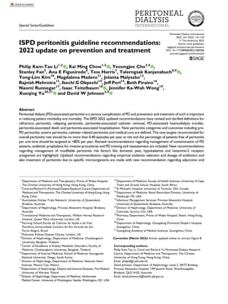
  • 10. observational and mechanistic studies of hypokalaemia in PD studies, the main contributory factor of hypokalaemia is low dietary potassium intake, rather than increased potas- sium excretion or intracellular shift.101,102 Dietary inter- vention is recommended to mitigate hypokalaemia. Observational data from a single-centre study suggested that regular lactulose use is associated with a lower rate of peritonitis.103 However, the benefit of lactulose to reduce peritonitis rate, compared with sennosides, has not been confirmed in a single-centre randomised controlled trial.104 There are emerging data to suggest that gastric acid suppression, especially with histamine-2 receptor antago- nists, is a modifiable risk factor for enteric peritonitis in PD patients. The hazard ratio for enteric peritonitis, as demon- strated in an observational cohort of 119 PD patients on histamine-2 receptor antagonists, was 1.67 (95% confi- dence interval 1.02–2.80). The increase in infectious mor- tality among histamine-2 receptor antagonist users further supported the burden of this risk.105 However, the risk of peritonitis associated with proton pump inhibitors is less consistently reported.106–108 A similar finding of heigh- tened risk conferred by the use of histamine-2 receptor antagonists, but not proton pump inhibitors, was found in a case series of peritonitis after gastroscopy. Of note, histamine-2 receptor antagonist users had a significantly higher post gastroscopy peritonitis rate (9.4%) compared to non-users (2.9%).59 A meta-analysis of six non- randomised studies involving pooled data of 829 PD patients showed that histamine-2 receptor antagonist use was associated with an increased odds of enteric peritonitis (OR 1.4, 95% CI 1.01–1.93).108 Notably, even though the association between proton pump inhibitor use and perito- nitis is less compelling, other concerns with proton pump inhibitors (including but not limited to Clostridioides infec- tion) do not justify a routine switching of histamine-2 receptor antagonist to proton pump inhibitor therapy. Secondary prevention To prevent fungal peritonitis, we recommend that anti-fungal prophylaxis be co-prescribed whenever PD patients receive an antibiotic course, regardless of the indication for that antibiotic course (1B). The majority of fungal peritonitis episodes are preceded by courses of antibiotics.109–112 A number of observational studies113–120 and randomised trials121,122 have examined the use of either oral nystatin (500,000 units qid) or fluco- nazole (200 mg every 48 h) as prophylaxis during antibiotic therapy. In essence, two randomised control trials121,122 and a systematic review43 showed a significant benefit. Most of the other reports on the prophylactic use of antifungals during antibiotic administration were non- randomised studies and have yielded mixed results. Unfortunately, nystatin is not available in some countries. Observational data118–120 and one randomised controlled trial122 showed that prophylactic fluconazole is effective. The randomised controlled trial of oral fluconazole included patients who received antibiotics for treating exit-site and tunnel infection, in addition to the treatment of peritonitis122 . However, there are potential problems (including drug interactions, emergence of resistant strains) with fluconazole prophylaxis. Overall, a Cochrane meta- analysis of the two randomised controlled studies on anti- fungal prophylaxis with oral nystatin or fluconazole showed a risk ratio of 0.28 (95% CI 0.12–0.63) for fungal peritonitis occurring after a patient has had an antibiotic course.43 Furthermore, each episode of peritonitis should be con- sidered a preventable event and hence evaluated.47 The CQI programme provides a means in secondary prevention. For each peritonitis episode, a root-cause analysis should be performed to determine the aetiology and, whenever possible, an intervention directed against any reversible risk factor should be made to prevent another episode. For example, Streptococcal viridans peritonitis could have indicated dental problems although such link is based on isolated case reports only.123,124 Peritonitis episodes caused by coagulase-negative staphylococcal species are associ- ated with touch contamination, while S. aureus infections have been associated with touch contamination or catheter infections. Identification of aetiology may involve review of the exchange technique. Retraining is sometimes neces- sary. Rarely, an outbreak of culture-negative peritonitis or peritonitis secondary to unusual organisms should trigger epidemiological investigation and field visit to look for environment risk factors such as PD fluid, hospital air or water contamination.125–127 Initial presentation and management of peritonitis The algorithm of initial management for PD patients pre- senting with a clinical diagnosis is summarised in Figure 1. We recommend that peritonitis always be diagnosed when at least two of the following are present: (1) clinical features consistent with peritonitis, that is, abdominal pain and/or cloudy dialysis effluent; (2) dialysis effluent white cell count 100/mL or 0.1 109 /L (after a dwell time of at least 2 h), with 50% PMN; and (3) positive dialysis effluent culture (1C). We recommend that PD effluent be tested for cell count, differential, gram stain and culture whenever peritonitis is suspected (1B). We recommend that PD patients presenting with cloudy effluent be presumed to have peritonitis and treated as such until the diagnosis can be confirmed or excluded (1C). Li et al. 119
  • 11. Patients with peritonitis usually present with cloudy PD effluent and abdominal pain. Cloudy effluent almost always represents infectious peritonitis, although there are other differential diagnoses classified according to cellular and non-cellular causes (Table 4).128 Some patients present with cloudy effluent but no or minimal abdominal pain. On the other hand, peritonitis should also be included in the differential diagnosis of the PD patient presenting with abdominal pain, even if the effluent is clear. In addition to the presenting symptoms, the patient should be ques- tioned about any recent contamination, accidental discon- nection, endoscopic or gynaecological procedures, as well as the presence of constipation or diarrhoea. In addition, the patient should be questioned about past history of peritoni- tis and exit-site infection. On physical examination, abdominal tenderness is typi- cally generalised and is less often associated with a rebound. Localised pain or tenderness should raise the sus- picion of underlying surgical pathology. Physical examina- tion should also include a careful inspection of the catheter tunnel and exit site. Any discharge from the exit site should be cultured. Erythema, tenderness and the presence of fluid collections (which may be confirmed with ultrasound) along the PD catheter tunnel may be indicative of a tunnel infection. The degrees of abdominal pain and tenderness are important factors in deciding whether a patient requires hospital admission. In general, patients with minimal pain could be treated on an outpatient basis with IP antibiotic therapy if this can be arranged. Follow-up within 3 days is advisable to confirm resolution and appropriateness of the antibiotic choice. When peritonitis is suspected, dialysis effluent should be drained, carefully inspected and sent for cell count with differential, Gram stain and culture.129 An effluent cell count with WBC 100/mL (after a dwell time of at least 2 h), with 50% PMN, is highly suggestive of peritoni- tis.130 Abdominal X-ray is generally not necessary and may be potentially misleading since pneumoperitoneum is com- mon (around one-third of CAPD patients)131 secondary to air entry into the peritoneal cavity via the PD catheter during exchanges. Peripheral blood cultures are usually negative132 and can be omitted unless the patient is clini- cally septic133 or on immunosuppression.134 Bacteraemia during peritonitis should raise the possibility of other intra-abdominal events.135,136 To prevent delay in treat- ment, antibiotic therapy (see below) should be initiated once the appropriate dialysis effluent specimens have been collected, without waiting for the results of laboratory testing. The WBC count in the effluent depends in part on the length of the dwell. For patients on APD with rapid cycle treatment, the clinician should use the percentage of PMN rather than the absolute WBC count to diagnose peritonitis, and a proportion above 50% PMN is strong evidence of peritonitis, even if the absolute WBC count is less than 100/mL.130 On the other hand, APD patients without a day- time exchange presenting with abdominal pain during the daytime may have no effluent to drain. In this case, 1 L of Figure 1. The algorithm of initial management for PD patients presenting with a clinical diagnosis of peritonitis. PD: peritoneal dialysis. 120 Peritoneal Dialysis International 42(2)
  • 12. dialysis solution should be infused, dwelled for 2 h, and then drained for inspection and laboratory testing. Identification of causative organisms We recommend that the blood culture bottle(s) be the preferred technique for bacterial culture of PD effluent (1C). We suggest that sampling and culture methods be reviewed and improved if more than 15% of perito- nitis episodes are culture negative (2C). Gram stain of the PD effluent should be performed even though the result is often negative.137,138 An additional benefit of Gram stain is its effectiveness in early detection of fungal elements, facilitating timely diagnosis and man- agement of fungal peritonitis.139 The diagnostic yield of the Gram stain is increased if it is performed on a centrifuged specimen. An appropriate method of culturing PD effluent is the most important step in establishing the causative organism. In some specialised centres, it has been possible to achieve a culture-negative peritonitis rate of less than 10%. Identification of the organism and subsequent anti- biotic sensitivities help to guide the choice of antibiotic, and the type of organism often indicates the possible source of infection. Bedside inoculation of 5–10 mL effluent in two (aerobic and anaerobic) blood-culture bottles has a reasonable sensitivity, and the culture-negative rate is typi- cally around 10–20%.140-143 The yield of peritoneal fluid culture is enhanced by inoculating the fluid directly into rapid blood-culture bottle kits (e.g. BACTEC, Kent, UK; Septi-Chek, Roche Diagnostics, Basel, Switzerland; BacT/ Alert, Biomerieux, Inc., Basingstoke, UK), centrifuging PD fluid and culturing the pellet or the lysis centrifugation technique as compared to inoculation into standard blood-culture bottles. Specifically, centrifugation of 50 mL PD effluent at 3000 g for 15 min, followed by resus- pension of the sediment in 3–5 mL supernatant and inocu- lation on solid culture media or standard blood-culture media, increases the yield by 5–10 times.39,142,143 The com- bination of water lysis, Tween-80 blood agar and Triton-X treatment of the PD effluent is also a sensitive culture method.144,145 The specimens should arrive at the labora- tory within 6 h. If immediate delivery to the laboratory is not possible, the inoculated culture bottles should ideally be incubated at 37 C. Inoculated bottles should not be refri- gerated or frozen, since it may kill or retard the growth of some microorganisms.146 The solid media should be incu- bated in aerobic, microaerophilic and anaerobic environ- ments. To fully assess yeast and filamentous fungal pathogens, appropriate fungal media should be selected; incubation of inoculated media under two temperature con- ditions (room temperature and 35–37 C) can increase the diagnostic yield.146 The speed with which bacteriological diagnosis can be established is very important. Concentration methods not only facilitate microbial identification, but also reduce the time needed for a positive culture. In over 75% of cases, microbiologic diagnosis can be established in less than 3 days. When the causative microorganism has been identi- fied, subsequent cultures for monitoring may be performed by only inoculating the effluent in blood-culture bottles. In a prospective study using facility-level data over 22 PD centres, immediate transfer of specimens or inoculated bottles to laboratories and the practice of PD effluent cen- trifugation are associated with lower culture-negative peri- tonitis rates.146 Notably, experience of the centre is important because culture-negative peritonitis rates fre- quently show an inverse relationship with the PD centre size.38,146 When cultures remain negative after 3–5 days of incu- bation, PD effluent should be sent for repeat cell count, differential count, fungal and mycobacterial culture. In addition, subculture on media with aerobic, anaerobic and microaerophilic incubation conditions for a further 3–4 days may help to identify slow-growing fastidious bacteria and yeasts that are undetectable in some automated culture systems. Furthermore, culture of the PD catheter can improve the diagnostic yield, especially for detection of fungi and enterococci.147 Other novel diagnostic techniques A number of novel diagnostic techniques have been explored for the early diagnosis of peritonitis, including leu- kocyte esterase reagent strips,148 biomarker assays (matrix metalloproteinase-8 and -9,149 neutrophil gelatinase- associated lipocalin150 and procalcitonin), polymerase chain Table 4. Differential diagnosis of cloudy effluent. Cellular causes PMN leucocytes Culture-positive infectious peritonitis Infectious peritonitis with sterile cultures Chemical peritonitis Eosinophils Dialysate eosinophilia Chemical peritonitis Monocyte/macrophages Specimen taken from ‘dry’ abdomen (after prolonged peritoneal rest) Red blood cells Hemoperitoneum Malignant cells Lymphoma Peritoneal metastasis Non-cellular causes Fibrin Triglycerides (milky white appearance of effluent) Calcium channel blockers Lymphatic obstruction Acute pancreatitis PMN: polymorphonuclear. Li et al. 121
  • 13. reaction (PCR) for bacterial-derived DNA fragments, PCR/ electrospray ionisation–mass spectrometry assay,151 16 S rRNA gene sequencing,152 matrix-assisted laser desorption ionisation-time of flight mass spectrometry153 and pathogen- specific ‘immune fingerprints’.154,155 However, none of them has been proved to be superior to conventional tech- niques. Immune fingerprint, for instance, by multicolour flow cytometry and multiplex enzyme-linked immunosor- bent assay, has been shown to discriminate between culture-negative, gram-positive, gram-negative episodes of peritonitis but provides no information on antibiotic resistance.155 Further refinement using mathematical machine-learning algorithms can characterise specific pathogens like streptococcal species and coagulase- negative staphylococci in a point-of-care manner.154 Utility of PD effluent phenotyping approach or immune fingerprint- ing remains to be validated before application to clinical use. In addition, a point-of-care device measuring levels of matrix metalloproteinase-8 and interleukin-6 has been tested to expedite diagnosis of peritonitis but is more useful to exclude peritonitis with a high negative predictive value over 98%.156 For rapid diagnosis of fungal peritonitis, PD effluent and serum galactomannan index might offer a faster turnaround time than the conventional culture method, but with a diag- nostic accuracy of 65.2% sensitivity, 85.0% specificity only.157,158 False-positive galactomannan results159 lead- ing to unnecessary use of antifungals is a definite concern. Empiric antibiotic selection We recommend that empirical antibiotic therapy be initiated as soon as possible, using either IP or sys- temic route, after appropriate microbiological speci- mens have been obtained (1B). We recommend that empirical antibiotic regimens be centre-specific and cover both gram-positive and gram-negative organisms (1C). We recommend that gram-positive organisms be covered by a first-generation cephalosporin or vancomycin and gram-negative organisms by a third-generation cephalosporin or an aminoglyco- side (1B). We suggest that cefepime monotherapy may be an acceptable alternative for empirical antibiotic regi- mens (2B). Once the diagnostic investigations have been com- pleted, empirical antibiotics should be started to achieve rapid resolution of inflammation, reduction of pain and preservation of the peritoneal membrane. No single anti- biotic regimen has been proven to be superior to others,160 and the choice should be centre-specific. There should be adequate coverage for both gram-positive and gram- negative organisms. A national registry confirmed that centres with higher proportions of peritonitis episodes receiving complete empirical coverage for both gram- positive and gram-negative organisms at presentation had higher odds of peritonitis cure by antibiotics.161 For the coverage of gram-positive organisms, vancomycin or first-generation cephalosporin is recommended. Cefazolin might be preferred to vancomycin when there is concern about emergence of organisms resistant to the latter. How- ever, vancomycin should be considered in centres with a high prevalence of methicillin-resistant organisms.162 The threshold prevalence of methicillin resistance that justifies empirical use of vancomycin remains controversial. No discernible difference in peritonitis cure rate was found between empirical cefazolin and vancomycin use for gram-positive or culture-negative peritonitis, according to observational data from PDOPPS.96 For the gram-negative coverage, third-generation cephalosporin or aminoglyco- side is suggested. Observational studies163,164 and one ran- domised controlled trial165 showed that aminoglycoside does not accelerate the loss of residual kidney function. However, repeated or prolonged aminoglycoside treatment was associated with a high incidence of vestibular toxicity or ototoxicity.166 It is also important to mention that treat- ment failure with ceftazidime is high with rising prevalence of extended-spectrum beta-lactamases (ESBL)-producing organisms. A recent analysis from PDOPPS reported that, for treatment of gram-negative peritonitis, empirical ami- noglycoside was associated with a higher likelihood of medical cure than ceftazidime.96 Monotherapy for empiri- cal treatment of peritonitis, instead of combination therapy, has now been accepted as an effective strategy. Two ran- domised controlled trials167,168 and one observational prospective study169 testing the use of IP cefepime mono- therapy have been published. Although there were differ- ences in cefepime dosing (intermittent, continuous, with and without adjustment for residual kidney function), all three studies showed primary response rates exceeding 80% on day 10.167–169 In particular, the largest study used a non-inferiority design and specified adjustment for resi- dual kidney function by increasing the loading and main- tenance doses of cefepime by 25% for urine volume more than 100 mL daily. Cefepime monotherapy was shown to be effective and non-inferior to standard dual therapy with cefazolin plus ceftazidime.168 In contrast, monotherapy with quinolones is not recommended because of the con- cern with emergence of resistant organisms and declining effectiveness.162,170 It is important to note that prompt administration of antibiotics has been consistently shown to be associated with better outcome of peritonitis treatment. In a prospec- tive multicentre study of 159 peritonitis episodes in West- ern Australia, the contact-to-treatment time was independently associated with treatment failure, defined as either catheter removal or death at 30 days. For each hour of delay in administering antibiotic therapy from the time of presentation to a hospital facility, the risk of PD failure or death was higher by 5.5%.171 In another 122 Peritoneal Dialysis International 42(2)
  • 14. retrospective study of 109 peritonitis episodes, a delay of starting IP or intravenous antibiotics treatment from the sign of peritonitis by 24 h conferred a threefold risk of peritoneal catheter removal by multivariate analysis.172 For logistics consideration, immediate IP antibiotic administration might not be feasible in the emergency department or wards in which staff are not familiar with PD. In order to avoid the adverse outcome of delayed peritonitis treatment, the systemic route should be started as a temporary measure when there is a foreseeable delay, such as long wait for dialysis unit bed or presentation outside the working hours of the ambulatory PD unit. However, the route of administrating antibiotics should still be switched to IP as soon as possible. Dosage of antibiotics We recommend that IP antibiotics be the preferred route of administration as long as the compatibility and stability of the IP antibiotics allow, unless the patient has features of systemic sepsis (1B). We suggest that IP aminoglycoside be administered as daily intermittent dosing (2B). We recommend that prolonged courses of IP amino- glycoside be avoided (1C). We suggest that adjunctive oral N-acetylcysteine therapy may help to prevent aminoglycoside oto- toxicity (2B). There is insufficient evidence to make a recommen- dation as to whether patients on APD should be temporarily switched to CAPD during treatment of peritonitis (Not Graded). The recommended dosage of antibiotics for the treatment of PD-related peritonitis is summarised in Table 5 (IP anti- biotics) and Table 6 (systemic antibiotics). However, the rec- ommended dosages of many antibiotics are based on published clinical experience rather than formal pharmacoki- netic studies. Most studies of IP antibiotics have been con- ducted in patients on CAPD rather than in patients on APD. The importance of adequate dosing of antibiotics was supported by an observational study of 339 episodes of gram-positive, gram-negative and culture-negative PD- related peritonitis, in which treatment failure was higher for patients with greater residual kidney function defined as urinary creatinine clearance more than 5 mL/min.247 The observation suggests that better clearance of antibiotics might lead to lower concentration of antibiotics, and hence the reduced time above the minimum inhibitory concentra- tion (MIC). Optimal dosing of antibiotics in patients with significant residual kidney function remains unknown, although fixed dosing irrespective of residual kidney func- tion might not be the best solution for antibiotics (such as cephalosporin) that exhibit time-dependent killing effects. Limited data are available to guide the adjustment of anti- biotics dosing, except a recent randomised controlled study advocating a 25% increase in the loading and main- tenance dose of cefepime, cefazolin and ceftazidime when PD patients have residual urine volumes of more than 100 mL daily.168 Vancomycin is the drug of choice in centres with a high prevalence of methicillin resistant gram-positive bacteria or for directed therapy in patients with relevant pathogens. IP administration is preferred because nearly 90% is absorbed in the presence of peritonitis.248 The superiority of treatment success rate with IP versus intravenous vanco- mycin is supported by Cochrane systematic review.160 Optimal dosing of IP vancomycin is unknown, and guide- line recommendations are variable regarding whether to prefer fixed dosing or target-guided dosing according to serum trough level. Although fixed dosing of IP vancomy- cin had been reported in a randomised controlled trial,234 it is unknown whether inter-individual variability of vanco- mycin bioavailability warrants adjustment of maintenance dose according to therapeutic drug monitoring of steady- state serum vancomycin concentration. A retrospective study reported that 60% of patients had subtherapeutic trough level following the loading dose after a fixed dosing of IP vancomycin 30 mg/kg every 5 days for CAPD and every 3 days for CCPD, irrespective of the residual renal function. However, all subsequent serum vancomycin lev- els were above 15 mg/L.249 Several observational studies did not show correlation between trough levels and cure rates of peritonitis.250,141 On the other hand, one observa- tional study reported a higher rate of peritonitis relapse with intravenous vancomycin use when the cumulative 4-week mean trough vancomycin levels were less than 12 mg/L.251 Another study of peritonitis due to methicillin-resistant coagulase-negative staphylococci showed that higher serum trough vancomycin levels achieved by IP vancomy- cin were associated with a lower relapse rate.252 Regarding the practice of trough-guided vancomycin dosing, there was no consensus on the preferred timing of obtaining trough vancomycin concentration. Based on a retrospective analysis of 61 episodes of gram-positive or culture- negative peritonitis, serum vancomycin levels lower than 10.1 mg/L on day 5, but not the level on day 3, were associated with worse outcomes (including transfer to hae- modialysis, death, persistent infection and relapse).208 Recently, trough-guided vancomycin dosing has been increasingly replaced by the area under the 24-h time- concentration curve (AUC)-guided dosing to optimise the management of severe S. aureus infection. Although the clinical significance of AUC pharmacokinetic parameters for monitoring vancomycin dosing in peritonitis treatment is incompletely understood, accumulating evidence sug- gests that trough level might not be the best option. A recent study of anuric patients on APD reported that peak serum concentration level (30 min after IP administration), but not trough vancomycin level, was associated with cure of gram-positive peritonitis.212 Li et al. 123
  • 15. Aminoglycosides remain useful for treating gram- negative peritonitis. Since aminoglycosides exhibit concentration-dependent activity, their maximal bacterial killing occurs at high peak drug concentrations. In addition, aminoglycosides continue to suppress bacterial growth even after drug concentration falls below MIC of the bac- teria, a characteristic known as the post-antibiotic effect.253 As a result of the post-antibiotic effect and concentration- dependent bactericidal characteristics, we favour intermit- tent daily dosing of IP aminoglycosides to minimise toxicity and adaptive resistance while maintaining drug efficacy. This has been confirmed in a randomised con- trolled trial comparing once-daily gentamicin dose versus continuous dosing; treatment success and relapse rate did not differ between the two regimens. The once-daily dosing strategy, nevertheless, was associated with lower trough Table 5. IP antibiotic dosing recommendations for treatment of peritonitis. Antibiotic Intermittent (1 exchange daily for at least 6 h) Continuous (all exchanges) Aminoglycosides Amikacin 2 mg/kg daily173 Not advised Gentamicin 0.6 mg/kg daily174,175 Not advised Netilmicin 0.6 mg/kg daily165 Not advised Tobramycin 0.6 mg/kg daily Not advised Cephalosporins Cefazolin 15 mg/kg daily (for long dwell)176,177 20 mg/kg daily (for short dwell)178,176 LD 500 mg/L, MD 125 mg/Ld 168,179 Cefepime 1000 mg daily LD 500 mg/L, MD 125 mg/Ld 168 Cefoperazone No data LD 500 mg/L, MD 62.5–125 mg/L180 Cefotaxime 500–1000 mg daily181 no data Ceftazidime 1000–1500 mg daily (for long dwell) 20 mg/kg daily (for short dwell)178 LD 500 mg/L, MD 125 mg/Ld 168,182 Ceftriaxone 1000 mg daily183 No data Penicillins Penicillin G No data LD 50,000 unit/L, MD 25,000 unit/L13 Amoxicillin No data MD 150 mg/L184 Ampicillina 4 gm daily185 MD 125 mg/L186 Ampicillin/ sulbactam LD 1000 mg/500 mg, MD 133.3 mg/66.7 mg187,188 Piperacillin/ tazobactam No data LD 4 gm/0.5 gm, MD 1 gm/0.125 gm189 Ticarcillin/clavulanic acid No data LD 3 gm/0.2 gm, MD 300 mg/20 mg/L190 Others Aztreonam 2 gm daily191 LD 500 mg/L192 , MD 250 mg/L192,193 Ciprofloxacin No data MD 50 mg/L194 Clindamycin No data MD 600 mg/bag195 Daptomycin 300 mg daily196 LD 100 mg/L197,198,199 , MD 20 mg/L197,200 Fosfomycin 4 g daily201,202 No data Imipenem/cilastatin 500 mg in alternate exchange203 LD 250 mg/L, MD 50 mg/L182 Ofloxacin No data LD 200 mg, MD 25 mg/L204 Polymyxin B No data MD 300,000 unit (30 mg)/bag188 Quinupristin/ dalfopristin 25 mg/L in alternate exchangesb205 No data Meropenem 500 mg daily (for long dwell in APD)207 1000 mg daily (for short dwell in CAPD)208,209 MD 125 mg/L206 Teicoplanin 15 mg/kg every 5 days210 LD 400 mg/bag, MD 20 mg/L211,140 Vancomycin 15–30 mg/kg every 5–7 daysc141,212 for CAPD 15 mg/kg every 4 days213 for APD LD 20–25 mg/kg, MD 25 mg/L214 Antifungal Fluconazole IP 150–200 mg every 24 to 48 h215,216 (oral route is preferred: see Table 6) No data Voriconazole IP 2.5 mg/kg daily217 (oral route is preferred: see Table 6) No data LD: loading dose in mg; MD: maintenance dose in mg; IP: intraperitoneal; APD: automated peritoneal dialysis. a IP ampicillin is not recommended for treatment of enterococcal peritonitis.218 b Given in conjunction with 500 mg intravenous twice daily. c Supplemental doses may be needed for APD patients and dwell time of at least 6 h is preferred. d Increase in doses by 25% may be needed for patients with significant residual kidney function.168 124 Peritoneal Dialysis International 42(2)
  • 16. serum gentamicin level.174 After the initiation of IP ami- noglycosides, a significant fraction of the drug can be absorbed into the systemic circulation, especially when the peritoneal solute transfer rate is increased during the acutely inflamed phase. High mass active transfer coeffi- cients for IP gentamicin and tobramycin were consistently reported in pharmacokinetic studies of patients with active peritonitis.175,254 In a case series of 24 PD patients with peritonitis, 76% of the IP gentamicin dose was absorbed into the systemic circulation and was higher among those with high and high average membrane solute transfer rates.175 Two studies in which outcomes were compared between patients with different gentamicin levels have not demonstrated any difference in gram-negative or culture- negative peritonitis cure rates.141,255 A major concern with aminoglycoside use in PD patients is ototoxicity. At the currently recommended peritonitis treatment dosage of aminoglycosides, ototoxicity could occur in PD patients, resulting in either vestibular or cochlear damage. Such oto- toxicity was reported even in the context of therapeutic serum concentrations.256,257 Not unexpectedly, ototoxicity occurs with IP aminoglycosides, similar to systemic admin- istration, as confirmed in both animal models258 and human.259,260 According to an observational study of PD patients, risk factors for hearing loss include older age, episodes of peritonitis and cumulative doses of amikacin and vancomycin.166 The mechanism of aminoglycoside ototoxicity is incompletely understood. Besides genetic predisposition, reactive oxygen species damage to the inner ear is the most accepted hypothesis. Based on three rando- mised controlled trials of N-acetylcysteine, the preventive approach with antioxidant protection of aminoglycoside- induced ototoxicity appears promising. The largest study involved 60 CAPD patients who received IP vancomycin and amikacin. Compared with the control group, patients randomly assigned to oral N-acetylcysteine 600 mg twice daily had significantly better protection from ototoxicity as measured by pure tone audiometry assessment of high- frequency hearing function at the first and fourth weeks.261 Similar findings were reported in two other randomised trials of N-acetylcysteine for PD patients receiving amikacin.262,263 Only one of the three trials included a control group with a placebo; the other two were open- label. A protective benefit using the same dose strategy of oral N-acetylcysteine on high tone frequency ototoxicity had also been demonstrated in haemodialysis patients receiving intravenous gentamicin for dialysis catheter- related bloodstream infection.264 None of these randomised Table 6. Systemic antibiotic dosing recommendations for treatment of peritonitis. Drug Dosing Antibacterial Amoxicillin Oral 500 mg thrice daily219 Ciprofloxacin Oral 500–750 mg daily220 Oral 750 mg BD for CCPD221 Clarithromycin Oral 250 mg BD222,223 Colistin IV 300 mg loading (for critically ill patients), then 60–200 mg dailyb224-226 Dalbavancin IV 1500 mg over 30 min single dose227 Daptomycin IV 4–6 mg/kg every 48 h228 Ertapenema IV 500 mg daily229 Levofloxacin Oral 250 mg daily230 or 500 mg every 48 h Linezolid IV or oral 600 mg BD231,232 for 48 h, then 300 mg BD233 Moxifloxacin Oral 400 mg daily234,235 Rifampicin Oral or IV 450 mg daily for BW 50 kg; 600 mg daily for BW 50 kg Ticarcillin/clavulanic acid IV 3 gm/0.2 gm every 12 h Tigecycline IV 100 mg loading, then 50 mg every 12 h236,237 Trimethoprim/sulfamethoxazole Oral 160 mg/800 mg BD238,239 Anti-fungal Amphotericin B desoxycholate IV 0.75–1.0 mg/kg/day over 4–6 h240 Amphotericin B (liposomal) IV 3–5 mg/kg/day241,242 Anidulafungin IV 200 mg loading, then 100 mg daily243,244 Caspofungin IV 70 mg loading, then 50 mg daily243 Fluconazole Oral 200 mg loading, then 100 mg daily240 Flucytosine Oral 1 gm daily240 Isavuconazole Oral or IV 200 mg every 8 h for 6 doses (48 h) loading, then 200 mg daily Micafungin IV 100 mg daily243,245 Posaconazole Oral tablet 300 mg every 12 h loading for two doses, then 300 mg daily246 Voriconazole Oral 200 mg every 12 h BD: twice a day; IV: intravenous; BW: body weight. a Ertapenem is not active against Pseudomonas or Acinetobacter species. b Expressed as colistin base activity in mg. Li et al. 125
  • 17. controlled trials assessed vestibular function. The pooled relative risk for otoprotection at 4–6 weeks was 0.14 (95% CI 0.05 to 0.45) according to meta-analysis.265 Notwith- standing the potential risks of bias of these trials with rel- atively small sample size, it is reasonable to consider co-administration of N-acetylcysteine at 600 mg twice daily for PD patients requiring aminoglycoside. In the absence of high-quality evidence to ameliorate potentially irreversible aminoglycoside ototoxicity, the best measure is to minimise prolonged or repeated administration. When an alternative drug of choice is available, early switch has been shown to have comparable clinical outcomes com- pared with continuing IP gentamicin.141 In other words, avoiding prolonged aminoglycoside should be advocated to prevent aminoglycoside ototoxicity. Fluoroquinolones, including ciprofloxacin266 and moxifloxacin,267 have been confirmed to be compatible with PD solutions and shown to be highly active and bactericidal in PD fluids with concentration-dependent activity.268 A small randomised controlled study sup- ported the safety and efficacy of IP vancomycin plus oral moxifloxaicin but was not powered to be a non-inferiority trial.234 Oral administration is an alternative and more convenient choice for susceptible organisms, as both ciprofloxacin and moxifloxacin can achieve adequate lev- els within the peritoneum.235,221 Oral ciprofloxacin should be administered in a once-daily dose of 500–750 mg instead of as a 250 mg twice daily dosing regimen,220 although higher dosing at 750 mg every 12 h has been suggested in CCPD patients.221 Patients should be instructed to avoid concomitant use of aluminium- containing antacids and oral phosphate binders (including calcium carbonate, lanthanum269 and sevelamer270 ) to avoid interference with absorption (and hence lower peak concentration) of fluoroquinolones.271 Antibiotic delivery and stability Stability and compatibility of antibiotics in PD solution (Table 7), as reviewed recently,272 is one of the factors which influences treatment success. Gentamicin is stable for 14 days both at room tempera- ture and under refrigeration in both dextrose-based and icodextrin-based PD solutions, but the duration of stability is reduced by admixture with heparin.13,273,274 Cefazolin is stable for 8 days at room temperature or for 14 days if refrigerated in dextrose-based PD solutions; addition of heparin has no adverse effect.13,275 In icodextrin-based PD solution, cefazolin is stable for 7 days at room temperature or for 14 days if refrigerated.273 Cef- tazidime is stable for 4 days at room temperature or 7 days if refrigerated in dextrose-based PD solutions. It is stable in icodextrin-based PD solution for 2 days at room tempera- ture or 14 days at refrigerated temperature.273 Cefepime is stable for 14 days in dextrose-based PD solutions when refrigerated.13,276 Vancomycin is stable for 28 days in dextrose-based PD solutions at room temperature, but the duration of stability is reduced at higher ambient temperatures.274 Stability of vancomycin in icodextrin-based PD solution has been con- firmed for 14 days at 4 C and 25 C.273 For compatibility of combined antibiotics in PD solu- tions, aminoglycosides and penicillins should not be added to the same bag due to chemical incompatibility.275 There are several antibiotics which can be mixed in the same PD bag; gentamicin is compatible with cefazolin or vancomy- cin, and ceftazidime is compatible with cefazolin or vancomycin.272,273,277 Emerging data of piperacillin/tazobactam showed that, when admixed with heparin in dextrose-based and icodextrin-based PD solutions, both drugs are stable for 7 days when refrigerated.278 Data on the stability of newer antibiotics and PD solu- tions are important to prepare the readiness for clinical use. Potential candidates include ceftolozane-tazobactam for gram-negative bacilli producing ESBL and Pseudomonas aeruginosa; the drug’s stability in PD solution has been confirmed.279 Special considerations for APD Extrapolation of antibiotic dosing from CAPD to APD is not recommended. First, patients on APD may have greater peritoneal antibiotic clearance. The implication of shorter antibiotic half-lives during the cycler exchanges is inadequate serum and dialysate drug concentrations throughout 24 h. An important concern for treating APD patients with peritonitis is the potential of underdosing, especially for antibiotics that exhibit time-dependent killing. Under such circumstances, it is important to use a dosing strategy that allows antibiotic concentrations to exceed the MIC for at least 50% of treatment time. Sufficient dwell time should be allowed for drug absorp- tion. Limited data are available to guide the optimal dwell time of antibiotics. A close correlation between vancomy- cin dwell time and bioavailability has been shown in phar- macokinetic study of APD patients.213 Minimal dwell time of 4 h should be used for vancomycin to achieve adequate peritoneal concentration according to previous APD experience,280 although dwelling for 6 h may be a more reasonable strategy.212 While conversion to CAPD is not always feasible for pragmatic reasons, this may be considered for antibiotics requiring continuous dosing. When the conversion to CAPD is difficult to implement, the treatment dose of IP antibiotics administered to short dwells should ideally be validated. For short-dwell automated cycling exchanges, cefazolin and ceftazidime can still be used based on phar- macokinetic studies on patients with281 and without peritonitis.178 126 Peritoneal Dialysis International 42(2)
  • 18. Adjunctive treatments We suggest that augmented peritoneal lavage should not be performed for the purpose of improving peri- tonitis cure (2B). We suggest that icodextrin be considered for volume overload which occurs during acute peritonitis (2C). Many patients with PD-related peritonitis could be man- aged on an outpatient basis. According to a PDOPPS anal- ysis of 1689 episodes of peritonitis internationally, only half of them had a hospitalisation within 14 days of peri- tonitis onset.31 The decision to hospitalise a patient depends on many factors, including social support, hemo- dynamic status of the patient, severity of signs and symp- toms and, for APD patients, the type of treatment schedule chosen as well as the ability to provide IP antibiotics as an outpatient and the reliability of the patient. The rationale for anti-fungal prophylaxis has been discussed in a previous section (see Secondary prevention section). Patients with cloudy effluent may benefit from the addi- tion of heparin 500 units/L IP to prevent occlusion of the catheter by fibrin. Depending on the severity of symptoms, some patients require analgesics for pain control. At the initial presentation and before IP antibiotics are initiated, one or two rapid PD exchanges are often performed for pain relief, although there are no data supporting this approach. Two randomised controlled trials showed that more extensive rapid-cycle peritoneal lavage, during the first 24 h of peritonitis282 or from day 3 to 5,283 did not improve the rate of complete cure or relapse. IP urokinase has been advocated for the treatment of bio- film, which may be the cause of refractory or relapsing peritonitis. A retrospective study found that IP urokinase and oral rifampicin, in addition to conventional antibiotics, could facilitate catheter salvage among patients with persisting asymptomatic infection following coagulase-negative staphy- lococcus peritonitis.284 However, three randomised controlled trials failed to show any benefit of IP urokinase for the treat- ment of refractory peritonitis.285–287 The rates of complete cure, catheter removal or relapsing episodes as well as overall mortality were not affected by adjunctive treatment with IP urokinase. In contrast, one randomised controlled study showed that simultaneous catheter removal and replacement was superior to IP urokinase in reducing relapsing peritonitis episodes.288 Peritoneal permeability to water and solutes typically increases during peritonitis. Reduced ultrafiltration is com- monly observed and may result in the complication of fluid overload. In addition to temporary use of hypertonic exchanges, management of fluid overload might require short dwell times, which can theoretically compromise local defence mechanisms (owing to decreased macrophage pha- gocytic capacity and immunoglobulin G concentration).289 Temporary use of icodextrin solution during acute peritonitis has been shown to be a better therapeutic option in one randomised controlled study.290 The primary cure rate of peri- tonitis was similar between the icodextrin and original glucose-based dialysis solution treatment groups in the study,290 although PDOPPS reported that icodextrin use was associated with a higher cure rate.96 Because of rapid glucose absorption, glycemic control may worsen in diabetic patients. Blood glucose monitoring with appropriate adjustments of insulin dosage may be needed. Protein loss during peritonitis is also increased. Screening for malnutrition should be Table 7. Summary of IP antibiotics stability. Antibiotics PD solutions Stability Storage conditions Remarksa Dextrose-based Icodextrin- based Room temperature Under refrigeration Tested for Stable for Gentamicin P 14 days P P 14 days P 14 days P P 14 days Cefazolin P 8 days P 8 days P 14 days P 14 days P 7 days P 7 days P 14 days P 14 days Ceftazidime P 4 days P 4 days P 7 days P 7 days P 2 days P 2 days P 14 days P 14 days Cefepime P 14 days P 14 days Vancomycin P 28 days P N/A P 14 days P P 14 days Piperacillin/ tazobactam þ Heparin P P 7 days P 7 days PD: peritoneal dialysis. a ’Stable for X days’ indicates that the antibiotic concentration retained at least 90% of its initial concentration up to day X. ‘Tested for X days’ indicates the antibiotic concentration retained at least 90% of its initial concentration up to the study duration set for X days only. Stability (Stable for X days) is interpreted according to the type of PD solutions and storage conditions specified. Li et al. 127
  • 19. undertaken in patients with prolonged peritoneal inflamma- tion. There are currently no high-quality, randomised studies that have examined the effects of dietary interventions or nutrition supplements in patients with peritonitis. Subsequent management of peritonitis We recommend that antibiotic therapy be adjusted once results and sensitivities are known (1C). The management algorithms for bacteria identified in dialysis effluent are summarised in Figures 2 to 4. Within 48 h of initiating therapy, most patients with PD-related peritonitis will show considerable clinical improvement. The effluent should be visually inspected regularly to deter- mine if clearing is occurring. Catheter lumen and exit site, tunnel should be re-examined. If there is no improvement after 48 h, cell counts and repeat cultures should be per- formed. In addition, monitoring of WBC count in PD efflu- ent may also predict treatment response. A retrospective study with a validation cohort showed that dialysis effluent WBC count 1090/mL on day 3 was an independent prog- nostic marker for treatment failure.291 Another retrospec- tive study further confirmed a prediction model incorporating a dialysis effluent WBC count 1000/mL on day 3–4 is associated with a substantially higher like- lihood of treatment failure.292 Nonfermenting gram-negative bacilli are important nosocomial pathogens contributing to serious peritonitis. Notably, P. aeruginosa, Acinetobacter baumannii and Ste- notrophomonas maltophilia are known to have a high intrinsic resistance and deserve special attention in relation to the choice of antimicrobial agents (see below). Refractory peritonitis We recommend that PD catheter be removed in refractory peritonitis episodes, defined as failure of the PD effluent to clear after 5 days of appropriate antibiotics (1D). We suggest that observation for antibiotic effect lon- ger than 5 days is appropriate if PD effluent white cell count is decreasing towards normal, instead of mandatory PD catheter removal if effluent does not clear up by day 5 (2C). After initiation of antibiotic treatment, there is usually clinical improvement in 72 h. Refractory peritonitis is defined as failure of the PD effluent to clear up after 5 days of appropriate antibiotics (Table 1). Catheter removal is indicated in cases of refractory peritonitis, or earlier if the patient’s clinical condition is deteriorating, in order to pre- serve the peritoneum for future PD as well as prevent mor- bidity and mortality. Prolonged attempts to treat refractory peritonitis by antibiotics without catheter removal are asso- ciated with extended hospital stay, peritoneal membrane damage, increased risk of fungal peritonitis and excessive mortality.293,294 The cut-off of 5 days in deciding PD catheter removal should be considered an arbitrary reference tool. Data to compare long-term outcomes between 5-day decision rule and longer wait for antibiotic effect are lacking. In a single- centre study including 190 consecutive peritonitis episodes, substantial variation of PD effluent white cell count was reported.295 The approach to less virulent organisms should probably be less aggressive to minimise premature or unne- cessary PD catheter removal. Instead of a ‘one-size-fits-all’ rule on day 5, the trajectory of effluent white cell count should be taken into consideration. A large observational study of 644 peritonitis episodes tracked the longitudinal change of effluent white cell count. Three patterns of treat- ment outcome were analysed: early response, delayed response (defined by gradual decline in effluent white cell count but still above 100/mL on day 5) and treatment failure (defined as peritonitis not cured by antibiotics, change to haemodialysis either temporarily or permanently or peritonitis-associated death).296 This study highlighted the varying rate or trajectory of effluent white cell count decline. In one-fifth of the cases, patients showed delayed response with 34% reduction of effluent white cell count by day 5, without the need for PD catheter removal.296 Thus, expectant of peritonitis episodes with longer antibiotic treatment duration without immediate catheter removal can be an option if the effluent white cell count is decreasing, albeit not reaching the nadir 100/mL by day 5. Relapsing, recurrent and repeat peritonitis We recommend timely PD catheter removal be con- sidered for relapsing, recurrent or repeat peritonitis episodes (1C). We suggest that simultaneous PD catheter removal and reinsertion be considered after culture of the PD effluent has become negative and the PD effluent white cell count is below 100/mL, in the absence of concomitant exit site or tunnel infection (2C). The definitions of relapsing, recurrent and repeat peri- tonitis are summarised in Table 1. Retrospective studies showed that relapsing, recurrent and repeat peritonitis epi- sodes are caused by different species of bacteria and prob- ably represent distinct clinical entities.297–301 When compared to non-relapsing episodes, relapsing ones are associated with a lower rate of cure, more ultrafiltration problems and higher rates of technique failure.297 Recur- rent peritonitis episodes had a worse prognosis than relap- sing ones.297,298 Centres with larger PD sizes are associated with lower rates of relapsing and recurrent peritonitis.161 To manage or reduce the risk of relapsing, recurrent or repeat peritonitis, simultaneous removal and reinsertion of PD catheters have been proposed.302 This allows a 128 Peritoneal Dialysis International 42(2)
  • 20. Figure 2. Management algorithm for Staphylococcus aureus peritonitis. Figure 3. Management algorithm for Streptococci identified in dialysis effluent. Li et al. 129
  • 21. continuation of PD without transfer to HD. Such a strategy should be considered only after culture of PD effluent has been confirmed negative following appropriate treatment, with a PD effluent white cell count lower than 100/mL and in the absence of concomitant exit-site or tunnel infection.27 Before the bacterial culture became negative, it would be inappropriate to attempt simultaneous removal and reinsertion of catheter because there could still be planktonic bacteria. To optimise the eradication success rate, we suggest deferring the procedure until the culture is negative, indicating absence of planktonic bacteria (when the bacteria are sequestered in biofilm). The simul- taneous removal and reinsertion of catheter procedure should be carried out under perioperative antibiotic coverage.27,303 The long-term benefit of simultaneous removal and reinsertion of PD catheters has been replicated in several series, with reported 1-year technique survival of 64%304 and median technique survival of more than 5 years.303 On the other hand, prolonged antibiotic treatment is not recommended. A randomised controlled study showed that extending antibiotic treatment duration for an additional week beyond that recommended by the ISPD is not advi- sable because such a strategy does not reduce the risk of relapsing, recurrent or repeat peritonitis and may increase the risk of repeat peritonitis. Another downside to pro- longed antibiotic use is the risk of developing secondary fungal peritonitis.305 A previous study suggested that bacterial DNA fragment levels in PD effluent are significantly higher 5 days before and on the date of completion of antibiotics amongst patients who subsequently develop relapsing or recurrent peritonitis.306 Despite the prognostic value of bacterial DNA fragments, a subsequent study showed that bacterial DNA levels do not decrease significantly with extended antibiotic therapy.305 Coagulase-negative Staphylococcus We suggest that coagulase-negative staphylococci be treated with IP cephalosporin or vancomycin, according to susceptibility, for a period of 2 weeks (2C). We suggest that retraining be considered for patients with coagulase-negative staphylococcal peritonitis (Not Graded). The leading cause of pathogenic coagulase-negative sta- phylococci peritonitis is Staphylococcus epidermidis, fol- lowed by Staphylococcus haemolyticus.307 Despite lower virulence properties than S. aureus, coagulase-negative staphylococci are more common, partly because host fibrinogen antimicrobial defences can elimi- nate the former but not coagulase-negative staphylococci.308 Coagulase-negative staphylococcal peritonitis is also challenging due to the large proportion of methicillin- resistant strains and biofilm formation. The methicillin resis- tance rate of coagulase-negative Staphylococcus causing peritonitis has been increasing to more than 50% in most centres309–311 and up to 70%.162,307 Such a high prevalence Figure 4. Management algorithm for other gram-positive organism including coagulase-negative Staphylococcus identified in dialysis effluent. 130 Peritoneal Dialysis International 42(2)