1. Breast Center
Digital Mammography:
Saving Time. Saving Lives.
“Digital mammography is one of the most
important diagnostic breakthroughs in
women’s health care.”
—James Werner, MD, designated radiologist at
the St. Francis Breast Center
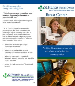
The St. Francis Breast Center uses digital
mammography, the latest in mammogram
technology. Digital mammography takes an
electronic image of the breast and stores it
directly into a computer. Digital equipment
provides clearer resolution and can penetrate
dense tissues for better results.
• Allows busy women to quickly get a
screening mammogram
• Allows the technologist to complete
mammograms in a shorter amount of time
• Allows the image to be electronically
enhanced, adjusted, magnified and stored for
further evaluation
• Results are back in a matter of days instead
of weeks
11/08
Providing high-tech care with a soft
touch because early detection
can save your life.
2. • Ultrasound-guided biopsy–high frequency
sound waves help guide the doctor’s
instruments to abnormal breast tissue during a
biopsy
Other Services
• Softer mammograms–a Mammopad
cushion providing comfort and warmth
during mammograms is available upon request
• Computer-Aided Detection (CAD)–a
radiologist uses this sophisticated software to
read images of the breast and surrounding
tissue. This provides a second pair of eyes for
better interpretation of results.
• Picture Archival and Communication
System (PACS)–an electronic system giving
physicians the capability to view X-ray images
from multiple locations resulting in faster care
For More Information
Contact the Breast Center at 785-295-8855 to
schedule an appointment at one of the Breast
Center’s three locations.
Description
The St. Francis Breast Center is a full-service
mammography facility approved by the Federal
Drug Administration and accredited by the
American College of Radiology.
Located on the second floor of the St. Francis
Health Center, it has convenient, designated
parking at Entrance A that provides quick access
to the lobby and the Breast Center’s designated
elevator.
Procedures
• Screening Mammography–an X-ray of the
breast is taken to detect breast changes in
women who have no signs or symptoms
• Diagnostic Mammography–an X-ray of the
breast is taken to check for abnormalities
after a lump or others signs or symptoms have
been found
• Bone densities–GE DEXA scanner uses special
X-ray to measure the mineral density such as
calcium in the bone to detect osteoporosis
• Breast exams–a certified MammaCare clinical
breast examiner performs clinical breast exams
upon request
• Breast ultrasound–high frequency sound
waves produce images of breast tissue
to determine if the tissue is normal
• Needle localization–a guide wire is placed
in abnormal breast tissue for surgical removal
• Stereotactic biopsy–a special mammography
machine uses ionized radiation to help guide
the biopsy needle to abnormal breast tissue
during the procedure