
Recommended
More Related Content
Similar to INFLAMMATION.pptx
Similar to INFLAMMATION.pptx (20)
Recently uploaded
Recently uploaded (20)
INFLAMMATION.pptx
- 2. INFLAMMATION Mrs Kalolekesha Msc. Pathology (Haematology )
- 3. Objectives By the end of this lecture you should be able to 1. Define inflammation 2. Describe the classification of inflammation 3. Understand the process of inflammation 4. Know the causes of inflammation 5. Learn the roles of various “chemical mediators” of acute inflammation 6. Know the three possible outcomes of acute inflammation 7. Visualize the morphologic patterns of acute inflammation
- 4. What is inflammation ? • Derived from latin word “inflammare”( to set on fire) • It is the local response of living mammalian tissues to injury arising from any agent(microbial, immunological, physical or chemical ) • It is the body’s immune response to harmful stimuli • It is the body’s defense mechanism to eliminate or limit the spread of an injurious agent , and remove the consequent necrosed cells and tissues. • In medical terms suffix –itis is used to denote inflammation e,g hepatitis, pharyngitis etc
- 5. History • Egyptian papyrus (3000 B.C.) • The Roman writer Celsus in 1st Century A.D. named the famous 4 Cardinal Signs of Inflammation. • Virchow – Fifth Clinical Sign i.e. Functio laesa • John Hunter (1973)– inflammation is not a disease but a non-specific response that has a salutary effect on its host. • Julius Cohnheim (1839-1884) – described the process of inflammation
- 6. Causes of inflammation 1.Infective agents like bacteria, viruses and their toxins, fungi, parasites. 2. Immunological agents like cell-mediated and antigenantibody reactions. 3. Physical agents like heat, cold, radiation, mechanical trauma. 4. Chemical agents like organic and inorganic poisons. 5. Inert materials such as foreign bodies
- 7. Advantages of Inflammation • It destroys microbes • Detoxification of toxins • Clearing of infections • Helps in healing process • Repair of damaged tissues
- 8. Disadvantages of Inflammation • May be life threatening e.g. in anaphylactic reactions • Obstruction of intestines by inflammation of peritoneum • Impaired cardiac function due to pericardial inflammation
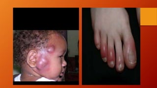
- 9. Cardinal Signs of Inflammation The classic signs of inflammation are described in latin as; • Rubor (Redness) • Tumor (Swelling) • Calor ( Heat ) • Dolor (Pain) • Functio laesa (Loss of Function)
- 10. Types of Inflammation • Inflammation can be classified as Acute Due to early response by the body Short duration Chronic Occurs after delay Longer duration Characterised by response by chronic inflammatory cells
- 11. ACUTE INFLAMMATION • Acute inflammatory response by the host to any agent is a continuous process but , it can be broadly divided into the following two events: I. Vascular events II. Cellular events • Intimately linked to these two processes is the release of mediators of acute inflammation
- 12. Acute Inflammation • It has three major components 1). Dilation of small vessels leading to increased blood flow 2) Increased permeability of microvasculature 3) Leukocyte emigration from microcirculation, accumulation in focus of injury and their activation to eliminate offending agent.
- 14. VASCULAR EVENTS • Alteration in the microvasculature (arterioles, capillaries and venules) is the earliest response to tissue injury • These alterations include: haemodynamic changes and changes in vascular permeability.
- 15. Vascular Events- Haemodynamic Changes • The earliest features of inflammatory response result from changes in the vascular flow and calibre of small blood vessels in the injured tissue Irrespective of the type of cell injury, immediate vascular response is of transient vasoconstriction of arterioles.
- 16. Haemodynamic Changes Next follows persistent progressive vasodilatation which involves mainly the arterioles, but to a lesser extent, affects other components of the microcirculation like venules and capillaries. Vasodilatation results in increased blood volume in microvascular bed of the area, which is responsible for redness and warmth at the site of acute inflammation.
- 17. Haemodynamic Changes Progressive vasodilatation, in turn, may elevate the local hydrostatic pressure resulting in transudation of fluid into the extracellular space. This is responsible for swelling at the local site of acute inflammation. Slowing or stasis of microcirculation follows which causes increased concentration of red cells, and thus, raised blood viscosity. Stasis or slowing is followed by leucocytic margination or peripheral orientation of leucocytes (mainly neutrophils) along the vascular endothelium. The leucocytes stick to the vascular endothelium briefly, and then move and migrate through the gaps between the endothelial cells into the extravascular space. • This process is known as emigration.
- 18. Vascular Events -Haemodynamic Changes • Vasodilation due to Histamine Nitric oxide • Increased permeability of microvasculature • Slowing of circulation /stasis due to Exudation Microcirculation packed with red cell Increased viscosity of blood
- 19. Vascular Events-Vascular leakages • In and around the inflamed tissue, there is accumulation of oedema fluid in the interstitial compartment which comes from blood plasma by its escape through the endothelial wall of peripheral vascular bed. • In the initial stage, the escape of fluid is due to vasodilatation and consequent elevation in hydrostatic pressure. This is transudate in nature. • But subsequently, the characteristic inflammatory oedema, exudate, appears by increased vascular permeability of microcirculation.
- 20. Vascular Events- Vascular leakages • Increased vascular permeability in acute inflammation by which normally non-permeable endothelial layer of microvasculature becomes leaky can have the following patterns and mechanisms which may be acting singly or more often in Combination;
- 21. Vascular Events-Patterns of increased permeability i. Contraction of endothelial cells:This is the most common mechanism of increased leakiness that affects venules exclusively while capillaries and arterioles remain unaffected. ii) Contraction or mild endothelial damage: In this mechanism, there is structural re-organisation of the cytoskeleton of endothelial cells that causes reversible retraction at the intercellular junctions or mild form of endothelial damage. iii) Direct injury to endothelial cells: Direct injury to the endothelium causes cell necrosis and appearance of physical gaps at the sites of detached endothelial cells. Process of thrombosis involving platelets and fibrin is initiated at the site of damaged endothelial cells.
- 22. Vascular Events-Patterns of increased permeability iv) Leucocyte-mediated endothelial injury: Adherence of leucocytes to the endothelium at the site of inflammation may result in activation of leucocytes. The activated leucocytes release proteolytic enzymes and toxic oxygen species which may cause endothelial injury and increased vascular leakiness. v) Leakiness in neovascularization: In addition, the newly formed capillaries under the influence of vascular endothelial growth factor (VEGF) during the process of repair and in tumours are excessively leaky.
- 24. Vascular Changes ( Vascular Leakages) • Hallmark of acute inflammation( escape of a protein rich fluid ) • Mechanisms of vascular permeability include; Contraction of endothelial cells Junctional retraction Direct endothelial cell damage Leukocyte mediated endothelial cell damage Transcytosis
- 28. Cellular Events • This phase consists of two processes ; 1. Exudation of leukocytes 2. Phagocytosis
- 29. Cellular Events • The escape of leucocytes from the lumen of microvasculature to the interstitial tissue is the most important feature of inflammatory response. In acute inflammation, polymorphonuclear neutrophils (PMNs) comprise the first line of body defense, followed later by monocytes and macrophages
- 30. Cellular Events 1. Changes in the formed elements of blood :In the early stage of inflammation, the rate of flow of blood is increased due to vasodilatation. But subsequently, there is slowing or stasis of bloodstream . 2. Rolling and Adhesion: Peripherally marginated and pavemented neutrophils slowly roll over the endothelial cells lining the vessel wall (rolling phase). This is followed by transient bond between the leucocytes and endothelial cells becoming firmer (adhesion phase). The following cell adhesion molecules (CAMs) bring about rolling and adhesion phases ; selectins, integrins and Immunoglobulin gene superfamily adhesion molecules.
- 31. Cellular Events 3. Emigration : After sticking of neutrophils to endothelium, the former move along the endothelial surface till a suitable site between the endothelial cells is found where the neutrophils throw out cytoplasmic pseudopods . The neutrophils lodged between the endothelial cells and basement membrane cross the basement membrane by damaging it locally with secreted collagenases and escape out into the extravascular space; this is known as emigration
- 32. Cellular Events • Simultaneous to emigration of leucocytes, escape of red cells through gaps between the endothelial cells, diapedesis, takes place. It is a passive phenomenon—RBCs being forced out either by raised hydrostatic pressure or may escape through the endothelial defects left after emigration of leucocytes. • Diapedesis gives haemorrhagic appearance to the inflammatory exudate
- 33. Cellular Events 4. Chemotaxis : The transmigration of leucocytes after crossing several barriers (endothelium, basement membrane, perivascular myofibroblasts and matrix) to reach the interstitial tissues is a chemotactic factor-mediated process called chemotaxis.
- 40. PHAGOCYTOSIS • Phagocytosis is defined as the process of engulfment of solid particulate material by the cells (cell-eating). The cells performing this function are called phagocytes. • There are 2 main types of phagocytic cells a. Polymorphonuclear neutrophils (PMNs) which appear early in acute inflammatory response, sometimes called as microphages. b. Circulating monocytes and fixed tissue mononuclear phagocytes, commonly called as macrophages.
- 41. PHAGOCYTOSIS • Neutrophils and macrophages on reaching the tissue spaces produce several proteolytic enzymes—lysozyme, protease, collagenase, elastase, lipase, proteinase, gelatinase, and acid hydrolases. • These enzymes degrade collagen and extracellular matrix. Phagocytosis of the microbe by polymorphs and macrophages involves the following 3 steps : 1. Recognition and attachment 2. Engulfment 3. Killing and degradation
- 42. PHAGOCYTOSIS 1. Recognition and Attachment ; Phagocytosis is initiated by the expression of cell surface receptors on macrophages which recognise microorganisms: mannose receptor and scavenger receptor. The process of phagocytosis is further enhanced when the microorganisms are coated with specific proteins, opsonins, from the serum and the process is called opsonisation (meaning preparing for eating).
- 43. PHAGOCYTOSIS 2. Engulfment :The opsonised particle or microbe bound to the surface of phagocyte is ready to be engulfed. This is accomplished by formation of cytoplasmic pseudopods around the particle due to activation of actin filaments beneath cell wall, enveloping it in a phagocytic vacuole
- 44. PHAGOCYTOSIS 3. Killing and Degradation : Next comes the stage of killing and degradation of microorganism to dispose it off which is the major function of phagocytes as scavenger cells. The microorganisms after being killed by antibacterial substances are degraded by hydrolytic Enzymes In general,the following mechanisms are involved in disposal of microorganisms: A. Intracellular Mechanisms: i) Oxidative bactericidal mechanism by oxygen free radicals a) MPO-dependent b) MPO-independent ii) Oxidative bactericidal mechanism by lysosomal granules iii) Non-oxidative bactericidal mechanism B. Extracellular Mechanisms : i) Granules Degranulation of macrophages and neutrophils continues to exert its effects of proteolysis outside the cells as well. ii) Immune mechanisms : immune-mediated lysis of microbes takes place outside the cells by mechanisms of cytolysis, antibodymediated lysis and by cell-mediated cytotoxicity
Editor's Notes
- Note: the suffix “itis” is added to the base word to to state the condition in inflammation eg appendix-appendicitis