More Related Content
Similar to Falkow_Nature_Medicine_2014.pdf
Similar to Falkow_Nature_Medicine_2014.pdf (12)
Falkow_Nature_Medicine_2014.pdf
- 1. L as k e r ~ Ko s h l a n d S p e c i a l Ac h i e v e m e n t i n
M e d i ca l s c i e n c e awa r d
commentary
nature medicine volume 14 | number 10 | october 2008 1053
I never met a microbe I didn’t like
Stanley Falkow
At the age of 11, I read Paul de Kruif’s Microbe
Hunters, which dramatized the discovery of
bacteria and viruses and their roles in human
disease.The heroes of Microbe Hunters—Louis
Pasteur,Robert Koch and others—became my
heroes, and I dreamed of becoming a bacte-
riologist, doing research on the bacteria that
cause disease. I was lucky enough to fulfill my
boyhood dream; however, I could never have
imagined the path I eventually followed, or
how much my views of microbes and disease
would change (and continue to do so) in the
process.Of course,I did not make this journey
alone. During the five decades I worked as an
active scientist,I helped train over 100 graduate
students, postdoctoral fellows and clinical fel-
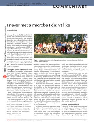
lows, and collaborated with 75 other scientists
(Fig.1).Each of us,in our own way,wondered,
“What is a pathogen?”
Entering the genetic and molecular world
I was a hospital bacteriologist and an autopsy
diener before I became a graduate student.
Thus, I learned about the world of microbes
from a practical standpoint before I learned the
tools to perform research. The medical bacte-
riology of the 1950s focused on differentiat-
ing the ‘good guys’ from the ‘bad guys’, and a
pathogen was simply defined as any organism
that caused disease.In basic bacteriology I was
taught that bacteria were Schizomycetes—
‘asexual primitive plants’. So, it was hard to
think of them as being inherently virulent.
When I entered Brown University as a grad-
uate student in 1957, I pestered my professors
asking what they thought made pathogens
different from non-pathogens. Professor C. A.
(‘Doc’) Stuart encouraged me to learn genet-
ics as a foundation for answering the question.
Professor Herman Chase, a mouse geneticist,
thought about my question and announced
that he had just the book to start me on my
voyage, the just-published compilation The
Chemical Basis of Heredity1. In this volume, I
learned for the first time about the structure
of DNA and embarked on my research during
the beginning of what Salvatore Luria called
the“Golden Age of Molecular Biology.”At the
time,bacterial genetics was basically restricted
to Escherichia coli K-12.
Shortly after I became a student, Lou Baron
at the Walter Reed Army Institute of Research
described for the first time the transfer of
genetic information from E. coli to Salmonella
typhi. I was anxious to use the tools of genet-
ics and molecular biology to establish the spe-
cific genes that defined the difference between
pathogens and non-pathogens. There were,
after all, E. coli that were clearly part of the
normal flora and E. coli that caused diarrhea
in infants. So, I immediately contacted Baron,
who supplied me with the necessary cultures
to study this problem.
I performed conjugation experiments using
E. coli K-12 donors and clinical isolates of
E. coli and Salmonella. I was excited when I
could actually detect E. coli surface antigens
in Salmonella. However, my conjugation
experiments revealed little about pathoge-
nicity. I was unable to transfer any gene from
Salmonella or Shigella that altered the measur-
able pathogenicity or host range of another
Salmonella species, or that made E. coli K-12
pathogenic2,3.
When I presented these results at a Cold
Spring Harbor meeting in 1963, the opinion
was almost unanimous that I was wasting my
time and ought to be concentrating on more
important biological questions. Indeed, at
that time there was a growing consensus that
infectious diseases were no longer of interest
for Western society. So, at age 30, I put my
dream of doing research on the meaning of
bacterial pathogenicity to rest for a time and
devoted my energy to examining the molecu-
lar nature of extrachromosomal elements—
now called plasmids—with help from Julius
Marmur (then at Brandeis University)4. I
focused especially on R plasmids, as these
mediators of antibiotic resistance were of
clear medical significance5.
Plasmids and pathogenicity
I discovered the joys of teaching in 1967 and
moved to Georgetown University. The follow-
ing year I met H. Williams Smith at a meeting
in London.He was a veterinarian who had used
basic bacterial mating experiments to show
that diarrhea in pigs and calves depended on
Stanley Falkow is the Robert W. and Vivian K.
Cahill Professor of Microbiology and Immunology
and of Medicine, Department of Microbiology
and Immunology, Stanford University School of
Medicine, 299 Campus Drive, D039, Stanford,
California 94305-5124, USA.
e-mail: falkow@stanford.edu
Figure 1 Lab alumni reunion in 2004, Falkow/Tompkins home, Hamilton, Montana, USA. Photo
courtesy of Manuel Amieva.
©
2008
Nature
Publishing
Group
http://www.nature.com/naturemedicine
- 2. com m e n ta ry
1054 volume 14 | number 10 | october 2008 nature medicine
E. coli strains that possessed two plasmids,one
encoding one or two enterotoxins, and a sec-
ond encoding an adherence factor that specifi-
cally recognized the epithelial cells of the small
bowel6. Willie asked me if my students and I
could use our molecular methods to examine
these virulence genes. By now we had become
adept at isolating plasmids from bacteria on
the basis of their circularity, and I was actively
engaged in using DNA hybridization to exam-
ine the relationships among plasmids.
Smith’s work became the foundation
for looking at whether certain E. coli from
humans possessed a similar plasmid arsenal
and caused traveler’s diarrhea.Our laboratory
showed that the plasmids encoding entero-
toxins from pigs had closely related counter-
parts in humans. Indeed, they were related to
the classic F factor of Joshua Lederberg and
to certain R plasmids7. Working with Naomi
Datta and Bob Hedges, we discovered that
there were many distinct groups of R plasmids
(Fig.2)8. Among some of them,the antibiotic-
resistance genes seemed to have a common
source, but the replication machinery, the
restriction-modification loci, and the pro-
teins that permit transmission of DNA from
a donor to a recipient were different. Yet
plasmids from a single group could carry
antibiotic resistance or have one or more
enterotoxin or adherence genes. It was as if
different gene cassettes could be inserted or
taken out of the same plasmid. It seemed
likely that these extrachromosomal elements
were an important part of bacterial evolution,
including the evolution of pathogenicity.
Recombinant DNA, gene transposition
and a return to understanding
pathogenicity
In June 1972 I moved to the University of
Washington in Seattle, where I would have a
more active role in teaching medical micro-
biology and directly participating in research
on infectious diseases. Soon after moving, I
attended a joint meeting of US and Japanese
plasmid researchers in Hawaii (Fig.3).
A major focus of the meeting was to gain
an understanding of how R plasmids acquired
resistance genes and whether R plasmids were
natural co-integrates of distinct replicons. A
concurrence of results on the origin of R plas-
mid resistance genes discussed one evening in
the unlikely setting of a Waikiki kosher deli-
catessen led to the idea of joining and splicing
DNA using restriction enzymes. My only con-
tribution was as a witness,occasional commen-
tator and donor of replicon RSF1010, known
to have a single EcoR1 cleavage site, for the
first pilot cloning experiments performed by
Stanley Cohen,Herb Boyer and their co-work-
ers9. However, I was aware of the implications
of the work. Indeed, I extracted from Herb
and Stan the promise that, if the experiment
succeeded and gene isolation and amplifica-
tion became a reality, I would send one of my
graduate students to Herb’s lab to pursue the
idea.Itworked,andweclonedthefirstvirulence
determinant of bacteria—the E.coli heat-stable
enterotoxin—in my laboratory10.
The implications of recombinant DNA tech-
nology were enormous, of course. Because of
my training in medical microbiology, their
impact on me was not just scientific. Indeed, I
participated in the historic Asilomar meeting
on the societal impact of recombinant DNA.
I served on the first I served on the first US
National Institutes of Health Recombinant
DNA Advisory Committee, established in
1974 in response to public concerns regarding
the public health and safety issues of manipu-
lating genetic material using recombinant
DNA techniques and the potential ethical
and social implications of the research. The
committee was initially charged with draft-
ing guidelines governing the safe conduct
of recombinant DNA research by outlining
appropriate biosafety practices and contain-
ment measures. These guidelines, now known
as the NIH Guidelines for Research Involving
Recombinant DNA Molecules, were first pub-
lished in 1976 and have evolved over time to
include other aspects of gene manipulation,
including genetic therapy. This was a time-
consuming and difficult task, and it was not
Figure 2 Photo taken on 25 May 1967 during my lecture at the Symposium on Infectious Multiple Drug
Resistance, held at the Georgetown University School of Medicine with support from the US Food and
Drug Administration. In the front row are, left to right, Arthur K. Saz (lighting his pipe), Piet A. Guinée,
Naomi Datta, David H. Smith (a Lasker Award Winner) and Tsutomu (Tom) Watanabe. The last four were
instrumental in discovering R plasmids and demonstrating their significance in clinical medicine.
Figure 3 The US–Japan plasmid meeting in
Honolulu, Hawaii, November 1972. Close-up
from a picture taken the day after the meeting
in the kosher deli, where we discussed the
experiment that led to gene splicing. Left to right:
Stanley Falkow, Robert H. Rownd, Herbert W.
Boyer, Stanley N. Cohen, Toshihiko Arai, Charles
C. Brinton Jr., Richard P. Novick (partly hidden)
and an unidentified Japanese scientist.
©
2008
Nature
Publishing
Group
http://www.nature.com/naturemedicine
- 3. com m e n ta ry
nature medicine volume 14 | number 10 | october 2008 1055
helpful to hear more than one scientist com-
plain that they had always poured their E. coli
cultures down the drain and “why is it now a
big deal?”It annoyed many scientists that there
were any restrictions concerning recombinant
DNA experiments that they deemed to be
harmless. Yet when scientists and physicians
participate in experiments that may have an
impact on society, society has the right of
informed consent.
Much of our subsequent research was con-
cerned with the application of genetic and
molecular tools, which now included recom-
binant DNA,to the study of infectious diseases.
Our laboratory and others discovered that the
antibiotic resistance genes of R plasmids were
transposable genetic elements, the ‘jumping
genes’ envisioned by Barbara McClintock
decades earlier11.
As happens in science, there was a juxtapo-
sition of what we learned about gene trans-
position and the sudden appearance of R
plasmids in Haemophilus influenzae and the
gonococcus12,13.We could say with reasonable
confidence that the penicillin-resistance genes
found in gonococci and E. coli isolated from
patients had a common ancestor, and trans-
position of tetracycline, chloramphenicol and
ampicillin antibiotic-resistance genes from
enteric species into commensal Haemophilus
wastheharbingerof theappearanceof thesame
resistance in pathogenic strains of Haemophilus
that cause meningitis. The agarose gel electro-
phoresis methods that we applied to help con-
struct the famous cloning vector pBR322 was
applied to characterize plasmids from clinical
specimens14: the first plasmid fingerprints—
and what has come to be known as molecular
epidemiology—was born15,16.A few years later
we could also show that a DNA sequence from
a specific or unique virulence gene could be
used for epidemiological investigations and
even for pathogen identification17.
The work on plasmid enterotoxins was
intriguing, and as I talked to those in the bac-
terial toxin field, I began to realize that the
dinucleotide-ribosylating enzymes secreted
by the E. coli enterotoxin-producing strains
resembled the toxins secreted by Vibrio chol-
erae,the diphtheria bacillus and Bordetella per-
tussis (the agent of whooping cough),and were
similar in function to the large heterotrimeric
G proteins of mammals18.It seemed to me that
microbes weren’t poisoning us as much as they
were undermining and subverting the normal
function of animal cells for their own survival.
Classical microbiologists often viewed toxins
as potential protective antigens that might be
used in vaccines. Clinicians viewed toxins as
the causative factors of disease.I was interested
in these facets, but thought that the toxin had
to be understood both in terms of the biology
of pathogenesis and of the utility of the toxin
for bacterial survival,persistence and transmis-
sion: what’s in it for the bug?
I decided that we now had the tools to
revisit my initial dream of understanding the
biology of pathogenicity. So, after a sabbatical
leave in England, I returned to the University
of Washington in 1978 and recruited a cadre
of students with the idea to examine bacterial
pathogenicity at the genetic and molecular
level.Perhaps because of my early background
in medical microbiology, I did not attempt to
focus the laboratory on a particular patho-
gen, but worked on many: gonococcus19,
Bordetella pertussis20, and the plague bacil-
lus21. The research ranged from clinical stud-
ies to molecular epidemiology of nosocomial
infection,but the major focus was on the basic
structure and function of virulence genes and
their regulation. A clinical investigation into
the phenotypic differences between commen-
sal E. coli and clinical isolates from urinary
tract infection led to the cloning of suspected
determinants of pathogenicity22,23, although
we argued a great deal about what exactly con-
stituted a ‘virulence gene’.
What is a pathogen?
I moved to Stanford University in the summer
of 1981. I spent my first years there isolating
and defining bacterial determinants that we
believed to be associated with pathogenicity.
I decided that one way to define virulence
genes was to apply some sort of ‘molecular
Koch’s postulates’: the specific inactivation
of the genes suspected to be associated with
virulence should lead to a measurable loss in
pathogenicity, and reversion or allelic replace-
ment of those genes should lead to restoration
of virulence24. It was an inadequate test, but if
we were to employ it at all, we needed a quan-
titative way to measure virulence.We could do
animal challenge experiments, but death is a
harsh end point.Instead,we learned cell culture
and adopted the methods of cell biologists.We
could now use different aspects of cell injury
and cell death instead of host death. Similarly,
we examined microbial numbers at different
sites in a host as a measure of invasiveness.Our
focus shifted from looking at the microbe only
to looking at the microbe–host interaction and
at the consequences for both parties25,26.
In 1987, we began to define what Catherina
Svanborg, one of our collaborators, called a
“pathogenic personality”27 (Box 1). The more
we studied pathogens, the more it became
apparent that there were common themes of
bacterial pathogenicity28. As we studied these
themes,we started learning as much about the
microbe as about the host. In 1987, we knew
that mobile genetic elements—plasmids,
bacteriophages and transposons—had been
central factors in the evolution of pathogenic
traits. Today, genomic analysis has revealed
that a wide range of bacteria, including plant
pathogens and obligate intracellular parasites
such as Chlamydia and Rickettsia, have related
blocks of genes that distinguish them from
their related commensal and nonpathogenic
brethren. These blocks of genes are ordinarily
found as contiguous,large DNA chromosomal
insertions called pathogenicity islands29.They
seem to have been transmitted by horizontal
gene transfer,as if the island DNA once resided
in a microbe distantly related to that in which
it now is found. In many cases, these gene
blocks encode a specialized secretory pathway
designed to dispense specific effector virulence
proteins to the bacterial surface or through a
protein structure into the host membrane and
cytoplasm. For pathogens such as Salmonella,
the proteinaceous delivery appendage viewed
in the microscope looks quite like a hypoder-
Box 1 Attributes shared by bacterial pathogens
• Entry into the host. Entry is not a random event but has selectively evolved to exploit
the host’s needs to breathe, eat, see, hear, eliminate waste and reproduce.
• Attainment of a unique niche. All pathogens have evolved a specific means of
association with at least one unique host cellular target shortly after entry. The specificity
of this molecular interaction may dictate the host–pathogen interaction for hours or even
days afterwards.
• The pathogenic signature. Pathogens avoid, circumvent, destroy or manipulate one or
more essential host defenses.
• Multiplication. The definitive goal of the pathogenic strategy is to produce sufficient
number of cells to persist in the host or to be transmitted to a new host.
• Exit from the host. It is likely that microbes have specialized determinants for leaving
their host, preparing for subsequent entry in a new host.
• Limited host range and the inherent ability to cross anatomical barriers and/or
breach other host defenses to establish themselves in areas usually devoid of other
microorganisms. This property is essential for their survival in nature.
©
2008
Nature
Publishing
Group
http://www.nature.com/naturemedicine
- 4. com m e n ta ry
1056 volume 14 | number 10 | october 2008 nature medicine
mic syringe and needle. Pathogens know their
cell biology! Depending on the pathogen, the
bacterial effector proteins delivered to the host
cell can lead to actin rearrangement (Fig. 4),
to the covalent modification of signaling mol-
ecules, or to the induction of apoptosis30.
If thereis satisfactionwiththerelativelyrapid
discovery of these common themes, we must
also be cautious,as we seem perilously close to a
situationinwhichmolecularsequencingdirects
the biologist, instead of the biologist directing
thesequencing31.Yetthemolecularfossilrecord
in the DNA of contemporary microorganisms
has revealed extraordinary information. It
suggests to me that pathogenicity is an ancient,
honorable microbial profession that has, at its
roots, the requirement for microbes to defeat
their predators—protozoa, nematodes and
otherorganismsthatusemicrobesastheirmain
food source.The microbes that infect us inher-
ited these principles, and evolution has finely
honed them not just to help bacteria avoid pre-
dation, but to take advantage of larger organ-
isms for their own survival.
Still … what is a pathogen?
The complete genome sequence of virtually
every important human and animal patho-
gen is at hand. The complete sequence of
their hosts is also at hand. Complete gene
arrays allow us to look at gene transcription
and genetic variability in both the host and
the pathogen. Yet, I still struggle with the
question, “What is a pathogen?” Those who
are concerned with infectious diseases must
adhere to the concept that any microbe that
causes disease is a pathogen. It matters not
whether an accidental or deliberate exposure
to bacteria leads to disease. Moreover, I have
been fond of invoking Walt Kelley’s Pogo and
declaring, “We have met the enemy and he is
us!” Human behavior has led to health crises
such as Legionnaire’s disease, toxic shock syn-
drome and an increase in food-borne disease
caused by international food-distribution net-
works32. It seems to me that a better term for
many emerging infectious diseases would be
‘diseases of human progress’.
Humans live with hundreds of commensal
species that reside in every inhabitable nook
and cranny and are present for our entire
lives but cause no harm. These commensal
species have become a focus of increasing
interest. In sheer number of cells they pre-
dominate in a human by a factor of 10 (ref.33).
Microbiologists are uncomfortably aware that
most species that inhabit us remain unknown,
except for a snippet of sequence that reveals
their presence. To what extent these members
of our bacterial flora influence our health and
disease is still an open question34.
The distinction between commensals and
pathogens can be blurred at times because
some commensals cause disease, albeit usu-
ally in immunocompromised hosts. Some
microbes could indeed be called ‘commensal
pathogens’35. For example, pneumococci and
meningococci regularly inhabit the human
nasopharynx and are mostly carried asymp-
tomatically, although they can cause life-
threatening diseases. Immunization against
these microbes not only protects against dis-
ease, but also prevents their ability to colonize
the host. From the bacterial viewpoint, the
production of a toxic protein might be seen
more precisely as colonization factors than as
virulence factors. Are these organisms simply
normal flora that evolved to live in a perilous
location, where they face less competition but
pay for it by coming into contact with deadly
elements of the immune system? A number of
the most frightening human pathogens, such
as Mycobacterium tuberculosis, the typhoid
bacillus and Helicobacter pylori, cause disease
only in a relatively small number of people
and can often persist asymptomatically for a
lifetime36. Might we say that organisms such
as Mycobacterium and H. pylori, which have
been with humans from the beginning, can be
considered indigenous flora?
After 50 years of study, I concede that there
is no simple definition that accounts for what
a pathogen is. It is important to have a medi-
cal definition of a pathogen and its relation to
disease. On the other hand, I would argue that
disease does not encompass all of the biological
aspects of pathogenicity and of the evolution
of the host–parasite relationship.For example,
CagA, a protein of H. pylori delivered by the
microorganism to host cells probably as a
means to loosen epithelial tight junctions and
gain nutrients, can cause gastric cancer over
decades in the right setting of diet and host
genetic determinants37,38. Good riddance to
H. pylori by antibiotic therapy, immunization
or increased sanitation! But the disappear-
ance of H. pylori from human flora may have
the equally important effect of predisposing
humans to esophageal cancer, asthma and
other diseases39. The biology is more complex
than we realized.
It really doesn’t matter how we define a
pathogen! To underestimate the evolutionary
potential of microorganisms and their abil-
ity to survive, even in the face of enormous
pressure to eradicate them, would be a mis-
take. Infectious agents will emerge as long as
there are microorganisms. Humans help the
evolutionary process, sometimes unwittingly,
and sometimes by arrogance or ignorance.
Fortunately,humans have evolved in wondrous
ways to avoid and repel microbial incursion.
Our immune system, both innate and adap-
tive,is a tribute to Nature’s skill in doing so,but
I confess the belief that microbes will always
have the last laugh (Box 2).
Box 2 Microbes may be smarter than you think
• They understand mathematics. They have mastered exponential equations and
understand biostatistics.
• They understand physics. They know that a small amount of energy applied to the right
point can ‘move’ a large object.
• They understand military tactics. They strike quickly with overwhelming numbers, cut
the lines of communication and wear camouflage.
• They are expert biologists. They have studied biology longer than any other living thing,
understood Darwin before he did, and also invented neo-Darwinism. They have mastered
genetics, cell biology and immunology.
• They always have the last laugh. They are generally the first living things we encounter
after birth and, when we die, they are the last living cells on our bodies. Then, they
devour us.
Figure 4 Electron micrograph of Salmonella
typhimurium entering into an M cell in the
mouse intestine. The organism causes ‘ruffling’
of the cell surface to gain entry. Underneath
the breached epithelial cell are cellular
elements of the immune system, including
macrophages and dendritic cells, which
can take up the invading Salmonella. This
particular photograph, which I took in 1988,
was important because it showed that the
phenomena we saw in cultured cells had their
counterparts during natural infection.
©
2008
Nature
Publishing
Group
http://www.nature.com/naturemedicine
- 5. com m e n ta ry
nature medicine volume 14 | number 10 | october 2008 1057
Infect. Immun. 41, 942–949 (1983).
24. Falkow, S. Molecular Koch’s postulates applied to
microbial pathogenicity. Rev. Infect. Dis. 10, S274–
S276 (1988).
25. Isberg, R.R. & Falkow, S. A single genetic locus
encoded by Yersinia pseudotuberculosis permits inva-
sion of cultured animal cells by Escherichia coli K-12.
Nature 317, 262–264 (1985).
26. Miller, V.L. & Falkow, S. Evidence for two genetic loci
in Yersinia enterocolitica that can promote invasion
of epithelial cells. Infect. Immun. 56, 1242–1248
(1988).
27. Falkow, S., Small, P., Isberg, R., Hayes, S.F. & Corwin, D.
A molecular strategy for the study of bacterial invasion.
Rev. Infect. Dis. 9, S450–S455 (1987).
28. Finlay, B.B. & Falkow, S. Common themes in microbial
pathogenicity. Microbiol. Rev. 53, 210–230 (1989).
29. Hacker, J., Blum-Oehler, G., Muhldorfer, I. & Tschape, H.
Pathogenicity islands of virulent bacteria: structure,
function and impact on microbial evolution. Mol.
Microbiol. 23, 1089–1097 (1997).
30. Finlay, B.B. & Falkow, S. Common themes in microbial
pathogenicity revisited. Microbiol. Mol. Biol. Rev. 61,
136–169 (1997).
31. Medini, D. et al. Microbiology in the post-genomic era.
Nat. Rev. Microbiol. 6, 419–430 (2008).
32. Falkow, S. Who speaks for the microbes? Emerg. Infect.
Dis. 4, 495–497 (1998).
33. Turnbaugh, P.J. et al. The human microbiome project.
Nature 449, 804–810 (2007).
34. Dethlefsen, L., McFall-Ngai, M. & Relman, D.A. An
ecological and evolutionary perspective on human-
microbe mutualism and disease. Nature 449, 811–
818 (2007).
35. Falkow, S. Is persistent bacterial infection good for
your health? Cell 124, 699–702 (2006).
36. Monack, D.M., Mueller, A. & Falkow, S. Persistent bac-
terial infections: the interface of the pathogen and the
host immune system. Nat. Rev. Microbiol. 2, 747–765
(2004).
37. Amieva, M.R. et al. Disruption of the epithelial apical-
junctional complex by Helicobacter pylori CagA.
Science 300, 1430–1434 (2003).
38. Mueller, A., Falkow, S. & Amieva, M.R. Helicobacter
pylori and gastric cancer: what can be learned by
studying the response of gastric epithelial cells to the
infection? Cancer Epidemiol. Biomarkers Prev. 14,
1859–1864 (2005).
39. Blaser, M.J., Chen, Y. & Reibman, J. Does Helicobacter
pylori protect against asthma and allergy? Gut 57,
561–567 (2008).
40. Falkow, S. The fortunate professor. Annu. Rev.
Microbiol. 62, 1–18 (2008).
10. So, M., Boyer, H.W., Betlach, M. & Falkow, S. Molecular
cloning of an Escherichia coli plasmid determinant
than encodes for the production of heat-stable entero-
toxin. J. Bacteriol. 128, 463–472 (1976).
11. Heffron, F., Sublett, R., Hedges, R.W., Jacob, A. &
Falkow, S. Origin of the TEM-beta-lactamase gene
found on plasmids. J. Bacteriol. 122, 250–256
(1975).
12. De Graaff, J., Elwell, L.P. & Falkow, S. Molecular
nature of two beta-lactamase-specifying plasmids iso-
lated from Haemophilus influenzae type b. J. Bacteriol.
126, 439–446 (1976).
13. Roberts, M., Elwell, L.P. & Falkow, S. Molecular char-
acterization of two beta-lactamase-specifying plasmids
isolated from Neisseria gonorrhoeae. J. Bacteriol. 131,
557–563 (1977).
14. Meyers, J.A., Sanchez, D., Elwell, L.P. & Falkow,
S. Simple agarose gel electrophoretic method for
the identification and characterization of plasmid
deoxyribonucleic acid. J. Bacteriol. 127, 1529–1537
(1976).
15. Tompkins, L.S., Plorde, J.J. & Falkow, S. Molecular
analysis of R-factors from multiresistant nosocomial
isolates. J. Infect. Dis. 141, 625–636 (1980).
16. Kaper, J.B., Bradford, H.B., Roberts, N.C. & Falkow, S.
Molecular epidemiology of Vibrio cholerae in the U.S.
gulf coast. J. Clin. Microbiol. 16, 129–134 (1982).
17. Moseley, S.L. et al. Detection of enterotoxigenic
Escherichia coli by DNA colony hybridization. J. Infect.
Dis. 142, 892–898 (1980).
18. Dallas, W.S. & Falkow, S. The molecular nature of heat-
labile enterotoxin (LT) of Escherichia coli. Nature 277,
406–407 (1979).
19. Koomey, J.M., Gill, R.E. & Falkow, S. Genetic and bio-
chemical analysis of gonococcal IgA1 protease: clon-
ing in Escherichia coli and construction of mutants of
gonococci that fail to produce the activity. Proc. Natl.
Acad. Sci. USA 79, 7881–7885 (1982).
20. Weiss, A.A., Hewlett, E.L., Myers, G.A. & Falkow, S.
Tn5-induced mutations affecting virulence factors
of Bordetella pertussis. Infect. Immun. 42, 33–41
(1983).
21. Portnoy, D.A., Moseley, S.L. & Falkow, S.
Characterization of plasmids and plasmid-associated
determinants of Yersinia enterocolitica pathogenesis.
Infect. Immun. 31, 775–782 (1981).
22. Hull, R.A., Gill, R.E., Hsu, P., Minshew, B.H. & Falkow, S.
Construction and expression of recombinant plasmids
encoding type 1 or d-mannose-resistant pili from a
urinary tract infection Escherichia coli isolate. Infect.
Immun. 33, 933–938 (1981).
23. Normark, S. et al. Genetics of digalactoside-binding
adhesin from a uropathogenic Escherichia coli strain.
ACKNOWLEDGMENTS
I wish to thank S. Fisher and L.S. Tompkins for
reading and commenting on this manuscript.
I was asked to write a commentary in conjunction
with my selection to receive the Lasker~Koshland
award. In science, the operative word is more often
‘we’ instead of ‘I’. Indeed, I was fortunate to have
worked with so many talented and genuinely nice
people during my career. The publication of this
article happens to coincide with the publication of
an autobiographical sketch The Fortunate Professor40,
which I dedicated to my mentors, former students and
colleagues. In that article, I wrote that my professional
life could be summarized simply by a statement from
the Talmud:
I learned much from my parents.
I learned more from my teachers.
I learned even more from my colleagues.
But I learned the most from my students.
Upon further reflection, I should add that all of us can
learn a lot from the microbes as well.
1. McElroy, W.D. & Glass, B. The Chemical Basis of Heredity
(Johns Hopkins Press, Baltimore, 1957).
2. Falkow, S., Rownd, R. & Baron, L.S. Genetic homol-
ogy between Escherichia coli K-12 and Salmonella.
J. Bacteriol. 84, 1303–1312 (1962).
3. Falkow, S., Schneider, H., Baron, L.S. & Formal, S.B.
Virulence of Escherichia-Shigella genetic hybrids for the
guinea pig. J. Bacteriol. 86, 1251–1258 (1963).
4. Falkow, S., Marmur, J., Carey, W.F., Spilman, W.M. &
Baron, L.S. Episomic transfer between Salmonella
typhosa and Serratia marcescens. Genetics 46, 703–
706 (1961).
5. Falkow, S., Citarella, R.V. & Wohlhieter, J.A. The molec-
ular nature of R factors. J. Mol. Biol. 17, 102–116
(1966).
6. Smith, H.W. & Halls, S. Studies on Escherichia coli
enterotoxin. J. Pathol. Bacteriol. 93, 531–543 (1967).
7. Guerry, P. & Falkow, S. Polynucleotide sequence relation-
ships among some bacterial plasmids. J. Bacteriol. 107,
372–374 (1971).
8. Falkow, S., Guerry, P., Hedges, R.W. & Datta, N.
Polynucleotide sequence relationships among plasmids
of the I compatibility complex. J. Gen. Microbiol. 85,
65–76 (1974).
9. Falkow, S. I’ll have the chopped liver please, or how
I learned to love the clone. ASM News 67, 555–559
(2001).
©
2008
Nature
Publishing
Group
http://www.nature.com/naturemedicine