
Recommended
More Related Content
What's hot
What's hot (20)
Similar to Presentation 3 uterine artery doppler .pdf
Similar to Presentation 3 uterine artery doppler .pdf (20)
Recently uploaded
Recently uploaded (20)
Presentation 3 uterine artery doppler .pdf
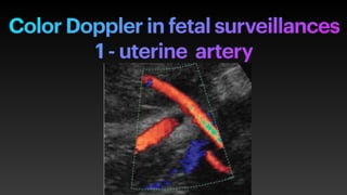
- 1. Color Doppler in fetal surveillances 1 - uterine artery
- 2. Uterine artery Doppler .uterine artery is a branch of internal iliac artery that is carry the blood to the uterus - ultrasound UTA. Measurement is used to check the blood f low between mother and her baby . ..enters the myometrium at the junction of uterine corpus and cervix Crossing the external lilac vessels (EIV). - measurement at the ascending branch 1-2 cm above crossing piont .
- 3. Uterine artery interpretation — predictive accuracy of f irst trimester uterine Doppler is better in the detection of early onset of preeclampsia than late onset diseases - mean UTAPI 11 - 13 weeks gestation measured trans abdominally was lower than that measured trans vaginally . —Mean UTAPI is measured at 16 - 22 weeks, — prediction of preeclampsia and FGR by 1- increasing mean uterine PI and RI of alue 2- persistent early diastolic notch after 24 weeks of gestation
- 4. uterine artery Doppler How to measure .Trans vaginal or trans abdominal abdominal approach . . The bladder is semi f illed (comfortable bladder) . .mid sagittal view of the uterus a n d c e r v i c a l c a n a l a r e obtained ( US/ prob at the level of iliac fossa right or left) then followed by introduction the color f low imaging at the level of internal os . .the prob tilted sideway laterally and maintaining its medial-- angulation ( lower paracervical area ) till the uterine artery is appear arise from internal iliac artery .it crosses the external iliac artery .
- 5. Uterine artery How to measure - Apply PW 1-2 cm above the point of cross over vessels، sample gate =2mm to cover the whole vessel ,and the angle of insinuation less than 30 . - Three symmetrical consecutive waves are obtained. - Peak systolic velocity (PSV) > 60cm /s to ensure that is uterine artery rather than arcuate artery. - Measure right and left uterine artery PI mean utAPI= uterine artery (R+L)/2 - UTA PI value shows progressive decrease until the late of pregnancy. - UTA indices have a clinical value in evaluation of placenta associated diseases - Prediction of Preeclampsea and IUGR
- 6. Normal uterine artery wave in non pregnant And early pregnancy Normal f inding of of uterine artery Doppler in non pregnant and early pregnancy —high resistive with low diastolic f low —presence of early diastolic notch If the notch persists after 24 weeks It is abnormal And associated with adverse outcome..
- 7. The normal uterine artery Doppler wave form During. pregnancy Normal impedance to f low of the uterine artery the f irst trimester Normal impedance to f low in late second and third trimester Normal impedance to f low in early second trimester
- 8. The normal Doppler indices Of uterine artery During pregnancy - At 11 -13 weeks the uterine artery indices that are measured by Trans abdominal us are less than measured by trans vaginal US . - Trans abdominal uterine PI 1.8 trans vaginal pi 1.9 and Ri 0.7 and 0.8 - .S/D. Are 2. 65 —2.57 - 16 — 22 weeks UAPI is 1.6or less ,UTA RI 0.58 or less were considered normal - the uterine artery indices are higher at f irst trimester of gestation than the second and third trimester - the uterine artery early diastolic notch had disappear at 22 weeks of gestation at more than half of the pregnant women. - -absence of the uterine artery early diastolic notch was related to decrease RI and PI.
- 9. Uterine artery Indices Early pregnancy Early second trimester indices Late seond trimester And third trimester
- 10. Abnormal uterine artery after 20- 24 weeks Abnormal pulsatile index (PI)> 95 centile Abnormal high resistance to f low of UTA With reversed diastolic f low Normal impedance of f low of UTA early diastolic notch Abnormal Increase impedance of f low of UTA With early diastolic notch
- 11. Role of uterine artery in recurrent pregnancy loss —High uterine artery resistance is accounted as one of of recurrent abortion reasons . —Current studies approved that high PI more than 2.5 in pts had recurrent abortion that’s means defect of the uterine blood f low. when we decrease the uterine A resistance and improve the uterine blood f low by impaction of aspirin and vitaminE Leads to better pregnancy outcome.