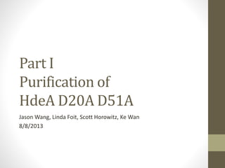
4. Problem that previously occurred
during purification of HdeA D20A
D51A from minimal medium
load
25
20
15
10
25
20
15
10
Increasing NaCl concentration during elution from HiTrap SP HP
HdeA
bla
5. PurificationsetupRT vs 4C
Cell Culture
Periplasmic
protein
extraction
Dialysis
Purification at
Room Temp/No
Glycerol
Purification at
4o C/ 5%
Glycerol
15. Future Directions Part I
Purification of the constitutively active
mutant HdeA D20A D51A
• Purify further with a Phenyl Column to attempt to remove
remaining contaminating proteins
• Redo the entire protocol using bacteria grown in 15N Labeled
media.
• Purify that protein and use it in NMR analysis
20. Variable 2: Variations of Im7
I22V
L18A
L19A
L37A
I54A
L53A
Largely destabilized Partially unfolded
Model for intermediate state
Model for unfolded state
21. Variable 3:
N- or C- Terminal 6xHis-tag
ss-HdeA – (GGGGS)2 – Im7 – GSG – 6xHis
Im7Linker
ss-6xHis-GSG-HdeA – (GGGGS)2 – Im7
HdeA
Im7Linker
His Tag
His Tag
HdeASS
SS
22. His Tag Rationale
• Protein was not completely pure after native
purification
• Still contaminating bands, probably degradation
products
23. His Tag Rationale
• Previous experiments demonstrated significant contamination
of the periplasmic extracts with potential degradation
products in addition to the fusion protein of interest
HdeA F35W-(GGGGS)2-Im7 L53A I54A ∆W
(LF 1434)
*
Full length
protein
Degradation
products?
*
*
20 ul
load
10 ul
load
24. His Tag Rationale
• Previous experiments demonstrated significant contamination
of the periplasmic extracts with potential degradation
products in addition to the fusion protein of interest
• Adding a His-tag creates a system designed to quickly screen
multiple types of proteins using one purification technique for
their suitability in NMR analysis
25. Screening for expression of the His-
tagged fusion proteins
1. Is the his-tagged fusion proteins expressed at all?
2. Is an N- or a C-terminal tag preferred?
• expression
• purification
27. Purification of His-Tagged HdeA-
Im7 Fusion Protein LF 1508
Im7Linker
His Tag
HdeASS
1508: Wild Type I22V His Tag
C Terminal His Tag
28. Increasing Concentration of Imidazole
35
25
15
kDa
35
25
15
kDa
ElutionFractionsof LF1508Purification
Im7Linker
His Tag
HdeASS
1508: Wild Type I22V His Tag
C Terminal
Fusion
Protein
*Pure Products
29. 1. NMR attemptwith a 15N HdeA-Im7 fusion
(HdeA WT-(GGGGS)2-Im7I22V-GSG-6xHis)
HiTrap Chelating
15N minimal medium
Purification Ke Wan
30. HSQC spectrum of
HdeA WT-(GGGGS)2-Im7 I22V-GSG-
6xHis
1H
15N
Expected # of peaks: 185
Buffer
50 mM KH2PO4
90 mM NaCl
1 mM DSS
0.5 mM EDTA
1 mM chloroacetic acid
5% D2O
pH 2.5
Scott
33. HSQC spectra 15N Im7 I22V +
HdeA
1H [ppm]
15N[ppm]
500 uM 15N Im7 I22V
500 uM 15N Im7 I22V + 571 uM HdeA
Linda + Loic
34. Im7 L18A L37A L38A might be a
better substrate for NMR
90% 1H2O
10% 2H2O
0.2 M
Na2SO4
10C
Pashley, C. L. et al. (2011): Journal of Molecular Biology (2011)
35. Purification of His-Tagged HdeA-
Im7 Fusion Protein LF 1510
Im7
HdeA Linker
His TagSS
N Terminal His Tag
1510: Wild Type L18A L19A L37AHis Tag
36. Elution Fractions of LF1510 Purification
Im7HdeA Linker
His TagSS
N Terminal His Tag
1510: Wild Type L18A L19A L37AHis Tag
Load B9 B8 B7 B6 B5 B4 B3 B2 B1 C1 C2 C3 C4 C5 C6 C7 C8
25
20
37
Kda
37. Purification of His-Tagged HdeA-
Im7 Fusion Protein LF 1507
Im7Linker
His Tag
HdeASS
1507: Wild Type L18A L19A L37A His Tag
C Terminal His Tag
38. Im7Linker
His Tag
HdeASS
1507: Wild Type L18A L19A L37A His Tag
C Terminal His Tag
Elution Fractions of LF1507 Purification
Load FT Wash A8 A9 A10 A11 A12 B12 B11 B10 B9 B8 B7
25
20
37
Kda
42. Degradation products (?) also
occur after purification I
HdeA F35W-(GGGGS)2-Im7 L53A I54A ∆W
(LF 1434)
*
Full length
protein
Degradation
products?
*
*
20 ul
load
10 ul
load
LINDA
43. Degradation products (?) also
occur after purification II
Selected elution fractions purification
HdeA WT-(GGGGS)2-
Im7 L18A L19A L37A-GSG-6xHis
(LF1507)
20
15
25
10
*
*
*
Full length
protein
Degradation
Product(s)?
20
15
25
10
Selected elution fractions purification
HdeA WT-(GGGGS)2-
Im7 I22V-GSG-6xHis
(LF1508)
*
*
44. C-terminal His-tag cannot be
detected for degradation products I
Selected elution fractions purification
HdeA WT-(GGGGS)2-
Im7 L18A L19A L37A-GSG-6xHis
(LF1507)
20
15
25
10
*
*
*
Anti 6xHis antibody
20
15
25
10
Full length
protein
Degradation
Product(s)?
*
47. N-terminal His-tag is present
however
Im7
HdeA Linker
His TagSS
N Terminal His Tag
1510: Wild Type L18A L19A L37AHis Tag
Elution fractions HiTrap Chelating column
48. Identify degradation products by mass
spectrometry (UofM Bioconsortium)
HdeA F35W-(GGGGS)2-
Im7 L53A I54A ∆W
(LF 1434)
*
Full length
protein
Degradation
products?
*
*
20 ul
load
10 ul
load
Cut out bands Proteolytic digest Identification of peptides
His-tag-GSG-HdeA WT-(GGGGS)2-
Im7 L18A L19A L37A
(LF1510)
49. Identify degradation products by mass
spectrometry (Indiana University)
LF 1434 HdeA F35W-link-Im7 L53A I54A W75F
LF 1508 HdeA WT -link-Im7 I22V-6xHis
LF 1510 6xHis-HdeA WT -link-Im7 L18A L19A L37A
Cut out bands Proteolytic digest
identification of peptides
Intact mass determination
50. Mass spec data turnaround
University of Michigan
Proteomics Core
Indiana University
52. Future experiments
• 15N labeling of HdeA D20A D51A
structural studies with NMR
• Identification of degradation products of the HdeA-Im7
fusions
use stable degradation products for structural studies
instead of full length protein
• Create fusion proteins between HdeA and peptides previously
identified as potential HdeA substrates in peptide array
Protein will not bind to anion exchange when employed as a first step
HdeA D20A D51A aggregates on the cation exchange column when purified from minimal medium
Problem does not occur when protein is purified from rich medium
Hypothesis: the protein is binding to the column
Hypothesis: the protein is binding to the column
This is showing that the first large UV vis peak is due to elution of mostly beta lactamase, and to show that the TINY amount of HdeA const. mutant we’re getting is coming in fractions after the end of the peak
Hypothesis: the protein is binding to the column
Hypothesis: the protein is binding to the column
Again showing at 4 degree C/5% glycerol the beta lactamase comes off mostly in the first peak
This now shows that the decrease in the first peak is because a lot of beta lactamase has already come off – yes it’s still there all the way through, but now the second peak is also do to a large amount of HdeA coming off
Linker artificially should create a 1:1 ratio of HdeA and Im7 to allow us to better study the interactions between these two proteins without the problem described earlier of precipitation of Im7
Different linker lengths still allow us to maintain a constant HdeA: Im7 ratio, but allow us to additionally study how distance might affect –
Kinetics of the interaction of the proteins
Also if the linker might be too short, and affect the ability of the proteins to interact at all
This will allow us to test the thermodynamics of the interactions between Im7 and HdeA
With the previous variable we’ll be able to determine how quickly it takes for Im7 and HdeA to bind
This variable will allow us to determine what mutations might weaken the intermolecular forces between the two proteins
Correction: we did the expression test to see
1 if the his-tagged fusion proteins would be expressed at all- to do this we collected proteins from bacteria transformed to express various constructs. The parameters were HdeA WT (variable a = linker length) (variable b = Im7 variant) (variable c = linker attaches Im7 to HdeA either on its
2 is N- or C-terminal tagged proteins had different or the same levels of expression
We used 4 different mutants of Im7
Show Im7 structure with mutations indicated here instead of table
Label different lanes, indicate which band we are interested in
Do we still have the samples and could run this gel again (longer?)
Can be purified to high purity
Show NMR spectrum here, Linda will provide the slide
HdeA gives a lot of info to us when we perform NMR spectra on it
Fig. 7. The 500-MHz 1H–15N HSQC spectrum of L18A–L19A–L37A. The spectrum was acquired in buffer A (90% 1H2O/10% 2H2O) with 0.2 M Na2SO4 at 10 °C. Sequential NOEs are observed for NH groups of i and i±1 residues for stretches of the protein involving residues 8–17, 18–37, 41–45, 51–52, 54–55, 60–63 and 67–69; however, no longer-range NOEs were observed in the three-dimensional HSQC-NOESY-HSQC experiment. The expanded region shows the assignments in crowded regions more clearly.
Since that variant (triple alanine) gave such a better spectra, we decided to try to purify the mutant created by Linda that expressed Im7 variant 3A in the fusion protein.