SlideShare a Scribd company logo
1 of 71
Download to read offline
1
Pediatric ocular trauma
Prepared by: Dr. Iddi Ndyabawe
Date: 25/08/2021
2
We broke down ocular injuries into the areas that are getting hurt.
3
Car barteries contain acid…. Acid injury
Paint can cause chemical injury.. paintball pellets implicated here. Some places in Uganda with
paintball game services; Kamooflage Paintball, Adonai adventure Park, Forest Park Paintball
Arena. Outside Kampala, in Busiika there is Extreme Adventure Park.
4
We shall start with the extraocular injuries… specifically the eyelid lacerations. This is a dog bite
injury. It’s a severe laceration. Often eyelid lacerations are very easily repaired by the emergency
physician in the ER. Most important thing is to make sure that the globe is not involved.
5
“The average age of pediatric dog bite patients with ophthalmic involvement is 3 years.
6
7
Any type of trauma involving the canaliculi area needs to be explored in the operating room.
This test assesses the canaliculi system.
8
You put a lot of fluorescein in that opening on the lid to allow some fluorescein to drain. And if
it’s draining properly and with enough fluid, this should go down into the Nasolacrimal duct.
And if you put a little swab into the nose for 10 seconds, you should be able to see that
fluorescein uptake on the swab using regular UV light.
Other extra ocular injuries we shall talk about are orbital fractures.
Orbital trauma is amongst the most common emergency department consultations and is often
accompanied by trauma of the facial bones and soft tissue.
• Additional injuries that may be observed in the setting of orbital trauma include orbital
hemorrhage, retained foreign body, and optic neuropathy.
• Ophthalmic manifestations of orbital trauma may include decreased vision, intraocular injury,
strabismus, and eyelid or globe malposition.
• An ophthalmic examination should be performed on all patients who have sustained orbital
trauma as ocular injuries may also be present
So, here is a nice diagram of a classic baseball to the eye causing a blow-out fracture. So you see
it… the globe is compressed a little bit… ultimately you are putting pressure to the surrounding
structures and it’s often this infraorbital ridge and the floor that breaks. This is an orbital floor
fracture that you can see here. The globe gets pushed back, provides extra pressure that pushes
down and …. This is the classic orbital floor fracture.
9
Le Fort fractures (lateral view). Note that all the fractures extend posteriorly through the
pterygoid plates (arrow).
10
Le Fort classification of midfacial fractures. Le Fort I, horizontal fracture of the maxilla, also
known as Guérin fracture. Le Fort II, pyramidal fracture of the maxilla. Le Fort III, craniofacial
disjunction.
This is a nice aaaah coronal view here… aaaah… where you can see pretty well,, I think these
are easiest to see on coronal view … aaamhh… you can see the nice disruption and the leaking
of fat or blood down… but something is leaking down to the maxillary sinus through this orbital
floor.
11
One of the characteristic features of the orbital floor fractures clinically is that if there is true
muscle entrapment.
Here is a nice photo of a child who Is asked to look up and … normal left eye can look up very
well… but the right affected eye can’t look up. This is entrapment of inferior rectus muscle
innervated by CN 3.
12
It can sometimes affect the infraorbital nerve where the kids will have decreased sensation on the
upper lip or that kind of nasolacrimal area.
In a patient with limited elevation, a positive forced-duction test indicates the presence of
restriction. Bradycardia, heart block, nausea, or syncope can occur as a vagal response to
entrapment.
A special presentation, the white-eyed blowout fracture, is characterized by marked restriction
(in both directions) of vertical ocular motility despite minimal signs of softtissue injury. This
restriction is due to entrapment of the inferior rectus muscle either beneath a trapdoor fracture or,
in the case of children, in a linear opening caused by flexion deformity of the floor. Early
surgery, rather than observation, is required in order to minimize permanent muscle and nerve
damage.
This is a zygomatic fracture.. usually lateral…not to many complications associated with it.
The one on the right shows aaa is an axial ct view and it's a nasal fracture so the nasal ethmoid is
a little more of a supporting structure and it can be more cosmetic as it can cause deformities of
the nose and septal deviation so it's associated with aesthetic impairment clinically you can see a
widened intracanthal distance, enophthalmos are some of the other clinical features.
When do you plan surgical repair of orbital floor fractures?
13
-When the patient has diplopia nd CT evidence of muscle entrapment plus unresolving Oculo
cardiac reflex. This is an indication for immediate surgery. Generally happens in children.
Otherwise early surgery is done at 2 weeks following initiation of antibiotics and steroids.
-Orbital fracture > 50% of orbital floor involved.
-Enophthalmos > 2mm
-Hypoglobus >2mm
-Infraorbital nerve hypoesthesia.
Principle of orbital floor fracture repair:
-Release of entrapment
-Placement of a barrier
-Repair of hypoglobus and enophthalmos
less commonly you see orbital roof fractures so this is you know this is again aaaa nice is aaaaa
coronal ct and you can see on this right image that there is a shatter to the roof of that orbit the
danger of these is that you know you can have communication with the CSF so it can be a
predisposing feature to cause a you know invasive CNS infection the refreshers usually occur in
14
kids less than 10 because the adult pneumatized frontal sinuses usually dissipate energy to the
orbital rim rather than this roof so we usually see it in younger kids you really want to look for
other signs of intracranial injury probably should extend a CT to include the the entire the head
to make sure there's not bleeding other signs and then we look clinically for CSF leak some
places that may or may not include neurosurgery or just follow up or close evaluation.
What do you look for on orbital CT scan?
-Bone windows
-Position and integrity of obirtal floor
-Orbital walls
-Optic nerve
-Optic
-Foreign bodies
What do you look for on the orbital floor?
-Tear drop sign
When the rim remains intact, this is termed blowout fracture.
15
so in general with orbital fractures if there's no globe rupture complex laceration signs of
intracranial disease or CSF leak usually the follow-up is with an ophthalmologist in about 24
hours if there is muscle entrapment, enophthalmos or orbital dystopia you know certainly those
are um you know other reasons to do this quickly otherwise i could often wait for a week so the
early intervention for most orbital fractures is is not that helpful unless you get those other signs
of entrapment and dystopia.
some folks consider antibiotics there's not great evidence for this but if there's substantial sinus
involvement or underlying sinusitis you know the potential for spread is there you can consider
adding oral antibiotics um you can also consider oral corticosteroids if there's nerve entrapment
but that probably should be done in consult with an ophthalmologist all right so that's the extra
ocular tissues…
16
now let's talk about the anterior segment and break this down into sort of sections one and two
the first is the cornea and the conjunctiva and the second is really the anterior chamber and the
iris in the lens
so let's talk about conjunctival injuries so… we see a lot of conjunctival foreign bodies you
know here's just a nice simple piece of debris you know here is an arrow injury i guess that's not
17
really a foreign body it's just penetration through the lid but i think the point here is that you only
know if there is a true foreign body if you can effectively evert the lid so this shows two
techniques of just grabbing the lid and inverting around your finger
so retraction is important there's a few different methods you know you can open manually from
the superior and inferior orbital ribs where you're basically just stretching the area like it's shown
in the figure on the right and then this sometimes can help easily just for simple things like dust
dirt sands to make sure if the contact's there or not.
18
sometimes you need to do a better true retraction reversion to really if there's that sensation that's
there and you need to look a little closer um this is using the cotton swab technique where you
can put a cotton swab you know at the bridge of of the upper orbital ridge and then roll the eyelid
over it to allow that true eversion of the lid this is an effective technique
there's other if you have these available things like lid retractors that can help open your area this
is often helpful especially if you want to have a better view of the sclera
19
there's a few other type of retractors that may be in a ophthalmology kit you have you know such
as this spring type one the the resource low method is to use paperclips i've absolutely used this
and it absolutely is effective if the parents will let you do it they don't think you're crazy coming
at them with a paperclip or a set of paperclips like is shown here but you can fold the edges of a
paper stuff so the non-sharp edges create a nice retractor and it's sometimes a nice way just to
open up your lids in a area where you might not have better tools
20
all right so when talking about the conjunctiva we're going to spread into the sub-conjunctival
area and this is a example of a sub-conjunctival hemorrhage and this is a very expensive one this
isn't really the typical one we see you can see these from non-trauma like a coughing illness that
can cause uh small subconscious hemorrhages and you can also see them from blunt trauma but i
think the important thing to know is when it's extensive or circumferential or you see going all
the way around um in the the the pupillary area um and around the iris then especially filling the
the cornea like it's shown here you definitely need to evaluate for globe rupture so you need to
ensure that the globe is intact in general some subconjunctival hemorrhages shouldn't affect
vision and it should be asymptomatic other than looking terrible like this poor eye does but it
does take a little while to go away like any soft tissue contusion where there's a hematoma it's
similar and it often takes 10-14 days to resolve.
21
Corneal abrasion is one of the most common ocular injuries in children and adults.
corneal abrasions we certainly see commonly from infants to adults that there are many reasons
and many in situations where kids can scratch their eyes so the a better term for this is probably a
corneal epithelial defect that you're creating some type of penetration of the epithelium of the
cornea and this is a superficial injury when we call it an abrasion. when we talk about ulceration
that's a deeper injury so these generally don't cause scarring because the bowman's membrane if
you remember uh epidermal if our dermatology pathology of the eye and some of the different
layers but if we're not breaking down the bowman's membrane um usually these can heal very
normally without any long-term effects uh corn abrasions can have a lot of different patterns so
it's important when you're looking at the abrasion to help to help look into what the potential
causes
22
you know sometimes the what's shown here in the bottom right is it kind of that's scratching and
that's sometimes when there's something trapped in the under lid that's scratching up and down
versus just a simple kind of blunt injury like a horizontal injury like it's shown here on the top
right
um there's a lot of misconceptions in um there's a lot of misconceptions in coronal abrasion you
do not need to pass it's never really been shown benefit of that in general accommodation can
make it hurt so ways to prevent accommodation can be helpful but true patching really isn't
topical anesthetics are not a good idea even though many of us use prepare cane or similar for the
evaluation it's not something the patient should go home with because there is evidence that that
will delay wound healing and topical steroids shouldn't be necessary because these do heal on
their own so the steroid can only promote infection and may be a predisposing factor for
infection like ulceration
23
Topical cycloplegic drops and antibiotic ointment may help reduce discomfort and risk of
infection, respectively. Traumatic corneal epithelial defects usually heal within 1-2 days.
uh in the emergency department or the urgent care center that you're working in the corner
abrasion should be managed with uh make sure tetanus is up to date exclude a corneal foreign
body or something that's causing it uh and then everything else is home management so uh
cycloplegia should be considered if the patient's having a lot of discomfort as a way to block that
accommodation reflex which often causes the pain when they're accommodating to light topical
antibiotics should be used absolutely until symptom free and topical NSAIDS are another pain
medicine that you can consider when patients are having a lot of discomfort.
topical antibiotics you know oftentimes our ophthalmologist will recommend a drop during the
day and an ointment at night because that ointment will add a little extra lubrication which can
be soothing at night and but i think both methods are effective in preventing progression to
ulceration.
Cigarette burns of the cornea are the most common thermal injuries to the ocular surface in
childhood. Usually, these occur in toddlers and are accidental, not manifestations of abuse. These
burns result from the child running into a cigarette held at eye level by an adult. Despite the
alarming initial white appearance of coagulated corneal epithelium, cigarette burns typically heal
in a few days and without scarring. Treatment is the same as for mechanical abrasions
24
so here's a a photo of that epithelium where you can see this is the the stroma of the eye and this
is the outer epithelium so when it is a deep penetration it can penetrate that bowman's membrane
and that's when we're talking about the the progression of an abrasion to an ulcer.
so an ulcer is usually uh a progression of an abrasion and not necessarily an injury all at once uh
and and then the science is so it's something that usually evolves over a little time and it's usually
from an effective component so the signs that you might be doing more than in a corneal
abrasion or when you see corneal infiltrates you i mean this is without a fluorescent lamp you
25
can see that whitening around it and those are infiltrates into the cornea or surrounding stromal
edema i i think those are signs you're dealing with something a little more significant than a
simple abrasion certainly if you see things like purulent discharge a particular fluorescence
without even using fluorescein which can be common in pseudomonas um or if you have that
persistent defect that doesn't go away and you know the signs that it's getting worse there's your
signs of ulceration
this is an example of an ulcer you can see that stromal defect and the white infiltrates that even
without any type of fluorescein intervention if you can see evidence of injury that's that's a sign
that it's more significant so when we're talking about corneal ulcers the management is more
intensive corneal cultures should be obtained by an ophthalmologist broad spectrum antibiotics
should be started.
26
corneal ulcers can as we're talking about infection uh develop other complications such as pasta
can accumulate in the anterior chamber like you see in this photo so oftentimes in kids when we
have these severe diseases and whether it's a high female or it's a corning ulcer when you need to
use q1 hour eye drops that is one of the indications that we do hospitalize to make sure that it can
get done because we know at home parents often especially if they have other kids you know
cannot keep up with the necessary number of antibiotic prophylaxis to make this heal and
prevent it from getting worse.
27
all right so the other corneal injury we commonly see is burns and these can be acid or base …
base tend to be more devastating so the alkali injuries are more frequent more severe and
penetrate deeper and almost all of these that we see in kids are caused by household products
28
so when we talk about alkali burns you know things like bleach we certainly talk about um
bleach isn't that dangerous to swallow but it can be dangerous in the eye and most of the
household products tend to be pretty dilute and mild but if we have more industrial products in
the house or in the garage that's where it can get more dangerous so bleach in the eye is is
usually pretty mild you know we talk eye is is usually pretty mild you know we talk about
ammonia you know ammonia is more severe that's sometimes in refrigerants and household
cleaners one of the most severe that we see is in drain cleaners and it's you know material called
something called lye which is a sodium hydroxide and it's often inherited straighteners or drain
cleaners that can be very dangerous and cause significant burns
29
um some other ones you'll see things like potassium hydroxide, magnesium hydroxide and then
again here's lime which is in cement and plaster not to become supply all those can cause
significant injury
30
so when we're talking about corneal burns you can also have acids and acids usually they usually
look worse initially but they're not as dangerous and they can denature corneal proteins
opacifying the cornea but providing but preventing the deep penetration that we see with bases.
31
so you know in battery explosions and things like that that's usually acid so some explosives
again the eye tend to be more acidic burnt um simple common things like acetic acid are very
mild um some of the more significant ones that we see are you know rust removers even gasoline
that have hydrofluoric acid can be more significant here's hydrochloric acid which is sometimes
found in pool cleaners and household cleaners.
32
chromic acid and certain glazings sulfuric acid which we talked about in batteries um and
sulfurous acid which is in some refrigerants though so you know before we had freon and some
other materials so some of these acids can be dangerous as well
33
if you ever want to look to see what it is and what's in it there you know it's a great database in
the department of health and human services household products where you can look up
particular material and learn a little bit more about its potential danger and also calling the poison
control center can be helpful.
34
when we're talking about burns we do try to grade burns in a grade of one through four on how
much corneal injury there is but i think any corneal burn needs an evaluation by an
ophthalmologist emergently um you know the initial management we'll talk about a second but
here's a grade one burn but there's really not much corneal opacity or limbal ischemia and then as
you progress a grade two shows that the cornea is hazy but the iris details are still intact
uh moving along that's a slit lamp that's lit lighting up through it but you can see the cornea is
increasingly hazy there's epithelial law of stromal haze and then progression where you you can't
distinguish the iris of the people so there's not that uh that fine differentiation between those two
structures because the cornea is completely pacified.
35
subacutely so usually you know a week or two later you start to see the corneovascularization
and the pacification that have alkali injuries and these are just the signs of devastating corneal
burns and what happens in the healing process over the next few weeks.
for us in the emergency department or urgent care center we like to use uh irrigation so it's
important to irrigate irritants for about 30 minutes if not more irrigating with alkali or alkali or
acid burns often take one to two liters per eye it's important to measure the ph of the cul-de-sac
before and during and after irrigation you know the goal should be to have to continue irrigating
36
until the ph is between seven and a half and eight after copious irrigation sometimes helps to
breed some of the necrotic coral epithelium
and the methods of irrigation there certainly are different ways to do it if i'm very hard on a
young toddler but this is a example of the morgan lens irrigation system which i think is a
popular one and it usually helps the they show using a prepare cane or similar topical anaesthetic
having the patient look down inserting and then providing some type of fluid collection or towels
so that when a liter of fluid is put in that eye that it's a place to irrigate to and go
37
all right so that's burns now let's talk about corneal foreign bodies we mentioned a little before
about conjunctival foreign bodies and inverting the lids certainly you can sometimes get corneal
foreign bodies where some and it's usually an industrial worker where something fires up maybe
a small piece of metal a small piece of debris and it just gets stuck on the cornea very difficult for
people to remove themselves so it's often metal or rusty objects.
38
you know using a bevel needle edge or a magnet can be very helpful it's very difficult to come in
a patient with a needle toward their eye and so you're going to touch their eye open their eye will
move still i find this particularly difficult in older people that we can use a split lamp i think it's
obviously a much better uh much better evaluation mechanism but I think depending on your
setting uh certainly i do try to get these out or at least you know do my my best bet to try to you
know take them off the corner either with a swab or a small magnet or smugly bevel needle
before phoning a friend or an ophthalmologist if i'm unable to or if it's a very young child.
uh for you know sometimes you can get rust rings so it looks like it's sitting on the cornea but
it's actually the uh it's it's corn it's metallic corneal foreign bodies from iron oxidation and it
forms a little rust ring like shown in the picture so that usually requires the aid of a slit lamp and
a burr drill to help get that out and that usually requires our friends in ophthalmology.
39
all right so that's anterior segment one where we talked about the cornea and conjunctiva let's
talk about the other aspects of the anterior segment so let's talk about the iris and i think one of
the ones we commonly see and it's sometimes misdiagnosed is traumatic iritis.
40
so this is a sequelae of blunt trauma and where you get inflammation of the iris and the ciliary
body and i think this kind of falls within the classification of uveitis when people get red eyes
and eye pain with sometimes irregular bordered irises and that could be like a rheumatologic or
infectious phenomenon when we see it in the setting of trauma usually especially a few days later
you know this is what we call traumatic iritis when you look on a slit lamp you can see those
inflammatory cells sitting in the anterior chamber unlike the corneal abrasion the pain isn't
relieved by a topical anesthetic and if you have a really bad corneal abrasion i think we we know
that when you put a few drops of a proparacaine it's a it's just it's a new it's a new kid and the the
that pain just gets rapidly taken away so when you have traumatic i write it that just doesn't
happen um this usually occurs within three days of an injury so it's not something that we see
right away or it's very uncommon too it's usually you know one to four days later where you get
increasing pain and redness and sometimes you get that iris irregularity like is shown here
common causes racquetball baseball for example. the treatment is a consultation with an
ophthalmologist doesn't have to be emergently it can be in the next one to two days they tend to
use topical steroids and cyclopedics to prevent the accommodation reflux
all right so here's an example of an iris tear so as opposed to iris inflammation the iris can tear
from trauma so this isn't a penetrating drum it's usually a blunt trauma that increases intraocular
pressure it expands the globe and it causes a small tear so this resultant stretch can tear the
concentric eye rings and cause a disinsertion of the iris root which is a separation of the iris and
the ciliary body this is a nice photo here with the arrows showing the area that we're talking
about you know in general this is not a globe rupture or an open globe it's an example of an iris
tear that needs to be followed slowly for evidence of increased intraocular pressure so you know
we're following to make sure this doesn't develop into glaucoma you know or you know other
you know potentially devastating diseases from increased pressure
41
all right so then we know you can get kids can get a laceration lacerations and we talk about
illustration to the lids you can also get lacerations to the cornea and the sclera and the first step is
just to make sure this isn't a penetration that can cause globe rupture um but you know like in
these two pictures the one on the left showing the corner on the right really showing sclera that
these lacerations sometimes do need to be repaired but may not be signs of globe rupture so
doing a ct scan looking for foreign bodies looking for other signs that there's not a rupture of the
globe is the first step but if they're more than two millimeters they often require antibiotics and
suturing.
42
all right here is a great picture of your hyphema certainly one of the things i look for in the
setting of blunt eye injury and so it's where blood is layering in the anterior chamber this occurs
from bleeding from the iris or the ciliary body.
hyphema can be graded on basically how much blood is accumulating in the anterior chamber.
so this is grade one where it's less than a third of the anterior chamber grade two or it's a third to
a half grade three where it's you know pushing more than half of the the uh the ac and then a
grade four which we sometimes call an eight ball or a black ball which is basically where the
entire anterior chamber is covered with blood if you look at that eye on the right it's highly
suspicious for a rupture there's a circumferential subconjuctival hemorrhage and there's blood
43
completely opacifying the anterior chamber but in that case that actually was not a global rupture
it was just a 100 percent hyphen
so the er management of a hyphema is it really is uhhhh it's it's a little different between some
centers just on how conservative folks want to be but the risk is these mostly is to watch the
intraocular pressure and watch for re-bleeding so the emergency management is anything you
can do to decrease the IOP is helpful so you would elevate the edge of the bed use antiemetics
like zofran or you know similar to prevent wretching and things that are increasing chocolate
pressure um decreasing ciliary spasms so we do this you either can use cycloplegics you know
like atropine um or an eye shield and i feel like I mean anything that's it's preventing that
accommodation to light reflex that can cause ciliary spasm can cause re-bleeding or make it
worse avoid agents that can contribute to re-bleeding so things like aspirin and NSAIDS. and
then consider adjunctive meds so anti-fiber analytics miotics to enhance reabsorption of the
blood and i think those are definitely done in consultation with ophthalmology you know in kids
you know probably perhaps more so in adults we do consider admission mostly to observe for re-
bleeding and really in kids we do it to decrease stimuli in activity because you know a five-year-
old no matter how much we say you know bed rest you know we know that's just impossible so
sometimes we will admit children more so than we do adults to watch for re-bleeding make sure
we're keeping stimulation too low make sure there's compliance with things like eye shields and
and ways to decrease cellular spasm and then also to measure the increasing the intraocular
pressure.
44
um okay so that's hyphema and let's talk a little bit about lens dislocation so lens dislocation can
occur from trauma we also you know certainly have heard of this as something that occurs
spontaneously specifically with certain systemic diseases like the ones mentioned below marfans
syndrome ehlers-danlos homocystinuria etc but in general you can usually see this pretty clearly
um in the pupil as a dislocation of that lens and often see the edge you can also sometimes see it
on a ct scan where you can see separation if there's in the setting of a significant trauma.
45
all right so that's finishing the anterior segment let's talk about globe.
and globe open globe and global rupture so this is a you know some of the terminology we use
is a globe laceration which is a penetrating or perforating injury and we can differentiate that
from a globe rupture which we tend to use more where there's a blunt eye injury and then uh the
eye opens at the site of the greatest structural weakness so it's important to differentiate a little
between the two whether the injury is caused by something going into the eye or a blunt injury
causing a the weakest point of the eye to open.
46
but all these are under the category of open globe when we talk about penetrating globe injuries
um you can either see a corectopia which is a displacement of the pupil or this peak pupil on a
peak pupil is an example of a penetration that will develop that will cause a corneal scleral defect
and the photo remember we look before a traumatic iritis it really showed a very irregular pupil
this is a very distinct area where there's a defect here causing the defect in the iris and the the
corneal skull defect in the iris that causes this peak pupil appearance and that's an example of a
penetrating globe injury.
47
certainly when you see this avulsion this is so this is a perforation of the cornea showing iris
tinting and you can see this peak pupil abnormally shaped with with a vols tissue is another sign
of a globe rupture again here is a limbus tear a prolapse of the iris that's coming out you know all
these examples of corneal scleral defects iris tears are examples of perforating or penetrating
globe injury in open globe .
you know here's another nice one so a nice peak people so when i'm evaluating eye injuries
specifically blunt injuries i mean two of the most important to me are evaluating for hyphema
and then the shape of the pupil are some of the things because you would that you want to do to
exclude any uh signs of open globe ……… all right so globe rupture how do i know and i think
this is the tricky thing especially when you're in a low resource area that you know some of the
subtle signs are if you have a relative afferent pupillary defect uh so a significant trauma where
there really just isn't the pupillary accommodation you expect if you have an eccentric pupil like
we talked about i mean i think to me that's really saying that a peak pupil increase anterior
chamber depth which is really what you need to see with the split lamp extrusion of the vitreous
prolapse of the iris or the ciliary body tempting at the puncture site a low intraocular pressure or
we talked about before that circumferential subconscious subconjunctival hemorrhage.
48
if you've ever had a seidel test this is pretty cool and you can watch this animation but this
shows slow leaking of fluorescein through the open globe so if you if you put fluorescein in the
eye and you see this continued leaking of fluid um that's an example of a potential open globe so
this usually requires a lot of fluorescein but it shouldn't be a dynamic process so if you see
something dynamic that's a suggestion that the fluorescent is traveling and it's an open globe.
49
globe rupture is usually not subtle uh you can see these photos here or you gotta i mean they
they you know the biggest feature to me is that soft globe instead of the hard round globe it just
immediately kind of deflates like a balloon here you see bullets some gun sub-conjunctival
hemorrhages and here you see that kind of proptotic soft globe and the iris is obscured and
there's a lot of blood so those are sort of not subtle signs.
ct imaging definitely helps you can compare the nice round contour to the loss of contour on the
right um the extrusion of the lens sometimes can be a sign so here not only do you see that soft
glow but the lens is dislocated and out of place those are sometimes things you can see on CT.
50
open glow the management in the er similar to when we talked about hyphema you know we
want to decrease the IOPS so anti-medics and upright band bed i think this is a painful injury so
narcotic pain control is important because it's an open and open area that is subject to infection
using iv antibiotics is important an eye shield i think is is a great method to prevent that
accommodation here's some nice photos of different examples of kids in general i think the ones
like this are actually a little better because if you hit this it still doesn't cause any pressure to the
eye this one that's very close to the eye if someone puts pressure on it they're still pushing on the
eye so ones like this even though they might look ridiculous or they come out a little protrusion
and they actually are very effective so this is the styrofoam cup method like we talked about
doing a CT to help you know identify and make sure there is a foreign body also to identify the
globe contour but also to look for foreign body um tetanus prophylaxis like any open wound and
then NPO status for operative correction and obviously an emergent Ophthalmology consult.
51
okay so here is an example of glass and a orbital foreign body so you know we can have a
penetrating injury where the globe appears to be intact and one of the rules of the CT is to look
for those foreign bodies so here is gun powder that sits in the orbit here's glass that fits in the
orbit in general you know even though this can occur through a very small penetrating opening if
it's not the the blunt injuries that cause a true rupture sometimes more dangerous than the ones
where there's a slight penetration uh and a foreign body accumulation because sometimes you
like when a bullet is embedded in the soft tissue there are certain situations where small bits can
stay in the orbit and some start staying in the globe so we sometimes will remove uh when we
we sometimes will not remove certain form bodies depending what they are but obviously it
should be in consultation with an ophthalmologist.
52
okay so let's move on to the posterior segment i think this is probably the most difficult for most
era providers because you need to have fairly good visualization of the posterior segment and
split lamp examination skills to be able to really see that posterior segment.
53
so this obvious but i think ultimately the posterior segment injuries are never hyper acute and
these are usually things that develop with time and not something you see necessarily on day
zero of injury so the the posterior segment injuries are not the emergent injuries that we've talked
about like open globe and globe rupture um here's an example when we talk about posterior
segment injuries there's a few categories one's a choroidal rupture the other is a vitreous
hemorrhage so you know these are very nice pictures that we won't necessarily see with our
limited endoscopic exams and the acute setting but then here's a choroidal rupture where you see
concentric streaks around the optic nerve here's that vitreous hemorrhage where you see similar
features and these are all areas of you know the posterior segment and the back of the retina
getting stretched or bleeding that's preventing um some of the visualization that's why the the
primary symptom of posterior segment injury is is vision and changes in your vision so this
usually occurs in blunt trauma vision loss and floaters are really what most patients will
complain about so it often doesn't occur the day of it could be several days or even weeks later
where as as that hemorrhage occurs or some swelling occurs in that area that patients can see
floaters they can have just some minor vision loss in that area very hard to assess a very young
child but an older child will tell you that this is going on i think the other classic classically
symptomatic the classic symptom is that these are often painless so as opposed to some of these
other things like traumatic iritis now this is a painless injury where it's mostly a vision
component and then on your exam you have a normal ac a normal pupil and a normal cornea and
that's when you suspect posterior segment.
um the most significant is the retinal detachment and this obviously is one of the important
reasons that ophthalmology needs to be followed up so floaters flashes of light photo which is
54
you know what we call photopsias uh having a visual field defect along with no pain and no red
eye you know are some of the symptoms of potential retinal detachment like we talked about in
the posterior segment so you can have a dulling of the red reflux or a relative afferent pupillary
defect those are some other more subtle signs ultimately you know if we're suspecting posterior
segment injury ophthalmology and one of the helpful tools is doing a b scan ultrasound to help
look for that detachment especially if there isn't an obvious hemorrhage or other source or if a
hemorrhage is blocking the ability to fully visualize the retina this is vision threatening and it
may require surgical corrections so this is you know on the the spectrum of the potentially
evolving emergent conditions in eye trauma although it's not one of the ones you see on day
zero.
all right so the last category is retrobulbar injury.
55
so retro bulbar we're talking about areas behind the globe and in the orbit so the first is traumatic
optic neuritis again this is a blunt nerve injury it's usually a little later than a traumatic iritis so it's
usually over three to six weeks following a injury and the optic nerve appears normally acutely
but swells over three to six weeks following a injury and the optic nerve appears normally
acutely but swells over three to six weeks so some of the presenting features include decreased
visual acuity and visual loss abnormalities also loss of color vision is is i think one of the big
distinguishing factors so when you have sort of that longer time period and the posterior segment
looks normal that's when we start to suspect some injury behind the eye to the optic nerve.
56
when we're talking about emergencies you know this is really the one at the top of the list so i
think open globes hyphemas are really and we're prioritizing some of these injuries and retro
bulbar hematomas are some of the ones that are potentially the most devastating the most acute
so retrobulbar hematomas is a nice photo showing a lot of blood accumulating behind the eye
causing increased pressure and increased intraocular pressure subsequently which can be vision
threatening so compression of the optic nerve compresses the vascular compartment that's why
we sometimes terminal term we sometimes use the term orbital compartment syndrome for a
retro bulb or hematoma this can cause irreversible vision loss and can develop in 90 minutes so it
is an ophthalmologic emergency.
there's a nice kind of sagittal view where you can see the globe and the optic nerve and a
retrobulbar hemorrhage from trauma blood can accumulate in this orbital space.
57
so the treatment for this is something called lateral canthotomy this is something that is in the
wheelhouse of the emergency procedures and it's something people should be aware of that if
you're in a resource low area that it's a time-sensitive condition and it may be something that's
very important to do so a canthotomy is where you cut all tissue layers along the lateral canthal
fold so this is using a small scissors and you're basically trying to make an incision to allow
blood to get out to be able to relieve the pressure behind the eye okay so cantholysis is where
you cut the lateral campus tendon and that's by doing that that's so that allows the blood to come
out so you make an incision to extend that that uh that eye opening area you cut the tendon.
58
you have access to the inside of the retrobulbar area and then blood can drain.
so this is an example it's the opposite eye but an incision is made along the edge and then blood
is draining and it relieves the pressure so this is absolutely indicated when there's an acute loss of
vision increased IOP there's proptosis if the IOP is greater than 40 an unconscious patient um it's
relatively contraindicated if there's suspected globe rupture you know this is where your globe is
intact and you're trying to protect the optic nerve so if you can consult ophthalmology that's
59
certainly the way to go but just remember this is a time-sensitive condition and if you're in a
resource poor area then a lateral canthotomy might be vision saving.
all right so that's all the layers of the eye just in summary uh when we prioritize eye injuries
they're the ones that are immediate and time sensitive the alkali burns and the retrobulbar
hematomas you have to act now i think there's the emergencies like the open globes the high
females the complex eye lacerations the corneal ulcers that may require admission and advanced
therapy but don't have that sort of 60 minute window like the uh the immediate conditions do and
then all the other ones i think the traumatic iritis some of the posterior segment injuries um are
injuries that can develop over the you know the days to weeks that follow a blunt eye injury that
need to be followed closely this is a great website iwiki put together by a host of
ophthalmologists that has a lot of nice information on eye trauma and it's a free open wiki source
that uh i think was written in conjunction with the eye physicians insurgents.
60
Although most eye injuries in childhood are accidental or innocently caused by other children, a
significant portion of them result from physical abuse by adults. The terms used for intentional
physical abuse of a child include nonaccidental trauma and child abuse. Child abuse includes
emotional abuse, sexual abuse, and neglect as well as physical abuse.
61
A reliable history is often difficult to obtain when nonaccidental trauma has occurred. Suspicion
of nonaccidental trauma should be aroused when repeated accounts of the circumstances of
injury or histories obtained from different individuals are inconsistent or when the events
described do not correlate with the injuries (eg, bruises on multiple aspects of the head after a
fall) or with the child's developmental level (eg, a 1-month-old rolling off a bed or a 4-month-old
climbing out of a high chair).
Any physician who suspects child abuse is required by law in every Ugandan district to report
the incident to a designated governmental agency. Once this obligation has been discharged, full
investigation of the situation by appropriate specialists and authorities is usually performed.
Physicians should be familiar with the regulations in their own country. If possible, ocular
abnormalities should be documented photographically or with a detailed drawing to use as
evidence in court.
62
63
Definition: A unique complex of ocular, intracranial, and sometimes other injuries occur in
infants who have been abused by violent shaking. This is recognized as one of the most
important manifestations of child abuse.
Patients with AHT are usually younger than 5 years and most often younger than 12 months.
When a reliable history is available, it typically involves a parent or other caregiver who shook
an inconsolably crying baby in anger or frustration. Often, however, the only information
provided is that the child's mental status deteriorated or that a seizure or respiratory difficulty
developed. The involved caregiver may relate that an episode of relatively minor trauma
occurred, such as a fall from a bed.
Even without a supporting history, the diagnosis of AHT can still be made with confidence
on the basis of characteristic clinical findings.
answers to important questions concerning the timing and circumstances of injury and the
identity of the perpetrator frequently cannot be inferred from medical evidence alone.
Intracranial injury in AHT frequently includes subdural hematoma (typically bilateral over the
cerebral convexities or in the interhemispheric fissure) and subarachnoid hemorrhage.
Displacement of the brain in relation to the skull and dura mater ruptures bridging vessels, and
compression against the cranial bones produces further damage.
Neuroimaging may also acutely show intracranial edema, ischemia, or contusion and in later
stages atrophy. These findings are thought to result from repetitive abrupt deceleration of the
child's head as it whiplashes back and forth during the shaking episode. The infant's head is
64
particularly vulnerable to the effects of repeated acceleration-deceleration because of its
relatively large mass in relation to the body and poor stabilization by neck muscles. Some
authorities, citing the frequency with which patients with AHT also show evidence of having
received blows to the head, think that impact is an essential component, although in many cases
no sign of impact is found.
65
66
67
68
69
70
Articles on Science Direct
71

More Related Content

What's hot

A case of odontogenic orbital cellulitis causing blindness in young male
A case of odontogenic orbital cellulitis causing blindness in young maleA case of odontogenic orbital cellulitis causing blindness in young male
A case of odontogenic orbital cellulitis causing blindness in young maleDr Laltanpuia Chhangte
 
Glaucoma and cataract include treatment
Glaucoma and cataract include treatmentGlaucoma and cataract include treatment
Glaucoma and cataract include treatmentvaisakhgopakumar
 
Complications of cataract surgery by Dr. Iddi.pptx
Complications of cataract surgery by Dr. Iddi.pptxComplications of cataract surgery by Dr. Iddi.pptx
Complications of cataract surgery by Dr. Iddi.pptxIddi Ndyabawe
 
THE PATIENT WITH DECREASED VISION Classification and Management by Iddi.pptx
THE PATIENT WITH DECREASED VISION Classification and Management by Iddi.pptxTHE PATIENT WITH DECREASED VISION Classification and Management by Iddi.pptx
THE PATIENT WITH DECREASED VISION Classification and Management by Iddi.pptxIddi Ndyabawe
 
CATARACT SURGERY COMPLICATIONS
CATARACT SURGERY COMPLICATIONSCATARACT SURGERY COMPLICATIONS
CATARACT SURGERY COMPLICATIONSSiva Wurity
 
Glaucoma in Aphakia and Pesudophakia
Glaucoma in Aphakia and PesudophakiaGlaucoma in Aphakia and Pesudophakia
Glaucoma in Aphakia and Pesudophakiadrvasant162
 
Surgeries for glaucoma An Overview by Dr. Iddi.pptx
Surgeries for glaucoma An Overview by Dr. Iddi.pptxSurgeries for glaucoma An Overview by Dr. Iddi.pptx
Surgeries for glaucoma An Overview by Dr. Iddi.pptxIddi Ndyabawe
 
Common Cases: Lens and Glaucoma
Common Cases: Lens and GlaucomaCommon Cases: Lens and Glaucoma
Common Cases: Lens and GlaucomaRiyad Banayot
 
Cataract surgery in special situations by Dr. Iddi.pptx
Cataract surgery in special situations by Dr. Iddi.pptxCataract surgery in special situations by Dr. Iddi.pptx
Cataract surgery in special situations by Dr. Iddi.pptxIddi Ndyabawe
 
Cataract surgery types and per, early post op and late post op complications
Cataract surgery types and per, early post op and late post op complicationsCataract surgery types and per, early post op and late post op complications
Cataract surgery types and per, early post op and late post op complicationsMuhammad Nabeel
 
Pigment dispersion syndrome
Pigment dispersion syndromePigment dispersion syndrome
Pigment dispersion syndromeSSSIHMS-PG
 
Blunt trauma & blow out fracture
Blunt trauma  & blow out fractureBlunt trauma  & blow out fracture
Blunt trauma & blow out fractureAnuraag Singh
 

What's hot (20)

A case of odontogenic orbital cellulitis causing blindness in young male
A case of odontogenic orbital cellulitis causing blindness in young maleA case of odontogenic orbital cellulitis causing blindness in young male
A case of odontogenic orbital cellulitis causing blindness in young male
 
Glaucoma and cataract include treatment
Glaucoma and cataract include treatmentGlaucoma and cataract include treatment
Glaucoma and cataract include treatment
 
Botox in Ophthalmology
Botox in OphthalmologyBotox in Ophthalmology
Botox in Ophthalmology
 
Cataract surgery complications
Cataract surgery complicationsCataract surgery complications
Cataract surgery complications
 
Complications of cataract surgery by Dr. Iddi.pptx
Complications of cataract surgery by Dr. Iddi.pptxComplications of cataract surgery by Dr. Iddi.pptx
Complications of cataract surgery by Dr. Iddi.pptx
 
THE PATIENT WITH DECREASED VISION Classification and Management by Iddi.pptx
THE PATIENT WITH DECREASED VISION Classification and Management by Iddi.pptxTHE PATIENT WITH DECREASED VISION Classification and Management by Iddi.pptx
THE PATIENT WITH DECREASED VISION Classification and Management by Iddi.pptx
 
CATARACT SURGERY COMPLICATIONS
CATARACT SURGERY COMPLICATIONSCATARACT SURGERY COMPLICATIONS
CATARACT SURGERY COMPLICATIONS
 
Glaucoma in Aphakia and Pesudophakia
Glaucoma in Aphakia and PesudophakiaGlaucoma in Aphakia and Pesudophakia
Glaucoma in Aphakia and Pesudophakia
 
Retinal disorders
Retinal disordersRetinal disorders
Retinal disorders
 
Keratoconus
KeratoconusKeratoconus
Keratoconus
 
glaucoma n cornea
glaucoma n corneaglaucoma n cornea
glaucoma n cornea
 
Surgeries for glaucoma An Overview by Dr. Iddi.pptx
Surgeries for glaucoma An Overview by Dr. Iddi.pptxSurgeries for glaucoma An Overview by Dr. Iddi.pptx
Surgeries for glaucoma An Overview by Dr. Iddi.pptx
 
Common Cases: Lens and Glaucoma
Common Cases: Lens and GlaucomaCommon Cases: Lens and Glaucoma
Common Cases: Lens and Glaucoma
 
Management of cataract
Management of cataractManagement of cataract
Management of cataract
 
Cataract surgery in special situations by Dr. Iddi.pptx
Cataract surgery in special situations by Dr. Iddi.pptxCataract surgery in special situations by Dr. Iddi.pptx
Cataract surgery in special situations by Dr. Iddi.pptx
 
keratoconus
keratoconuskeratoconus
keratoconus
 
Cataract surgery types and per, early post op and late post op complications
Cataract surgery types and per, early post op and late post op complicationsCataract surgery types and per, early post op and late post op complications
Cataract surgery types and per, early post op and late post op complications
 
Pigment dispersion syndrome
Pigment dispersion syndromePigment dispersion syndrome
Pigment dispersion syndrome
 
Blunt trauma & blow out fracture
Blunt trauma  & blow out fractureBlunt trauma  & blow out fracture
Blunt trauma & blow out fracture
 
Keratoconus
KeratoconusKeratoconus
Keratoconus
 

Similar to Pediatric ocular trauma presentation repository.pdf

Similar to Pediatric ocular trauma presentation repository.pdf (20)

Blow out fractures
Blow out fracturesBlow out fractures
Blow out fractures
 
Ocular emergencies
Ocular emergenciesOcular emergencies
Ocular emergencies
 
Orbital ct and mr
Orbital ct and mrOrbital ct and mr
Orbital ct and mr
 
Keratitis
KeratitisKeratitis
Keratitis
 
Corneal injury
Corneal injuryCorneal injury
Corneal injury
 
Eye Injuries
Eye InjuriesEye Injuries
Eye Injuries
 
Eye & ent emergencies
Eye & ent emergenciesEye & ent emergencies
Eye & ent emergencies
 
Dysphotopsia
DysphotopsiaDysphotopsia
Dysphotopsia
 
retinal detachment.pptx
retinal detachment.pptxretinal detachment.pptx
retinal detachment.pptx
 
Good To Know!.pptx
Good To Know!.pptxGood To Know!.pptx
Good To Know!.pptx
 
Lecture on Clinical Methods; Anterior Segment Proptosis & Ptosis examination...
Lecture on Clinical Methods; Anterior Segment Proptosis & Ptosis  examination...Lecture on Clinical Methods; Anterior Segment Proptosis & Ptosis  examination...
Lecture on Clinical Methods; Anterior Segment Proptosis & Ptosis examination...
 
Head and neck
Head and neckHead and neck
Head and neck
 
Retinal tears and detachment
Retinal tears and detachmentRetinal tears and detachment
Retinal tears and detachment
 
Periorbital Ecchymosis
Periorbital EcchymosisPeriorbital Ecchymosis
Periorbital Ecchymosis
 
Medical
MedicalMedical
Medical
 
Mbb 2 b
Mbb 2 bMbb 2 b
Mbb 2 b
 
Ocularemerg
OcularemergOcularemerg
Ocularemerg
 
Cataract
Cataract Cataract
Cataract
 
all about glaucoma
all about glaucomaall about glaucoma
all about glaucoma
 
OSCE ophthalmology examination & answers
OSCE ophthalmology examination & answersOSCE ophthalmology examination & answers
OSCE ophthalmology examination & answers
 

More from Iddi Ndyabawe

Depositions and Degenerations of Conjuctiva and Cornea.docx
Depositions and Degenerations of Conjuctiva and Cornea.docxDepositions and Degenerations of Conjuctiva and Cornea.docx
Depositions and Degenerations of Conjuctiva and Cornea.docxIddi Ndyabawe
 
Corneal ulcers management essay.docx
Corneal ulcers management essay.docxCorneal ulcers management essay.docx
Corneal ulcers management essay.docxIddi Ndyabawe
 
Conjuctivitis Bacterial and Viral.docx
Conjuctivitis Bacterial and Viral.docxConjuctivitis Bacterial and Viral.docx
Conjuctivitis Bacterial and Viral.docxIddi Ndyabawe
 
Smartphone guided wide-field imaging for retinopathy of prematurity in neonat...
Smartphone guided wide-field imaging for retinopathy of prematurity in neonat...Smartphone guided wide-field imaging for retinopathy of prematurity in neonat...
Smartphone guided wide-field imaging for retinopathy of prematurity in neonat...Iddi Ndyabawe
 
The science of refractive surgery by Dr. Iddi.pptx
The science of refractive surgery by Dr. Iddi.pptxThe science of refractive surgery by Dr. Iddi.pptx
The science of refractive surgery by Dr. Iddi.pptxIddi Ndyabawe
 
Orbital surgery by Dr. Iddi.pptx
Orbital surgery by Dr. Iddi.pptxOrbital surgery by Dr. Iddi.pptx
Orbital surgery by Dr. Iddi.pptxIddi Ndyabawe
 
Lasers in glaucoma.pptx
Lasers in glaucoma.pptxLasers in glaucoma.pptx
Lasers in glaucoma.pptxIddi Ndyabawe
 
Introduction to ocular trauma Dr. Iddi Slides.pptx
Introduction to ocular trauma Dr. Iddi Slides.pptxIntroduction to ocular trauma Dr. Iddi Slides.pptx
Introduction to ocular trauma Dr. Iddi Slides.pptxIddi Ndyabawe
 
Collagen crosslinking by Dr. Iddi.pptx
Collagen crosslinking by Dr. Iddi.pptxCollagen crosslinking by Dr. Iddi.pptx
Collagen crosslinking by Dr. Iddi.pptxIddi Ndyabawe
 
Approach to a patient with ectropion, entropion, symblepharon.pptx
Approach to a patient with ectropion, entropion, symblepharon.pptxApproach to a patient with ectropion, entropion, symblepharon.pptx
Approach to a patient with ectropion, entropion, symblepharon.pptxIddi Ndyabawe
 
Enucleation pro forma 1.docx
Enucleation pro forma 1.docxEnucleation pro forma 1.docx
Enucleation pro forma 1.docxIddi Ndyabawe
 
Care for surgical instruments.docx
Care for surgical instruments.docxCare for surgical instruments.docx
Care for surgical instruments.docxIddi Ndyabawe
 
Conjuctival Lymphoma.docx
Conjuctival Lymphoma.docxConjuctival Lymphoma.docx
Conjuctival Lymphoma.docxIddi Ndyabawe
 
Herpetic Keratitis.docx
Herpetic Keratitis.docxHerpetic Keratitis.docx
Herpetic Keratitis.docxIddi Ndyabawe
 
Episcleritis and Scleritis.docx
Episcleritis and Scleritis.docxEpiscleritis and Scleritis.docx
Episcleritis and Scleritis.docxIddi Ndyabawe
 
Ophthalmia neonatorum.docx
Ophthalmia neonatorum.docxOphthalmia neonatorum.docx
Ophthalmia neonatorum.docxIddi Ndyabawe
 
Ocular Surface Squamos Neoplasia.docx
Ocular Surface Squamos Neoplasia.docxOcular Surface Squamos Neoplasia.docx
Ocular Surface Squamos Neoplasia.docxIddi Ndyabawe
 
Vernal Keratoconjuctivitis.docx
Vernal Keratoconjuctivitis.docxVernal Keratoconjuctivitis.docx
Vernal Keratoconjuctivitis.docxIddi Ndyabawe
 

More from Iddi Ndyabawe (20)

Depositions and Degenerations of Conjuctiva and Cornea.docx
Depositions and Degenerations of Conjuctiva and Cornea.docxDepositions and Degenerations of Conjuctiva and Cornea.docx
Depositions and Degenerations of Conjuctiva and Cornea.docx
 
Corneal ulcers management essay.docx
Corneal ulcers management essay.docxCorneal ulcers management essay.docx
Corneal ulcers management essay.docx
 
Conjuctivitis Bacterial and Viral.docx
Conjuctivitis Bacterial and Viral.docxConjuctivitis Bacterial and Viral.docx
Conjuctivitis Bacterial and Viral.docx
 
Smartphone guided wide-field imaging for retinopathy of prematurity in neonat...
Smartphone guided wide-field imaging for retinopathy of prematurity in neonat...Smartphone guided wide-field imaging for retinopathy of prematurity in neonat...
Smartphone guided wide-field imaging for retinopathy of prematurity in neonat...
 
The science of refractive surgery by Dr. Iddi.pptx
The science of refractive surgery by Dr. Iddi.pptxThe science of refractive surgery by Dr. Iddi.pptx
The science of refractive surgery by Dr. Iddi.pptx
 
Orbital surgery by Dr. Iddi.pptx
Orbital surgery by Dr. Iddi.pptxOrbital surgery by Dr. Iddi.pptx
Orbital surgery by Dr. Iddi.pptx
 
Lasers in glaucoma.pptx
Lasers in glaucoma.pptxLasers in glaucoma.pptx
Lasers in glaucoma.pptx
 
Introduction to ocular trauma Dr. Iddi Slides.pptx
Introduction to ocular trauma Dr. Iddi Slides.pptxIntroduction to ocular trauma Dr. Iddi Slides.pptx
Introduction to ocular trauma Dr. Iddi Slides.pptx
 
Collagen crosslinking by Dr. Iddi.pptx
Collagen crosslinking by Dr. Iddi.pptxCollagen crosslinking by Dr. Iddi.pptx
Collagen crosslinking by Dr. Iddi.pptx
 
Approach to a patient with ectropion, entropion, symblepharon.pptx
Approach to a patient with ectropion, entropion, symblepharon.pptxApproach to a patient with ectropion, entropion, symblepharon.pptx
Approach to a patient with ectropion, entropion, symblepharon.pptx
 
Enucleation pro forma 1.docx
Enucleation pro forma 1.docxEnucleation pro forma 1.docx
Enucleation pro forma 1.docx
 
Care for surgical instruments.docx
Care for surgical instruments.docxCare for surgical instruments.docx
Care for surgical instruments.docx
 
Conjuctival Lymphoma.docx
Conjuctival Lymphoma.docxConjuctival Lymphoma.docx
Conjuctival Lymphoma.docx
 
Herpetic Keratitis.docx
Herpetic Keratitis.docxHerpetic Keratitis.docx
Herpetic Keratitis.docx
 
Episcleritis and Scleritis.docx
Episcleritis and Scleritis.docxEpiscleritis and Scleritis.docx
Episcleritis and Scleritis.docx
 
Blepharitis.docx
Blepharitis.docxBlepharitis.docx
Blepharitis.docx
 
Ophthalmia neonatorum.docx
Ophthalmia neonatorum.docxOphthalmia neonatorum.docx
Ophthalmia neonatorum.docx
 
Xerophthalmia.docx
Xerophthalmia.docxXerophthalmia.docx
Xerophthalmia.docx
 
Ocular Surface Squamos Neoplasia.docx
Ocular Surface Squamos Neoplasia.docxOcular Surface Squamos Neoplasia.docx
Ocular Surface Squamos Neoplasia.docx
 
Vernal Keratoconjuctivitis.docx
Vernal Keratoconjuctivitis.docxVernal Keratoconjuctivitis.docx
Vernal Keratoconjuctivitis.docx
 

Recently uploaded

Call Girls Visakhapatnam Just Call 8250077686 Top Class Call Girl Service Ava...
Call Girls Visakhapatnam Just Call 8250077686 Top Class Call Girl Service Ava...Call Girls Visakhapatnam Just Call 8250077686 Top Class Call Girl Service Ava...
Call Girls Visakhapatnam Just Call 8250077686 Top Class Call Girl Service Ava...Dipal Arora
 
Russian Call Girls Lucknow Just Call 👉👉7877925207 Top Class Call Girl Service...
Russian Call Girls Lucknow Just Call 👉👉7877925207 Top Class Call Girl Service...Russian Call Girls Lucknow Just Call 👉👉7877925207 Top Class Call Girl Service...
Russian Call Girls Lucknow Just Call 👉👉7877925207 Top Class Call Girl Service...adilkhan87451
 
The Most Attractive Hyderabad Call Girls Kothapet 𖠋 9332606886 𖠋 Will You Mis...
The Most Attractive Hyderabad Call Girls Kothapet 𖠋 9332606886 𖠋 Will You Mis...The Most Attractive Hyderabad Call Girls Kothapet 𖠋 9332606886 𖠋 Will You Mis...
The Most Attractive Hyderabad Call Girls Kothapet 𖠋 9332606886 𖠋 Will You Mis...chandars293
 
Independent Call Girls In Jaipur { 8445551418 } ✔ ANIKA MEHTA ✔ Get High Prof...
Independent Call Girls In Jaipur { 8445551418 } ✔ ANIKA MEHTA ✔ Get High Prof...Independent Call Girls In Jaipur { 8445551418 } ✔ ANIKA MEHTA ✔ Get High Prof...
Independent Call Girls In Jaipur { 8445551418 } ✔ ANIKA MEHTA ✔ Get High Prof...parulsinha
 
Top Rated Hyderabad Call Girls Chintal ⟟ 9332606886 ⟟ Call Me For Genuine Se...
Top Rated  Hyderabad Call Girls Chintal ⟟ 9332606886 ⟟ Call Me For Genuine Se...Top Rated  Hyderabad Call Girls Chintal ⟟ 9332606886 ⟟ Call Me For Genuine Se...
Top Rated Hyderabad Call Girls Chintal ⟟ 9332606886 ⟟ Call Me For Genuine Se...chandars293
 
Premium Bangalore Call Girls Jigani Dail 6378878445 Escort Service For Hot Ma...
Premium Bangalore Call Girls Jigani Dail 6378878445 Escort Service For Hot Ma...Premium Bangalore Call Girls Jigani Dail 6378878445 Escort Service For Hot Ma...
Premium Bangalore Call Girls Jigani Dail 6378878445 Escort Service For Hot Ma...tanya dube
 
Call Girl In Pune 👉 Just CALL ME: 9352988975 💋 Call Out Call Both With High p...
Call Girl In Pune 👉 Just CALL ME: 9352988975 💋 Call Out Call Both With High p...Call Girl In Pune 👉 Just CALL ME: 9352988975 💋 Call Out Call Both With High p...
Call Girl In Pune 👉 Just CALL ME: 9352988975 💋 Call Out Call Both With High p...chetankumar9855
 
Best Rate (Patna ) Call Girls Patna ⟟ 8617370543 ⟟ High Class Call Girl In 5 ...
Best Rate (Patna ) Call Girls Patna ⟟ 8617370543 ⟟ High Class Call Girl In 5 ...Best Rate (Patna ) Call Girls Patna ⟟ 8617370543 ⟟ High Class Call Girl In 5 ...
Best Rate (Patna ) Call Girls Patna ⟟ 8617370543 ⟟ High Class Call Girl In 5 ...Dipal Arora
 
Best Rate (Guwahati ) Call Girls Guwahati ⟟ 8617370543 ⟟ High Class Call Girl...
Best Rate (Guwahati ) Call Girls Guwahati ⟟ 8617370543 ⟟ High Class Call Girl...Best Rate (Guwahati ) Call Girls Guwahati ⟟ 8617370543 ⟟ High Class Call Girl...
Best Rate (Guwahati ) Call Girls Guwahati ⟟ 8617370543 ⟟ High Class Call Girl...Dipal Arora
 
(Low Rate RASHMI ) Rate Of Call Girls Jaipur ❣ 8445551418 ❣ Elite Models & Ce...
(Low Rate RASHMI ) Rate Of Call Girls Jaipur ❣ 8445551418 ❣ Elite Models & Ce...(Low Rate RASHMI ) Rate Of Call Girls Jaipur ❣ 8445551418 ❣ Elite Models & Ce...
(Low Rate RASHMI ) Rate Of Call Girls Jaipur ❣ 8445551418 ❣ Elite Models & Ce...parulsinha
 
Night 7k to 12k Chennai City Center Call Girls 👉👉 7427069034⭐⭐ 100% Genuine E...
Night 7k to 12k Chennai City Center Call Girls 👉👉 7427069034⭐⭐ 100% Genuine E...Night 7k to 12k Chennai City Center Call Girls 👉👉 7427069034⭐⭐ 100% Genuine E...
Night 7k to 12k Chennai City Center Call Girls 👉👉 7427069034⭐⭐ 100% Genuine E...hotbabesbook
 
Call Girls Service Jaipur {9521753030} ❤️VVIP RIDDHI Call Girl in Jaipur Raja...
Call Girls Service Jaipur {9521753030} ❤️VVIP RIDDHI Call Girl in Jaipur Raja...Call Girls Service Jaipur {9521753030} ❤️VVIP RIDDHI Call Girl in Jaipur Raja...
Call Girls Service Jaipur {9521753030} ❤️VVIP RIDDHI Call Girl in Jaipur Raja...Sheetaleventcompany
 
Top Rated Hyderabad Call Girls Erragadda ⟟ 9332606886 ⟟ Call Me For Genuine ...
Top Rated  Hyderabad Call Girls Erragadda ⟟ 9332606886 ⟟ Call Me For Genuine ...Top Rated  Hyderabad Call Girls Erragadda ⟟ 9332606886 ⟟ Call Me For Genuine ...
Top Rated Hyderabad Call Girls Erragadda ⟟ 9332606886 ⟟ Call Me For Genuine ...chandars293
 
Top Quality Call Girl Service Kalyanpur 6378878445 Available Call Girls Any Time
Top Quality Call Girl Service Kalyanpur 6378878445 Available Call Girls Any TimeTop Quality Call Girl Service Kalyanpur 6378878445 Available Call Girls Any Time
Top Quality Call Girl Service Kalyanpur 6378878445 Available Call Girls Any TimeCall Girls Delhi
 
Manyata Tech Park ( Call Girls ) Bangalore ✔ 6297143586 ✔ Hot Model With Sexy...
Manyata Tech Park ( Call Girls ) Bangalore ✔ 6297143586 ✔ Hot Model With Sexy...Manyata Tech Park ( Call Girls ) Bangalore ✔ 6297143586 ✔ Hot Model With Sexy...
Manyata Tech Park ( Call Girls ) Bangalore ✔ 6297143586 ✔ Hot Model With Sexy...vidya singh
 
Model Call Girls In Chennai WhatsApp Booking 7427069034 call girl service 24 ...
Model Call Girls In Chennai WhatsApp Booking 7427069034 call girl service 24 ...Model Call Girls In Chennai WhatsApp Booking 7427069034 call girl service 24 ...
Model Call Girls In Chennai WhatsApp Booking 7427069034 call girl service 24 ...hotbabesbook
 
Call Girls Shimla Just Call 8617370543 Top Class Call Girl Service Available
Call Girls Shimla Just Call 8617370543 Top Class Call Girl Service AvailableCall Girls Shimla Just Call 8617370543 Top Class Call Girl Service Available
Call Girls Shimla Just Call 8617370543 Top Class Call Girl Service AvailableDipal Arora
 
Call Girls Hosur Just Call 9630942363 Top Class Call Girl Service Available
Call Girls Hosur Just Call 9630942363 Top Class Call Girl Service AvailableCall Girls Hosur Just Call 9630942363 Top Class Call Girl Service Available
Call Girls Hosur Just Call 9630942363 Top Class Call Girl Service AvailableGENUINE ESCORT AGENCY
 
Call Girls Rishikesh Just Call 8250077686 Top Class Call Girl Service Available
Call Girls Rishikesh Just Call 8250077686 Top Class Call Girl Service AvailableCall Girls Rishikesh Just Call 8250077686 Top Class Call Girl Service Available
Call Girls Rishikesh Just Call 8250077686 Top Class Call Girl Service AvailableDipal Arora
 
Top Rated Bangalore Call Girls Ramamurthy Nagar ⟟ 9332606886 ⟟ Call Me For G...
Top Rated Bangalore Call Girls Ramamurthy Nagar ⟟  9332606886 ⟟ Call Me For G...Top Rated Bangalore Call Girls Ramamurthy Nagar ⟟  9332606886 ⟟ Call Me For G...
Top Rated Bangalore Call Girls Ramamurthy Nagar ⟟ 9332606886 ⟟ Call Me For G...narwatsonia7
 

Recently uploaded (20)

Call Girls Visakhapatnam Just Call 8250077686 Top Class Call Girl Service Ava...
Call Girls Visakhapatnam Just Call 8250077686 Top Class Call Girl Service Ava...Call Girls Visakhapatnam Just Call 8250077686 Top Class Call Girl Service Ava...
Call Girls Visakhapatnam Just Call 8250077686 Top Class Call Girl Service Ava...
 
Russian Call Girls Lucknow Just Call 👉👉7877925207 Top Class Call Girl Service...
Russian Call Girls Lucknow Just Call 👉👉7877925207 Top Class Call Girl Service...Russian Call Girls Lucknow Just Call 👉👉7877925207 Top Class Call Girl Service...
Russian Call Girls Lucknow Just Call 👉👉7877925207 Top Class Call Girl Service...
 
The Most Attractive Hyderabad Call Girls Kothapet 𖠋 9332606886 𖠋 Will You Mis...
The Most Attractive Hyderabad Call Girls Kothapet 𖠋 9332606886 𖠋 Will You Mis...The Most Attractive Hyderabad Call Girls Kothapet 𖠋 9332606886 𖠋 Will You Mis...
The Most Attractive Hyderabad Call Girls Kothapet 𖠋 9332606886 𖠋 Will You Mis...
 
Independent Call Girls In Jaipur { 8445551418 } ✔ ANIKA MEHTA ✔ Get High Prof...
Independent Call Girls In Jaipur { 8445551418 } ✔ ANIKA MEHTA ✔ Get High Prof...Independent Call Girls In Jaipur { 8445551418 } ✔ ANIKA MEHTA ✔ Get High Prof...
Independent Call Girls In Jaipur { 8445551418 } ✔ ANIKA MEHTA ✔ Get High Prof...
 
Top Rated Hyderabad Call Girls Chintal ⟟ 9332606886 ⟟ Call Me For Genuine Se...
Top Rated  Hyderabad Call Girls Chintal ⟟ 9332606886 ⟟ Call Me For Genuine Se...Top Rated  Hyderabad Call Girls Chintal ⟟ 9332606886 ⟟ Call Me For Genuine Se...
Top Rated Hyderabad Call Girls Chintal ⟟ 9332606886 ⟟ Call Me For Genuine Se...
 
Premium Bangalore Call Girls Jigani Dail 6378878445 Escort Service For Hot Ma...
Premium Bangalore Call Girls Jigani Dail 6378878445 Escort Service For Hot Ma...Premium Bangalore Call Girls Jigani Dail 6378878445 Escort Service For Hot Ma...
Premium Bangalore Call Girls Jigani Dail 6378878445 Escort Service For Hot Ma...
 
Call Girl In Pune 👉 Just CALL ME: 9352988975 💋 Call Out Call Both With High p...
Call Girl In Pune 👉 Just CALL ME: 9352988975 💋 Call Out Call Both With High p...Call Girl In Pune 👉 Just CALL ME: 9352988975 💋 Call Out Call Both With High p...
Call Girl In Pune 👉 Just CALL ME: 9352988975 💋 Call Out Call Both With High p...
 
Best Rate (Patna ) Call Girls Patna ⟟ 8617370543 ⟟ High Class Call Girl In 5 ...
Best Rate (Patna ) Call Girls Patna ⟟ 8617370543 ⟟ High Class Call Girl In 5 ...Best Rate (Patna ) Call Girls Patna ⟟ 8617370543 ⟟ High Class Call Girl In 5 ...
Best Rate (Patna ) Call Girls Patna ⟟ 8617370543 ⟟ High Class Call Girl In 5 ...
 
Best Rate (Guwahati ) Call Girls Guwahati ⟟ 8617370543 ⟟ High Class Call Girl...
Best Rate (Guwahati ) Call Girls Guwahati ⟟ 8617370543 ⟟ High Class Call Girl...Best Rate (Guwahati ) Call Girls Guwahati ⟟ 8617370543 ⟟ High Class Call Girl...
Best Rate (Guwahati ) Call Girls Guwahati ⟟ 8617370543 ⟟ High Class Call Girl...
 
(Low Rate RASHMI ) Rate Of Call Girls Jaipur ❣ 8445551418 ❣ Elite Models & Ce...
(Low Rate RASHMI ) Rate Of Call Girls Jaipur ❣ 8445551418 ❣ Elite Models & Ce...(Low Rate RASHMI ) Rate Of Call Girls Jaipur ❣ 8445551418 ❣ Elite Models & Ce...
(Low Rate RASHMI ) Rate Of Call Girls Jaipur ❣ 8445551418 ❣ Elite Models & Ce...
 
Night 7k to 12k Chennai City Center Call Girls 👉👉 7427069034⭐⭐ 100% Genuine E...
Night 7k to 12k Chennai City Center Call Girls 👉👉 7427069034⭐⭐ 100% Genuine E...Night 7k to 12k Chennai City Center Call Girls 👉👉 7427069034⭐⭐ 100% Genuine E...
Night 7k to 12k Chennai City Center Call Girls 👉👉 7427069034⭐⭐ 100% Genuine E...
 
Call Girls Service Jaipur {9521753030} ❤️VVIP RIDDHI Call Girl in Jaipur Raja...
Call Girls Service Jaipur {9521753030} ❤️VVIP RIDDHI Call Girl in Jaipur Raja...Call Girls Service Jaipur {9521753030} ❤️VVIP RIDDHI Call Girl in Jaipur Raja...
Call Girls Service Jaipur {9521753030} ❤️VVIP RIDDHI Call Girl in Jaipur Raja...
 
Top Rated Hyderabad Call Girls Erragadda ⟟ 9332606886 ⟟ Call Me For Genuine ...
Top Rated  Hyderabad Call Girls Erragadda ⟟ 9332606886 ⟟ Call Me For Genuine ...Top Rated  Hyderabad Call Girls Erragadda ⟟ 9332606886 ⟟ Call Me For Genuine ...
Top Rated Hyderabad Call Girls Erragadda ⟟ 9332606886 ⟟ Call Me For Genuine ...
 
Top Quality Call Girl Service Kalyanpur 6378878445 Available Call Girls Any Time
Top Quality Call Girl Service Kalyanpur 6378878445 Available Call Girls Any TimeTop Quality Call Girl Service Kalyanpur 6378878445 Available Call Girls Any Time
Top Quality Call Girl Service Kalyanpur 6378878445 Available Call Girls Any Time
 
Manyata Tech Park ( Call Girls ) Bangalore ✔ 6297143586 ✔ Hot Model With Sexy...
Manyata Tech Park ( Call Girls ) Bangalore ✔ 6297143586 ✔ Hot Model With Sexy...Manyata Tech Park ( Call Girls ) Bangalore ✔ 6297143586 ✔ Hot Model With Sexy...
Manyata Tech Park ( Call Girls ) Bangalore ✔ 6297143586 ✔ Hot Model With Sexy...
 
Model Call Girls In Chennai WhatsApp Booking 7427069034 call girl service 24 ...
Model Call Girls In Chennai WhatsApp Booking 7427069034 call girl service 24 ...Model Call Girls In Chennai WhatsApp Booking 7427069034 call girl service 24 ...
Model Call Girls In Chennai WhatsApp Booking 7427069034 call girl service 24 ...
 
Call Girls Shimla Just Call 8617370543 Top Class Call Girl Service Available
Call Girls Shimla Just Call 8617370543 Top Class Call Girl Service AvailableCall Girls Shimla Just Call 8617370543 Top Class Call Girl Service Available
Call Girls Shimla Just Call 8617370543 Top Class Call Girl Service Available
 
Call Girls Hosur Just Call 9630942363 Top Class Call Girl Service Available
Call Girls Hosur Just Call 9630942363 Top Class Call Girl Service AvailableCall Girls Hosur Just Call 9630942363 Top Class Call Girl Service Available
Call Girls Hosur Just Call 9630942363 Top Class Call Girl Service Available
 
Call Girls Rishikesh Just Call 8250077686 Top Class Call Girl Service Available
Call Girls Rishikesh Just Call 8250077686 Top Class Call Girl Service AvailableCall Girls Rishikesh Just Call 8250077686 Top Class Call Girl Service Available
Call Girls Rishikesh Just Call 8250077686 Top Class Call Girl Service Available
 
Top Rated Bangalore Call Girls Ramamurthy Nagar ⟟ 9332606886 ⟟ Call Me For G...
Top Rated Bangalore Call Girls Ramamurthy Nagar ⟟  9332606886 ⟟ Call Me For G...Top Rated Bangalore Call Girls Ramamurthy Nagar ⟟  9332606886 ⟟ Call Me For G...
Top Rated Bangalore Call Girls Ramamurthy Nagar ⟟ 9332606886 ⟟ Call Me For G...
 

Pediatric ocular trauma presentation repository.pdf

  • 1. 1 Pediatric ocular trauma Prepared by: Dr. Iddi Ndyabawe Date: 25/08/2021
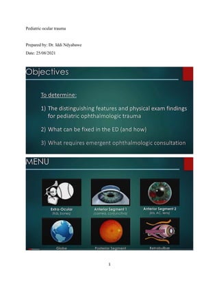
  • 2. 2 We broke down ocular injuries into the areas that are getting hurt.
  • 3. 3 Car barteries contain acid…. Acid injury Paint can cause chemical injury.. paintball pellets implicated here. Some places in Uganda with paintball game services; Kamooflage Paintball, Adonai adventure Park, Forest Park Paintball Arena. Outside Kampala, in Busiika there is Extreme Adventure Park.
  • 4. 4 We shall start with the extraocular injuries… specifically the eyelid lacerations. This is a dog bite injury. It’s a severe laceration. Often eyelid lacerations are very easily repaired by the emergency physician in the ER. Most important thing is to make sure that the globe is not involved.
  • 5. 5 “The average age of pediatric dog bite patients with ophthalmic involvement is 3 years.
  • 6. 6
  • 7. 7 Any type of trauma involving the canaliculi area needs to be explored in the operating room. This test assesses the canaliculi system.
  • 8. 8 You put a lot of fluorescein in that opening on the lid to allow some fluorescein to drain. And if it’s draining properly and with enough fluid, this should go down into the Nasolacrimal duct. And if you put a little swab into the nose for 10 seconds, you should be able to see that fluorescein uptake on the swab using regular UV light. Other extra ocular injuries we shall talk about are orbital fractures. Orbital trauma is amongst the most common emergency department consultations and is often accompanied by trauma of the facial bones and soft tissue. • Additional injuries that may be observed in the setting of orbital trauma include orbital hemorrhage, retained foreign body, and optic neuropathy. • Ophthalmic manifestations of orbital trauma may include decreased vision, intraocular injury, strabismus, and eyelid or globe malposition. • An ophthalmic examination should be performed on all patients who have sustained orbital trauma as ocular injuries may also be present So, here is a nice diagram of a classic baseball to the eye causing a blow-out fracture. So you see it… the globe is compressed a little bit… ultimately you are putting pressure to the surrounding structures and it’s often this infraorbital ridge and the floor that breaks. This is an orbital floor fracture that you can see here. The globe gets pushed back, provides extra pressure that pushes down and …. This is the classic orbital floor fracture.
  • 9. 9 Le Fort fractures (lateral view). Note that all the fractures extend posteriorly through the pterygoid plates (arrow).
  • 10. 10 Le Fort classification of midfacial fractures. Le Fort I, horizontal fracture of the maxilla, also known as Guérin fracture. Le Fort II, pyramidal fracture of the maxilla. Le Fort III, craniofacial disjunction. This is a nice aaaah coronal view here… aaaah… where you can see pretty well,, I think these are easiest to see on coronal view … aaamhh… you can see the nice disruption and the leaking of fat or blood down… but something is leaking down to the maxillary sinus through this orbital floor.
  • 11. 11 One of the characteristic features of the orbital floor fractures clinically is that if there is true muscle entrapment. Here is a nice photo of a child who Is asked to look up and … normal left eye can look up very well… but the right affected eye can’t look up. This is entrapment of inferior rectus muscle innervated by CN 3.
  • 12. 12 It can sometimes affect the infraorbital nerve where the kids will have decreased sensation on the upper lip or that kind of nasolacrimal area. In a patient with limited elevation, a positive forced-duction test indicates the presence of restriction. Bradycardia, heart block, nausea, or syncope can occur as a vagal response to entrapment. A special presentation, the white-eyed blowout fracture, is characterized by marked restriction (in both directions) of vertical ocular motility despite minimal signs of softtissue injury. This restriction is due to entrapment of the inferior rectus muscle either beneath a trapdoor fracture or, in the case of children, in a linear opening caused by flexion deformity of the floor. Early surgery, rather than observation, is required in order to minimize permanent muscle and nerve damage. This is a zygomatic fracture.. usually lateral…not to many complications associated with it. The one on the right shows aaa is an axial ct view and it's a nasal fracture so the nasal ethmoid is a little more of a supporting structure and it can be more cosmetic as it can cause deformities of the nose and septal deviation so it's associated with aesthetic impairment clinically you can see a widened intracanthal distance, enophthalmos are some of the other clinical features. When do you plan surgical repair of orbital floor fractures?
  • 13. 13 -When the patient has diplopia nd CT evidence of muscle entrapment plus unresolving Oculo cardiac reflex. This is an indication for immediate surgery. Generally happens in children. Otherwise early surgery is done at 2 weeks following initiation of antibiotics and steroids. -Orbital fracture > 50% of orbital floor involved. -Enophthalmos > 2mm -Hypoglobus >2mm -Infraorbital nerve hypoesthesia. Principle of orbital floor fracture repair: -Release of entrapment -Placement of a barrier -Repair of hypoglobus and enophthalmos less commonly you see orbital roof fractures so this is you know this is again aaaa nice is aaaaa coronal ct and you can see on this right image that there is a shatter to the roof of that orbit the danger of these is that you know you can have communication with the CSF so it can be a predisposing feature to cause a you know invasive CNS infection the refreshers usually occur in
  • 14. 14 kids less than 10 because the adult pneumatized frontal sinuses usually dissipate energy to the orbital rim rather than this roof so we usually see it in younger kids you really want to look for other signs of intracranial injury probably should extend a CT to include the the entire the head to make sure there's not bleeding other signs and then we look clinically for CSF leak some places that may or may not include neurosurgery or just follow up or close evaluation. What do you look for on orbital CT scan? -Bone windows -Position and integrity of obirtal floor -Orbital walls -Optic nerve -Optic -Foreign bodies What do you look for on the orbital floor? -Tear drop sign When the rim remains intact, this is termed blowout fracture.
  • 15. 15 so in general with orbital fractures if there's no globe rupture complex laceration signs of intracranial disease or CSF leak usually the follow-up is with an ophthalmologist in about 24 hours if there is muscle entrapment, enophthalmos or orbital dystopia you know certainly those are um you know other reasons to do this quickly otherwise i could often wait for a week so the early intervention for most orbital fractures is is not that helpful unless you get those other signs of entrapment and dystopia. some folks consider antibiotics there's not great evidence for this but if there's substantial sinus involvement or underlying sinusitis you know the potential for spread is there you can consider adding oral antibiotics um you can also consider oral corticosteroids if there's nerve entrapment but that probably should be done in consult with an ophthalmologist all right so that's the extra ocular tissues…
  • 16. 16 now let's talk about the anterior segment and break this down into sort of sections one and two the first is the cornea and the conjunctiva and the second is really the anterior chamber and the iris in the lens so let's talk about conjunctival injuries so… we see a lot of conjunctival foreign bodies you know here's just a nice simple piece of debris you know here is an arrow injury i guess that's not
  • 17. 17 really a foreign body it's just penetration through the lid but i think the point here is that you only know if there is a true foreign body if you can effectively evert the lid so this shows two techniques of just grabbing the lid and inverting around your finger so retraction is important there's a few different methods you know you can open manually from the superior and inferior orbital ribs where you're basically just stretching the area like it's shown in the figure on the right and then this sometimes can help easily just for simple things like dust dirt sands to make sure if the contact's there or not.
  • 18. 18 sometimes you need to do a better true retraction reversion to really if there's that sensation that's there and you need to look a little closer um this is using the cotton swab technique where you can put a cotton swab you know at the bridge of of the upper orbital ridge and then roll the eyelid over it to allow that true eversion of the lid this is an effective technique there's other if you have these available things like lid retractors that can help open your area this is often helpful especially if you want to have a better view of the sclera
  • 19. 19 there's a few other type of retractors that may be in a ophthalmology kit you have you know such as this spring type one the the resource low method is to use paperclips i've absolutely used this and it absolutely is effective if the parents will let you do it they don't think you're crazy coming at them with a paperclip or a set of paperclips like is shown here but you can fold the edges of a paper stuff so the non-sharp edges create a nice retractor and it's sometimes a nice way just to open up your lids in a area where you might not have better tools
  • 20. 20 all right so when talking about the conjunctiva we're going to spread into the sub-conjunctival area and this is a example of a sub-conjunctival hemorrhage and this is a very expensive one this isn't really the typical one we see you can see these from non-trauma like a coughing illness that can cause uh small subconscious hemorrhages and you can also see them from blunt trauma but i think the important thing to know is when it's extensive or circumferential or you see going all the way around um in the the the pupillary area um and around the iris then especially filling the the cornea like it's shown here you definitely need to evaluate for globe rupture so you need to ensure that the globe is intact in general some subconjunctival hemorrhages shouldn't affect vision and it should be asymptomatic other than looking terrible like this poor eye does but it does take a little while to go away like any soft tissue contusion where there's a hematoma it's similar and it often takes 10-14 days to resolve.
  • 21. 21 Corneal abrasion is one of the most common ocular injuries in children and adults. corneal abrasions we certainly see commonly from infants to adults that there are many reasons and many in situations where kids can scratch their eyes so the a better term for this is probably a corneal epithelial defect that you're creating some type of penetration of the epithelium of the cornea and this is a superficial injury when we call it an abrasion. when we talk about ulceration that's a deeper injury so these generally don't cause scarring because the bowman's membrane if you remember uh epidermal if our dermatology pathology of the eye and some of the different layers but if we're not breaking down the bowman's membrane um usually these can heal very normally without any long-term effects uh corn abrasions can have a lot of different patterns so it's important when you're looking at the abrasion to help to help look into what the potential causes
  • 22. 22 you know sometimes the what's shown here in the bottom right is it kind of that's scratching and that's sometimes when there's something trapped in the under lid that's scratching up and down versus just a simple kind of blunt injury like a horizontal injury like it's shown here on the top right um there's a lot of misconceptions in um there's a lot of misconceptions in coronal abrasion you do not need to pass it's never really been shown benefit of that in general accommodation can make it hurt so ways to prevent accommodation can be helpful but true patching really isn't topical anesthetics are not a good idea even though many of us use prepare cane or similar for the evaluation it's not something the patient should go home with because there is evidence that that will delay wound healing and topical steroids shouldn't be necessary because these do heal on their own so the steroid can only promote infection and may be a predisposing factor for infection like ulceration
  • 23. 23 Topical cycloplegic drops and antibiotic ointment may help reduce discomfort and risk of infection, respectively. Traumatic corneal epithelial defects usually heal within 1-2 days. uh in the emergency department or the urgent care center that you're working in the corner abrasion should be managed with uh make sure tetanus is up to date exclude a corneal foreign body or something that's causing it uh and then everything else is home management so uh cycloplegia should be considered if the patient's having a lot of discomfort as a way to block that accommodation reflex which often causes the pain when they're accommodating to light topical antibiotics should be used absolutely until symptom free and topical NSAIDS are another pain medicine that you can consider when patients are having a lot of discomfort. topical antibiotics you know oftentimes our ophthalmologist will recommend a drop during the day and an ointment at night because that ointment will add a little extra lubrication which can be soothing at night and but i think both methods are effective in preventing progression to ulceration. Cigarette burns of the cornea are the most common thermal injuries to the ocular surface in childhood. Usually, these occur in toddlers and are accidental, not manifestations of abuse. These burns result from the child running into a cigarette held at eye level by an adult. Despite the alarming initial white appearance of coagulated corneal epithelium, cigarette burns typically heal in a few days and without scarring. Treatment is the same as for mechanical abrasions
  • 24. 24 so here's a a photo of that epithelium where you can see this is the the stroma of the eye and this is the outer epithelium so when it is a deep penetration it can penetrate that bowman's membrane and that's when we're talking about the the progression of an abrasion to an ulcer. so an ulcer is usually uh a progression of an abrasion and not necessarily an injury all at once uh and and then the science is so it's something that usually evolves over a little time and it's usually from an effective component so the signs that you might be doing more than in a corneal abrasion or when you see corneal infiltrates you i mean this is without a fluorescent lamp you
  • 25. 25 can see that whitening around it and those are infiltrates into the cornea or surrounding stromal edema i i think those are signs you're dealing with something a little more significant than a simple abrasion certainly if you see things like purulent discharge a particular fluorescence without even using fluorescein which can be common in pseudomonas um or if you have that persistent defect that doesn't go away and you know the signs that it's getting worse there's your signs of ulceration this is an example of an ulcer you can see that stromal defect and the white infiltrates that even without any type of fluorescein intervention if you can see evidence of injury that's that's a sign that it's more significant so when we're talking about corneal ulcers the management is more intensive corneal cultures should be obtained by an ophthalmologist broad spectrum antibiotics should be started.
  • 26. 26 corneal ulcers can as we're talking about infection uh develop other complications such as pasta can accumulate in the anterior chamber like you see in this photo so oftentimes in kids when we have these severe diseases and whether it's a high female or it's a corning ulcer when you need to use q1 hour eye drops that is one of the indications that we do hospitalize to make sure that it can get done because we know at home parents often especially if they have other kids you know cannot keep up with the necessary number of antibiotic prophylaxis to make this heal and prevent it from getting worse.
  • 27. 27 all right so the other corneal injury we commonly see is burns and these can be acid or base … base tend to be more devastating so the alkali injuries are more frequent more severe and penetrate deeper and almost all of these that we see in kids are caused by household products
  • 28. 28 so when we talk about alkali burns you know things like bleach we certainly talk about um bleach isn't that dangerous to swallow but it can be dangerous in the eye and most of the household products tend to be pretty dilute and mild but if we have more industrial products in the house or in the garage that's where it can get more dangerous so bleach in the eye is is usually pretty mild you know we talk eye is is usually pretty mild you know we talk about ammonia you know ammonia is more severe that's sometimes in refrigerants and household cleaners one of the most severe that we see is in drain cleaners and it's you know material called something called lye which is a sodium hydroxide and it's often inherited straighteners or drain cleaners that can be very dangerous and cause significant burns
  • 29. 29 um some other ones you'll see things like potassium hydroxide, magnesium hydroxide and then again here's lime which is in cement and plaster not to become supply all those can cause significant injury
  • 30. 30 so when we're talking about corneal burns you can also have acids and acids usually they usually look worse initially but they're not as dangerous and they can denature corneal proteins opacifying the cornea but providing but preventing the deep penetration that we see with bases.
  • 31. 31 so you know in battery explosions and things like that that's usually acid so some explosives again the eye tend to be more acidic burnt um simple common things like acetic acid are very mild um some of the more significant ones that we see are you know rust removers even gasoline that have hydrofluoric acid can be more significant here's hydrochloric acid which is sometimes found in pool cleaners and household cleaners.
  • 32. 32 chromic acid and certain glazings sulfuric acid which we talked about in batteries um and sulfurous acid which is in some refrigerants though so you know before we had freon and some other materials so some of these acids can be dangerous as well
  • 33. 33 if you ever want to look to see what it is and what's in it there you know it's a great database in the department of health and human services household products where you can look up particular material and learn a little bit more about its potential danger and also calling the poison control center can be helpful.
  • 34. 34 when we're talking about burns we do try to grade burns in a grade of one through four on how much corneal injury there is but i think any corneal burn needs an evaluation by an ophthalmologist emergently um you know the initial management we'll talk about a second but here's a grade one burn but there's really not much corneal opacity or limbal ischemia and then as you progress a grade two shows that the cornea is hazy but the iris details are still intact uh moving along that's a slit lamp that's lit lighting up through it but you can see the cornea is increasingly hazy there's epithelial law of stromal haze and then progression where you you can't distinguish the iris of the people so there's not that uh that fine differentiation between those two structures because the cornea is completely pacified.
  • 35. 35 subacutely so usually you know a week or two later you start to see the corneovascularization and the pacification that have alkali injuries and these are just the signs of devastating corneal burns and what happens in the healing process over the next few weeks. for us in the emergency department or urgent care center we like to use uh irrigation so it's important to irrigate irritants for about 30 minutes if not more irrigating with alkali or alkali or acid burns often take one to two liters per eye it's important to measure the ph of the cul-de-sac before and during and after irrigation you know the goal should be to have to continue irrigating
  • 36. 36 until the ph is between seven and a half and eight after copious irrigation sometimes helps to breed some of the necrotic coral epithelium and the methods of irrigation there certainly are different ways to do it if i'm very hard on a young toddler but this is a example of the morgan lens irrigation system which i think is a popular one and it usually helps the they show using a prepare cane or similar topical anaesthetic having the patient look down inserting and then providing some type of fluid collection or towels so that when a liter of fluid is put in that eye that it's a place to irrigate to and go
  • 37. 37 all right so that's burns now let's talk about corneal foreign bodies we mentioned a little before about conjunctival foreign bodies and inverting the lids certainly you can sometimes get corneal foreign bodies where some and it's usually an industrial worker where something fires up maybe a small piece of metal a small piece of debris and it just gets stuck on the cornea very difficult for people to remove themselves so it's often metal or rusty objects.
  • 38. 38 you know using a bevel needle edge or a magnet can be very helpful it's very difficult to come in a patient with a needle toward their eye and so you're going to touch their eye open their eye will move still i find this particularly difficult in older people that we can use a split lamp i think it's obviously a much better uh much better evaluation mechanism but I think depending on your setting uh certainly i do try to get these out or at least you know do my my best bet to try to you know take them off the corner either with a swab or a small magnet or smugly bevel needle before phoning a friend or an ophthalmologist if i'm unable to or if it's a very young child. uh for you know sometimes you can get rust rings so it looks like it's sitting on the cornea but it's actually the uh it's it's corn it's metallic corneal foreign bodies from iron oxidation and it forms a little rust ring like shown in the picture so that usually requires the aid of a slit lamp and a burr drill to help get that out and that usually requires our friends in ophthalmology.
  • 39. 39 all right so that's anterior segment one where we talked about the cornea and conjunctiva let's talk about the other aspects of the anterior segment so let's talk about the iris and i think one of the ones we commonly see and it's sometimes misdiagnosed is traumatic iritis.
  • 40. 40 so this is a sequelae of blunt trauma and where you get inflammation of the iris and the ciliary body and i think this kind of falls within the classification of uveitis when people get red eyes and eye pain with sometimes irregular bordered irises and that could be like a rheumatologic or infectious phenomenon when we see it in the setting of trauma usually especially a few days later you know this is what we call traumatic iritis when you look on a slit lamp you can see those inflammatory cells sitting in the anterior chamber unlike the corneal abrasion the pain isn't relieved by a topical anesthetic and if you have a really bad corneal abrasion i think we we know that when you put a few drops of a proparacaine it's a it's just it's a new it's a new kid and the the that pain just gets rapidly taken away so when you have traumatic i write it that just doesn't happen um this usually occurs within three days of an injury so it's not something that we see right away or it's very uncommon too it's usually you know one to four days later where you get increasing pain and redness and sometimes you get that iris irregularity like is shown here common causes racquetball baseball for example. the treatment is a consultation with an ophthalmologist doesn't have to be emergently it can be in the next one to two days they tend to use topical steroids and cyclopedics to prevent the accommodation reflux all right so here's an example of an iris tear so as opposed to iris inflammation the iris can tear from trauma so this isn't a penetrating drum it's usually a blunt trauma that increases intraocular pressure it expands the globe and it causes a small tear so this resultant stretch can tear the concentric eye rings and cause a disinsertion of the iris root which is a separation of the iris and the ciliary body this is a nice photo here with the arrows showing the area that we're talking about you know in general this is not a globe rupture or an open globe it's an example of an iris tear that needs to be followed slowly for evidence of increased intraocular pressure so you know we're following to make sure this doesn't develop into glaucoma you know or you know other you know potentially devastating diseases from increased pressure
  • 41. 41 all right so then we know you can get kids can get a laceration lacerations and we talk about illustration to the lids you can also get lacerations to the cornea and the sclera and the first step is just to make sure this isn't a penetration that can cause globe rupture um but you know like in these two pictures the one on the left showing the corner on the right really showing sclera that these lacerations sometimes do need to be repaired but may not be signs of globe rupture so doing a ct scan looking for foreign bodies looking for other signs that there's not a rupture of the globe is the first step but if they're more than two millimeters they often require antibiotics and suturing.
  • 42. 42 all right here is a great picture of your hyphema certainly one of the things i look for in the setting of blunt eye injury and so it's where blood is layering in the anterior chamber this occurs from bleeding from the iris or the ciliary body. hyphema can be graded on basically how much blood is accumulating in the anterior chamber. so this is grade one where it's less than a third of the anterior chamber grade two or it's a third to a half grade three where it's you know pushing more than half of the the uh the ac and then a grade four which we sometimes call an eight ball or a black ball which is basically where the entire anterior chamber is covered with blood if you look at that eye on the right it's highly suspicious for a rupture there's a circumferential subconjuctival hemorrhage and there's blood
  • 43. 43 completely opacifying the anterior chamber but in that case that actually was not a global rupture it was just a 100 percent hyphen so the er management of a hyphema is it really is uhhhh it's it's a little different between some centers just on how conservative folks want to be but the risk is these mostly is to watch the intraocular pressure and watch for re-bleeding so the emergency management is anything you can do to decrease the IOP is helpful so you would elevate the edge of the bed use antiemetics like zofran or you know similar to prevent wretching and things that are increasing chocolate pressure um decreasing ciliary spasms so we do this you either can use cycloplegics you know like atropine um or an eye shield and i feel like I mean anything that's it's preventing that accommodation to light reflex that can cause ciliary spasm can cause re-bleeding or make it worse avoid agents that can contribute to re-bleeding so things like aspirin and NSAIDS. and then consider adjunctive meds so anti-fiber analytics miotics to enhance reabsorption of the blood and i think those are definitely done in consultation with ophthalmology you know in kids you know probably perhaps more so in adults we do consider admission mostly to observe for re- bleeding and really in kids we do it to decrease stimuli in activity because you know a five-year- old no matter how much we say you know bed rest you know we know that's just impossible so sometimes we will admit children more so than we do adults to watch for re-bleeding make sure we're keeping stimulation too low make sure there's compliance with things like eye shields and and ways to decrease cellular spasm and then also to measure the increasing the intraocular pressure.
  • 44. 44 um okay so that's hyphema and let's talk a little bit about lens dislocation so lens dislocation can occur from trauma we also you know certainly have heard of this as something that occurs spontaneously specifically with certain systemic diseases like the ones mentioned below marfans syndrome ehlers-danlos homocystinuria etc but in general you can usually see this pretty clearly um in the pupil as a dislocation of that lens and often see the edge you can also sometimes see it on a ct scan where you can see separation if there's in the setting of a significant trauma.
  • 45. 45 all right so that's finishing the anterior segment let's talk about globe. and globe open globe and global rupture so this is a you know some of the terminology we use is a globe laceration which is a penetrating or perforating injury and we can differentiate that from a globe rupture which we tend to use more where there's a blunt eye injury and then uh the eye opens at the site of the greatest structural weakness so it's important to differentiate a little between the two whether the injury is caused by something going into the eye or a blunt injury causing a the weakest point of the eye to open.
  • 46. 46 but all these are under the category of open globe when we talk about penetrating globe injuries um you can either see a corectopia which is a displacement of the pupil or this peak pupil on a peak pupil is an example of a penetration that will develop that will cause a corneal scleral defect and the photo remember we look before a traumatic iritis it really showed a very irregular pupil this is a very distinct area where there's a defect here causing the defect in the iris and the the corneal skull defect in the iris that causes this peak pupil appearance and that's an example of a penetrating globe injury.
  • 47. 47 certainly when you see this avulsion this is so this is a perforation of the cornea showing iris tinting and you can see this peak pupil abnormally shaped with with a vols tissue is another sign of a globe rupture again here is a limbus tear a prolapse of the iris that's coming out you know all these examples of corneal scleral defects iris tears are examples of perforating or penetrating globe injury in open globe . you know here's another nice one so a nice peak people so when i'm evaluating eye injuries specifically blunt injuries i mean two of the most important to me are evaluating for hyphema and then the shape of the pupil are some of the things because you would that you want to do to exclude any uh signs of open globe ……… all right so globe rupture how do i know and i think this is the tricky thing especially when you're in a low resource area that you know some of the subtle signs are if you have a relative afferent pupillary defect uh so a significant trauma where there really just isn't the pupillary accommodation you expect if you have an eccentric pupil like we talked about i mean i think to me that's really saying that a peak pupil increase anterior chamber depth which is really what you need to see with the split lamp extrusion of the vitreous prolapse of the iris or the ciliary body tempting at the puncture site a low intraocular pressure or we talked about before that circumferential subconscious subconjunctival hemorrhage.
  • 48. 48 if you've ever had a seidel test this is pretty cool and you can watch this animation but this shows slow leaking of fluorescein through the open globe so if you if you put fluorescein in the eye and you see this continued leaking of fluid um that's an example of a potential open globe so this usually requires a lot of fluorescein but it shouldn't be a dynamic process so if you see something dynamic that's a suggestion that the fluorescent is traveling and it's an open globe.
  • 49. 49 globe rupture is usually not subtle uh you can see these photos here or you gotta i mean they they you know the biggest feature to me is that soft globe instead of the hard round globe it just immediately kind of deflates like a balloon here you see bullets some gun sub-conjunctival hemorrhages and here you see that kind of proptotic soft globe and the iris is obscured and there's a lot of blood so those are sort of not subtle signs. ct imaging definitely helps you can compare the nice round contour to the loss of contour on the right um the extrusion of the lens sometimes can be a sign so here not only do you see that soft glow but the lens is dislocated and out of place those are sometimes things you can see on CT.
  • 50. 50 open glow the management in the er similar to when we talked about hyphema you know we want to decrease the IOPS so anti-medics and upright band bed i think this is a painful injury so narcotic pain control is important because it's an open and open area that is subject to infection using iv antibiotics is important an eye shield i think is is a great method to prevent that accommodation here's some nice photos of different examples of kids in general i think the ones like this are actually a little better because if you hit this it still doesn't cause any pressure to the eye this one that's very close to the eye if someone puts pressure on it they're still pushing on the eye so ones like this even though they might look ridiculous or they come out a little protrusion and they actually are very effective so this is the styrofoam cup method like we talked about doing a CT to help you know identify and make sure there is a foreign body also to identify the globe contour but also to look for foreign body um tetanus prophylaxis like any open wound and then NPO status for operative correction and obviously an emergent Ophthalmology consult.
  • 51. 51 okay so here is an example of glass and a orbital foreign body so you know we can have a penetrating injury where the globe appears to be intact and one of the rules of the CT is to look for those foreign bodies so here is gun powder that sits in the orbit here's glass that fits in the orbit in general you know even though this can occur through a very small penetrating opening if it's not the the blunt injuries that cause a true rupture sometimes more dangerous than the ones where there's a slight penetration uh and a foreign body accumulation because sometimes you like when a bullet is embedded in the soft tissue there are certain situations where small bits can stay in the orbit and some start staying in the globe so we sometimes will remove uh when we we sometimes will not remove certain form bodies depending what they are but obviously it should be in consultation with an ophthalmologist.
  • 52. 52 okay so let's move on to the posterior segment i think this is probably the most difficult for most era providers because you need to have fairly good visualization of the posterior segment and split lamp examination skills to be able to really see that posterior segment.
  • 53. 53 so this obvious but i think ultimately the posterior segment injuries are never hyper acute and these are usually things that develop with time and not something you see necessarily on day zero of injury so the the posterior segment injuries are not the emergent injuries that we've talked about like open globe and globe rupture um here's an example when we talk about posterior segment injuries there's a few categories one's a choroidal rupture the other is a vitreous hemorrhage so you know these are very nice pictures that we won't necessarily see with our limited endoscopic exams and the acute setting but then here's a choroidal rupture where you see concentric streaks around the optic nerve here's that vitreous hemorrhage where you see similar features and these are all areas of you know the posterior segment and the back of the retina getting stretched or bleeding that's preventing um some of the visualization that's why the the primary symptom of posterior segment injury is is vision and changes in your vision so this usually occurs in blunt trauma vision loss and floaters are really what most patients will complain about so it often doesn't occur the day of it could be several days or even weeks later where as as that hemorrhage occurs or some swelling occurs in that area that patients can see floaters they can have just some minor vision loss in that area very hard to assess a very young child but an older child will tell you that this is going on i think the other classic classically symptomatic the classic symptom is that these are often painless so as opposed to some of these other things like traumatic iritis now this is a painless injury where it's mostly a vision component and then on your exam you have a normal ac a normal pupil and a normal cornea and that's when you suspect posterior segment. um the most significant is the retinal detachment and this obviously is one of the important reasons that ophthalmology needs to be followed up so floaters flashes of light photo which is
  • 54. 54 you know what we call photopsias uh having a visual field defect along with no pain and no red eye you know are some of the symptoms of potential retinal detachment like we talked about in the posterior segment so you can have a dulling of the red reflux or a relative afferent pupillary defect those are some other more subtle signs ultimately you know if we're suspecting posterior segment injury ophthalmology and one of the helpful tools is doing a b scan ultrasound to help look for that detachment especially if there isn't an obvious hemorrhage or other source or if a hemorrhage is blocking the ability to fully visualize the retina this is vision threatening and it may require surgical corrections so this is you know on the the spectrum of the potentially evolving emergent conditions in eye trauma although it's not one of the ones you see on day zero. all right so the last category is retrobulbar injury.
  • 55. 55 so retro bulbar we're talking about areas behind the globe and in the orbit so the first is traumatic optic neuritis again this is a blunt nerve injury it's usually a little later than a traumatic iritis so it's usually over three to six weeks following a injury and the optic nerve appears normally acutely but swells over three to six weeks following a injury and the optic nerve appears normally acutely but swells over three to six weeks so some of the presenting features include decreased visual acuity and visual loss abnormalities also loss of color vision is is i think one of the big distinguishing factors so when you have sort of that longer time period and the posterior segment looks normal that's when we start to suspect some injury behind the eye to the optic nerve.
  • 56. 56 when we're talking about emergencies you know this is really the one at the top of the list so i think open globes hyphemas are really and we're prioritizing some of these injuries and retro bulbar hematomas are some of the ones that are potentially the most devastating the most acute so retrobulbar hematomas is a nice photo showing a lot of blood accumulating behind the eye causing increased pressure and increased intraocular pressure subsequently which can be vision threatening so compression of the optic nerve compresses the vascular compartment that's why we sometimes terminal term we sometimes use the term orbital compartment syndrome for a retro bulb or hematoma this can cause irreversible vision loss and can develop in 90 minutes so it is an ophthalmologic emergency. there's a nice kind of sagittal view where you can see the globe and the optic nerve and a retrobulbar hemorrhage from trauma blood can accumulate in this orbital space.
  • 57. 57 so the treatment for this is something called lateral canthotomy this is something that is in the wheelhouse of the emergency procedures and it's something people should be aware of that if you're in a resource low area that it's a time-sensitive condition and it may be something that's very important to do so a canthotomy is where you cut all tissue layers along the lateral canthal fold so this is using a small scissors and you're basically trying to make an incision to allow blood to get out to be able to relieve the pressure behind the eye okay so cantholysis is where you cut the lateral campus tendon and that's by doing that that's so that allows the blood to come out so you make an incision to extend that that uh that eye opening area you cut the tendon.
  • 58. 58 you have access to the inside of the retrobulbar area and then blood can drain. so this is an example it's the opposite eye but an incision is made along the edge and then blood is draining and it relieves the pressure so this is absolutely indicated when there's an acute loss of vision increased IOP there's proptosis if the IOP is greater than 40 an unconscious patient um it's relatively contraindicated if there's suspected globe rupture you know this is where your globe is intact and you're trying to protect the optic nerve so if you can consult ophthalmology that's
  • 59. 59 certainly the way to go but just remember this is a time-sensitive condition and if you're in a resource poor area then a lateral canthotomy might be vision saving. all right so that's all the layers of the eye just in summary uh when we prioritize eye injuries they're the ones that are immediate and time sensitive the alkali burns and the retrobulbar hematomas you have to act now i think there's the emergencies like the open globes the high females the complex eye lacerations the corneal ulcers that may require admission and advanced therapy but don't have that sort of 60 minute window like the uh the immediate conditions do and then all the other ones i think the traumatic iritis some of the posterior segment injuries um are injuries that can develop over the you know the days to weeks that follow a blunt eye injury that need to be followed closely this is a great website iwiki put together by a host of ophthalmologists that has a lot of nice information on eye trauma and it's a free open wiki source that uh i think was written in conjunction with the eye physicians insurgents.
  • 60. 60 Although most eye injuries in childhood are accidental or innocently caused by other children, a significant portion of them result from physical abuse by adults. The terms used for intentional physical abuse of a child include nonaccidental trauma and child abuse. Child abuse includes emotional abuse, sexual abuse, and neglect as well as physical abuse.
  • 61. 61 A reliable history is often difficult to obtain when nonaccidental trauma has occurred. Suspicion of nonaccidental trauma should be aroused when repeated accounts of the circumstances of injury or histories obtained from different individuals are inconsistent or when the events described do not correlate with the injuries (eg, bruises on multiple aspects of the head after a fall) or with the child's developmental level (eg, a 1-month-old rolling off a bed or a 4-month-old climbing out of a high chair). Any physician who suspects child abuse is required by law in every Ugandan district to report the incident to a designated governmental agency. Once this obligation has been discharged, full investigation of the situation by appropriate specialists and authorities is usually performed. Physicians should be familiar with the regulations in their own country. If possible, ocular abnormalities should be documented photographically or with a detailed drawing to use as evidence in court.
  • 62. 62
  • 63. 63 Definition: A unique complex of ocular, intracranial, and sometimes other injuries occur in infants who have been abused by violent shaking. This is recognized as one of the most important manifestations of child abuse. Patients with AHT are usually younger than 5 years and most often younger than 12 months. When a reliable history is available, it typically involves a parent or other caregiver who shook an inconsolably crying baby in anger or frustration. Often, however, the only information provided is that the child's mental status deteriorated or that a seizure or respiratory difficulty developed. The involved caregiver may relate that an episode of relatively minor trauma occurred, such as a fall from a bed. Even without a supporting history, the diagnosis of AHT can still be made with confidence on the basis of characteristic clinical findings. answers to important questions concerning the timing and circumstances of injury and the identity of the perpetrator frequently cannot be inferred from medical evidence alone. Intracranial injury in AHT frequently includes subdural hematoma (typically bilateral over the cerebral convexities or in the interhemispheric fissure) and subarachnoid hemorrhage. Displacement of the brain in relation to the skull and dura mater ruptures bridging vessels, and compression against the cranial bones produces further damage. Neuroimaging may also acutely show intracranial edema, ischemia, or contusion and in later stages atrophy. These findings are thought to result from repetitive abrupt deceleration of the child's head as it whiplashes back and forth during the shaking episode. The infant's head is
  • 64. 64 particularly vulnerable to the effects of repeated acceleration-deceleration because of its relatively large mass in relation to the body and poor stabilization by neck muscles. Some authorities, citing the frequency with which patients with AHT also show evidence of having received blows to the head, think that impact is an essential component, although in many cases no sign of impact is found.
  • 65. 65
  • 66. 66
  • 67. 67
  • 68. 68
  • 69. 69
  • 71. 71