2. H. Duzkale et al.
cardiomyopathy from 5 to 46 genes in our laboratory
resulted in a threefold increase in clinical sensitivity
but an even more dramatic increase in inconclusive
cases, many with multiple VUSs. In addition, exome
and genome sequencing tests add a new layer of com-
plexity, as the genes interrogated may not have been
carefully assessed for their role in disease until variants
are identified.
Although molecular genetic testing has a unique
place in the diagnosis, management, and prevention
of genetic disorders, the field is compromised by the
absence of a standard, comprehensive, and efficient
variant assessment protocol approved and shared by
the community. However, guidelines for variant inter-
pretation are available and being updated as variant-
level knowledge expands, including those from the
American College of Medical Genetics and Genomics
(ACMG) (4–7). To supplement these guidelines and
capture the evolving state of the field, we developed a
variant assessment tool (VAT) that systematically evalu-
ates multiple parameters for each variant and facilitates
the capture of new knowledge in the literature and
databases (Appendix S1).
The clinical significance of a variant in relation to a
disease or phenotype can be determined by answering
three core questions. (i) Does the variant alter the
function of the gene [i.e. loss-of-function (LOF) or
gain-of-function (GOF)]? (ii) Can the functional change
result in disease or another phenotype? (iii) Is the
associated disease or phenotype relevant to the specific
clinical condition present in the tested individual? In
some cases variant assessment in a clinical laboratory
may only be focused on the first two questions;
however, for maximal benefit to the patient, a careful
assessment of the third dimension can be highly
informative, particularly for VUSs. Here, we share
our decade-long experience with variant assessment,
highlighting key points and challenges of clinical inter-
pretation. We have evaluated 245 genes associated with
53 diseases while testing greater than 22,000 cases. We
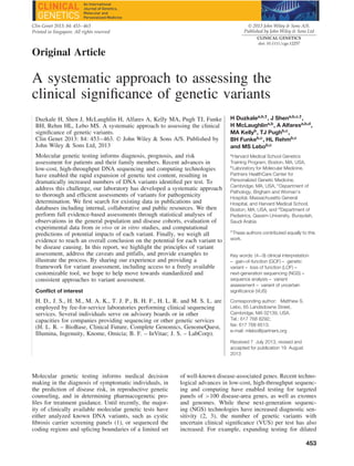
Fig. 1. Variant assessment workflow. Genetic variants identified by laboratory testing are annotated with information from various sources including
publications, computational prediction algorithms, and public, collaborative and internal databases. After evaluation of all pertinent information in
conjunction with patient specific clinical and family information, a professionally trained individual will classify the variant into one of the five
clinical categories and combine all variants for a clinical report.
have iteratively developed a framework through clinical
assessments of over 17,000 variants, including >8000
that have been validated and reported in patients. Using
our semi-automated tool, it takes on average 40 min
to perform a thorough evidence-based clinical variant
assessment for variants being returned after disease-
targeted testing. When assessments with literature are
excluded, the average time decreases to 22 min. An
overview of the process is presented in Fig. 1 and useful
online resources are presented in Table 1. In addition,
the approaches described below refer to the provided
VAT with corresponding SOP available for download
through Appendices S1–S3.
Linking genes to disease
As a first step in variant assessment, it is necessary
to determine which disease phenotypes are associated
with a gene and which types of variation may result
in clinically relevant consequences. This includes the
types of variants that are known to cause disease
in the gene (truncating/LOF, non-truncating, etc.), the
inheritance patterns observed for variants in the gene,
the protein domains that are implicated in disease, and
any genotype–phenotype correlations described.
To characterize disease phenotypes, it is important
to review the literature for common clinical features as
well as phenotypic variation among affected individ-
uals. Large cohort studies may provide expressivity,
age-of-onset, penetrance, and prevalence information,
while detailed reports of families with multiple affected
individuals help determine the mode of inheritance
and strength of association. Comparison of the variant
spectrum in affected individuals against that in the
general population may be useful in identifying the
types of mutations that are disease-causing. For
instance, heterozygous LOF variants in MYBPC3
have been reported in 14% (311/2302) of patients
with hypertrophic cardiomyopathy (HCM) tested
in our laboratory, but in <0.1% (6/6500) of the
454
3. A systematic approach to assessing variant clinical significance
Table 1. Useful online resources for variant assessment
Usage Online tools Url
Computational
prediction for
missense variants
Align GVGD (42) http://agvgd.iarc.fr/agvgd_input.php/
CONDEL http://bg.upf.edu/condel/analysis/
MutationAssessor http://mutationassessor.org/
MutationTaster http://www.mutationtaster.org/
PolyPhen2 (43) http://genetics.bwh.harvard.edu/pph2/
SIFT (44) http://sift.jcvi.org/
Computational
prediction for
splicing variants
GeneSplicer http://ccb.jhu.edu/software/genesplicer/
Human Splicing Finder http://www.umd.be/HSF/
MaxEntScan http://genes.mit.edu/burgelab/maxent/Xmaxentscan_scoreseq.html
NNSplice http://www.fruitfly.org/seq_tools/splice.html
Disease curation GeneReviews http://www.ncbi.nlm.nih.gov/books/NBK1116/
OMIM http://omim.org/
Domain database NCBI conserved domain
database
http://www.ncbi.nlm.nih.gov/Structure/cdd/wrpsb.cgi
Genome Browser Ensembl http://www.ensembl.org/index.html
UCSC Genome Browser http://genome.ucsc.edu/
Literature database PubMed http://www.ncbi.nlm.nih.gov/pubmed
Variant database 1000 Genomes Project http://browser.1000genomes.org
ClinVar http://www.ncbi.nlm.nih.gov/clinvar/
dbSNP http://www.ncbi.nlm.nih.gov/projects/SNP/
Exome Variant Server (9) http://evs.gs.washington.edu/EVS/
HGMD http://www.hgmd.cf.ac.uk/ac/index.php
Variant validation HGVS nomenclature http://www.hgvs.org/mutnomen/
Mutalyzer https://mutalyzer.nl/
general population per the NHLBI Exome Sequenc-
ing Project (ESP), supporting that LOF MYBPC3
variants are a common mechanism in HCM (8).
However, external information must be carefully
vetted. An apparent frameshift variant in MYBPC3
NM_000256:c.2854_2858del reported to occur in 7%
of the general population in ESP is likely a technical
artifact, as we have never observed it sequencing the
region by NGS and/or Sanger in over 2000 cases.
Different types of variants in the same gene may be
associated with distinct phenotypes or inheritance pat-
terns. For example, missense GOF variants in PTPN11
cause RASopathies, such as Noonan syndrome, whereas
LOF variants lead to an entirely different phenotype, a
cartilage tumor syndrome (metachondromatosis) char-
acterized by enchondromas and exostoses (9). Certain
missense variants in TECTA lead to autosomal dominant
hearing loss (10), whereas LOF variants result in auto-
somal recessive hearing loss (11). Similarly, variants in
different regions or domains of a gene may cause differ-
ent phenotypes (10, 12). Important questions to consider
when analyzing gene–disease associations and specific
variants within a gene can be found in Table 2.
Validating variants to ensure accuracy
As test complexity has increased, so has the need to
ensure variants identified and included on a clinical
report are technically accurate. This is especially
important for sequencing tests where the variants are
not part of a pre-defined list. It is essential to review
raw assay results (e.g. chromatographs of Sanger
sequencing traces or NGS reads) to verify the variants
and their nomenclature. Prior to variant assessment,
the laboratory should predefine the genome build,
gene name, and reference transcript that will be used
in interpretation and reporting, along with a method
linking genomic coordinates to cDNA and amino acid
level annotations. Laboratories should also be aware
of homologous and repetitive regions particularly
from pseudogenes and segmental duplications, which
may result in lack of coverage, alignment difficulties,
and incorrect variant calls. These steps will enable
validation of the correct variant call, zygosity and
nomenclature according to the Human Genome Vari-
ation Society (HGVS) guidelines (13). Validation
information is captured in the ‘Variant’ tab of the VAT.
Because standards for variant nomenclature have
only recently been widely adopted and still do not
address all modifications, variants may have differing
names in publications and databases. The amino acid
position may not be numbered according to the start
codon to be consistent with current recommendations.
For example, TTR variants were originally numbered
according to the position within the mature protein
lacking the 20 amino acid signal peptide (14). Partial
cloning of a gene may have led to inconsistent
nomenclature in early publications (15, 16). Nucleotide
gene numbering may have been determined using the
transcription start site instead of the translation start site,
455
4. H. Duzkale et al.
Table 2. Variant assessment checklist
Gene-level information
Confirm gene is implicated in disease with sufficient evidence, including human genetic data and functional data
Determine inheritance pattern, age-of-onset, penetrance and prevalence for each gene-disease association, if
possible
Determine types of disease-associated variants in gene (gain-of-function, loss-of-function, etc.)
Variant validation
Review raw sequence data to confirm the variant call
Determine zygosity of the variant
Associate genome build, genomic coordinate, and reference transcript to the variant
Confirm variant nomenclature
Genetic data
Determine frequency of variant in large population studies, parsed by race
Determine if population frequency is consistent with disease inheritance, age-of-onset, penetrance and prevalence
Evaluate whether variant segregates with disease in affected family members
If disease is inherited in a recessive manner, determine if the variant is found in trans with a pathogenic variant
If applicable, determine if there is a statistically significant difference in variant frequency between cases and
controls
Functional data
Evaluate available in vivo functional data
Confirm type of animal model is relevant for human disease
Evaluate available in vitro functional data
Confirm assays used reflect disease-associated cellular mechanisms
Computational data
Evaluate nucleotide alignment data and assess evolutionary conservation (for all variants)
Evaluate amino acid alignment data and assess evolutionary conservation (for missense variants)
Eliminate any poor species alignments
Determine if computational tools predict an effect on protein structure or splicing (for all variants)
which was particularly challenging given transcriptional
start site variability. Furthermore, for many small
insertions and deletions, it is not possible to determine
the exact location of the inserted or deleted base(s).
This can lead to multiple potential names for the
same variant, highlighting the importance for following
standard HGVS nomenclature rules such as attributing
alternations within a repetitive stretch to the most 3
possible position. Legacy terms and alternative aliases
are useful to maintain association with the correctly
named variant both to facilitate searching the literature
and databases, as well as communicating with ordering
physicians and other laboratories.
Genes may have multiple transcripts, some of
which are tissue-specific and associated with distinct
phenotypes. For example, the shorter USH1C transcript
(NM_005709) is expressed in both the retina and inner
ear, whereas the longer transcript (NM_153676) is
expressed exclusively in the inner ear (17). Accord-
ingly, variants in exons of shorter transcript lead to
Usher syndrome type 1C, characterized by profound
deafness, retinitis pigmentosa, and vestibular dys-
function, whereas variants in additional exons unique
to NM_153676 lead to non-syndromic hearing loss
(18). Variants should be reported according to a single
primary transcript. The reported reference is typically
the major transcript unless a more severe impact is
predicted on an alternative transcript, in which case the
variant should be defined according to the alternative
transcript, noting an alias to the primary transcript
(Fig. 2a).
When multiple variants in the same gene are
identified, the phase of the variants (i.e. on the
same chromosome – in cis – or on homologous
chromosomes – in trans) may influence the interpre-
tation, especially for autosomal recessive traits. If
variants are within the same NGS fragment, the phase
may be determined without parental samples (Fig. 2b).
Collecting evidence to determine the likelihood of
pathogenicity
Once the variant call is validated, literature, variant
databases, and population control studies should be
evaluated. This information is used to determine
whether and under what context the variant has been
previously observed. The population, literature and
internal case data are captured in the ‘Control_Freq’,
‘DB’ and ‘Publ+Internal_data’ tabs of the VAT.
Recent large-scale population studies such as the
NHLBI Exome Sequencing Project (9), the 1000
Genomes Project (19), the ClinSeq Project (20) and
others found in dbSNP (21) have catalogued large
amounts of sequence variation (Table 1). Because these
populations may include presymptomatic individuals
with late onset diseases, asymptomatic individuals
with low penetrance diseases or younger than typical
age-of-onset, and heterozygous carriers of recessive
456
5. A systematic approach to assessing variant clinical significance
Fig. 2. (a) Reference transcript selection. Two transcripts for OTOF are shown: NM_194248, the longest transcript selected as the primary
transcript, and NM_194322, a shorter alternate transcript. Position g.26700697 (grey box) is non-coding in NM_194248 (c.2215-80) but coding
in NM_194322 (c.65); therefore NM_194322 should be selected while evaluating this variant. Adapted from Alamut® software (Interactive
Biosoftware, http://www.interactive-biosoftware.com) (b) Phasing multiple variants. Two variants are present at positions c.2401 and c.2402 in
OTOF (NM_194248). The top traces show the chromatographs from Sanger sequencing with the consensus reference sequence shown underneath.
Representative aligned NGS reads are shown below. Grey bars represent reference sequence with variants highlighted in red. The bottom schematic
shows associated OTOF coding exons (rectangles) and the reference amino acid sequence. The arrow indicates the 5 to 3 direction. The c.2401G>T
(p.Glu801*) is listed in dbSNP (rs75624587) and ESP as a nonsense variant but with an allele frequency of 10% in African Americans. However,
NGS reads reveal that the variants are in cis and should therefore be named c.2401_2402delinsTT (p.Glu801Leu). (c) Segregation analysis with
incomplete penetrance. A family with hypertrophic cardiomyopathy is shown. Affected individuals are indicated by filled squares (males) or circles
(females). Mutation-positive individuals are indicated by a ‘+’, while mutation-negative individuals are indicated by a ‘−’. All mutation-positive
individuals are affected, with the exception of individual II-1. Because HCM can display reduced penetrance, individual II-1 would not be considered
a non-segregation. (d) Conservation based on multiple species alignment. An example of alignment of TNNC1 in UCSC Genome Browser is shown.
Tree shrew sequence shows poor alignment (red box). Arrows point to non-conserved residues (45). Adapted from http://genome.ucsc.edu using
hg19 reference sequence. (e) Conflicting computational predictions of a missense variant. The results of multiple computational tools are captured
in the VAT. They provide conflicting predictions for the NM_000366:c.688G>A (p.Asp230Asn) variant in TPM1, suggesting that at least some
tools are not reliable. (f) Predicted splicing effect of a coding variant. Splicing prediction tools indicate that the NM_022124:c.5712G>A variant in
CDH23, which affects the last base in exon 43, may impact splicing. However, not all programs agree in the potential effect on splicing, and they
cannot predict whether it would lead to exon skipping, intron retention or use of cryptic splice sites. Adapted from Alamut® software (Interactive
Biosoftware, http://www.interactive-biosoftware.com)
457
6. H. Duzkale et al.
Fig. 2. Continued.
traits, variants should not be assumed benign simply
because of their presence in large population studies.
Information on affected individuals with the variant can
be obtained from internal and public variant databases
(e.g. ClinVar, HGMD (22) or locus-specific databases),
as well as from the literature. Public variant databases
are of varying quality and may be outdated or contain
contradictory data. Recent studies have demonstrated
a large number of false-positive variants incorrectly
identified as clinically relevant in these databases
(23–27). Therefore, databases available today should
be used to identify relevant primary literature rather
than directly reference a variant classification.
For Mendelian disorders, the pathogenicity of
a variant can be ruled out if its frequency in the
general population exceeds what can be accounted
for by inheritance pattern, age-of-onset, prevalence,
penetrance, and heterogeneity. Large sample sizes
without selection bias towards individuals with disease
phenotypes are required to achieve confidence in
estimating the population allele frequency. Moreover,
disease prevalence is not always known, accurate, or
applicable across all populations. Because one affected
allele is sufficient to cause an autosomal dominant trait,
a pathogenic allele must present at a frequency lower
than the disease prevalence in the general population.
HCM is primarily an autosomal dominant condition
occurring in 1 in 500 individuals (1/1000 chromosomes
or 0.1% allele frequency) (28). We consider a variant
likely benign if the allele frequency is >0.3% which is
a conservative 1.5 times above the highest frequency
expected even if penetrance was only 50% and the
disease was due to one pathogenic variant. In contrast,
both paternal and maternal alleles need to be affected
to cause an autosomal recessive disorder. The heterozy-
gous carrier frequency of any pathogenic allele must be
less than twice the square root of the disease prevalence
(which is the hypothetical allele frequency if only one
disease allele accounts for all cases). For example, the
prevalence of congenital hearing loss with a genetic
etiology is roughly 1 in 1000 and half of these cases
are due to GJB2 variants. Therefore, the estimated
458
7. A systematic approach to assessing variant clinical significance
prevalence of GJB2-related hearing loss is 1 in 2000.
Accordingly, pathogenic variants in GJB2 are expected
to occur no more than 4% in the general population.
It is not surprising that the carrier frequency for the
c.35delG variant in GJB2 could be as high as 2% (29).
Population data pertaining to a specific ethnic composi-
tion are particularly useful. The 1000 Genomes Project
has revealed many variants common in certain ethnic
groups, but rare in general (30). If a subpopulation does
not have an increased occurrence of the associated dis-
ease and affected individuals are not under-diagnosed,
variant classification based on the allele frequency in
the subpopulation can be applied more broadly.
While a high allele frequency in the general popula-
tion may rule out pathogenicity of a variant for a rare
disorder, absence or a very low frequency of a vari-
ant in the broad population cannot be used to assume
pathogenicity. While coding variants below 1% allele
frequency in the seven populations examined by the
1000 Genomes Project are enriched for functional vari-
ants (31), lack of a variant from population datasets
cannot be used to assume absence from the popula-
tion unless it is determined that the study technically
interrogated the position sufficiently to rule out a poten-
tial false-negative result. A variant is statistically more
likely pathogenic if it occurs in affected individuals
more than expected by chance. The likelihood of ran-
dom occurrence can be calculated as the probability of
co-incidence of rare events, as the logarithm of odds
(LOD) score through linkage analysis or as p-values
through case–control studies using a Fisher’s exact or
chi-square test. Low probabilities of co-incidence sta-
tistically demonstrate non-random occurrences of the
variant in affected individuals.
The presence of de novo variants may support dis-
ease association due to their rarity. The de novo point
mutation rate is ∼1 per exome (32), consistent with
an average rate of 1.2 × 10⁻⁸
per nucleotide per gen-
eration in human genome (33). Therefore, confirmed
de novo status of a variant in a disease-associated
gene strongly increases the likelihood of pathogenic-
ity in rare conditions when the patient’s disease is
de novo and matches the associated phenotypes. Testing
of biological parents and excluding the possibilities of
non-paternity and sample swap (e.g. genotyping with
microsatellite markers) are necessary for confirmation
of de novo variants. Similarly, for rare recessive
disorders, if a rare variant is confirmed in trans with
another pathogenic variant in the same disease gene, it
is more likely pathogenic.
Significant co-segregation of a variant with disease
provides strong genetic linkage evidence to support
pathogenicity. Linkage analysis programs can be used
to calculate the LOD scores, but a simple count of
informative segregations can provide an estimate. As
a rule of thumb, 10 informative segregations would
achieve a LOD score > 3.0, necessary to establish
linkage between a genetic locus and a disease. For
established disease genes, given the a priori proba-
bility of disease association, fewer informative segre-
gations may be acceptable in combination with other
supporting evidence. Because genotype-phenotype cor-
relation may be masked by incomplete penetrance, vari-
able expressivity, and late age-of-onset in genotype-
positive individuals, unaffected family members should
not contribute segregation information under these cir-
cumstances (34) (Fig. 2c). Additional evidence may still
be required to establish pathogenicity, as any variant in
linkage disequilibrium with the causative variant will
segregate with the disease. For example, the Ile148Thr
variant in CFTR was removed from the original cystic
fibrosis carrier-testing panel because it was later deter-
mined its association with the disease was due to tight
linkage with another pathogenic variant (35, 36).
Functional evidence that links the variant to disease
phenotypes is important to establish causality. How-
ever, this information is typically unavailable for indi-
vidual variants in routine diagnostic testing. When stud-
ies regarding a specific variant have been published, it
is important to determine the type of assay used and
whether the results and conclusions drawn are applica-
ble to the mechanism and presentation of the disease.
In general, direct assays on patient tissues provide the
strongest functional evidence because they reveal true
biological consequences of a variant within a human
individual. In vivo studies in mammals may add more
evidence at the system level. In vitro studies can be use-
ful, especially in cases where the in vitro assay directly
tests an established molecular mechanism of disease
[e.g. structural proteins or ion channels (37)], but may
not accurately represent the biological environment or
directly prove causation of disease.
In summary, population, statistical and functional
evidence need to be carefully evaluated to determine
the clinical significance of a variant. Table 2 lists some
important considerations when collecting this data.
Predicting disease association using bioinformatics
tools
If the evidence for disease association from existing
data is not strong or the mechanism of gene function is
unclear, a number of bioinformatics tools may be used
to predict the possible impact of the variant on the gene
or protein. Computational predictions are generally
based on the type of change, the domain structure,
sequence conservation, and biochemical properties
of the affected amino acid residues. Computational
information is captured in the ‘Conserv_Biochem’ and
‘Splicing’ tabs of the VAT.
At both the nucleotide and amino acid level, sequence
conservation may indicate regions and positions of
functional importance, as negative selection removes
changes that are deleterious to proper biological func-
tion, leading to high evolutionary conservation (38).
Computationally derived alignments can indicate when
a specific sequence is important to the underlying gene
or protein function (Fig. 2d). Conversely, presence of
the variant amino acid in other species, particularly
primates and other mammals, may indicate a tolerance
to that change.
459
10. H. Duzkale et al.
Many algorithms are available to classify missense
substitutions and potential splicing alterations (Fig. 2e).
Use of multiple prediction algorithms is recommended.
Because most of the programs use similar underlying
datasets and assumptions, they should not be regarded
as independent evidence, though some may include
additional features. The datasets used for training the
algorithms are mostly from non-clinical grade databases
that may not be accurate or comprehensive. Disease
specific algorithms can be applied to a specific set of
genes with significantly enhanced performance (39, 40),
though these are limited in availability. Predicting the
effect of variants occurring near the splice region can
be particularly challenging as it is often unclear what
kind of abnormal transcript may be produced (Fig. 2e).
In summary, although computational predictions are
useful in guiding classification, they are not able to
determine or rule out pathogenicity. Table 2 addresses
specific questions for consideration when examining
computational data.
Combining multiple lines of evidence to reach an
overall interpretation
Final interpretation of the clinical significance of
a variant requires examination of all the available
evidence. While some data can be strong enough to
determine or rule out pathogenicity, most information
only moderately influences final conclusions and is
valuable in combination. Table 3 lists examples of how
a laboratory may combine different types of available
evidence into a clinical classification scheme.
For instance, a synonymous variant in exon 16
of TECTA, NM_005422.2: c.5331G>A p.Leu1777Leu,
may not be expected to be pathogenic because it does
not alter the amino acid. However, it is predicted
to lead to loss of an exonic splice enhancer binding
site, has not been reported in large population studies,
and has been reported to segregate with disease in
10 affected family members with autosomal dominant
hearing loss (41). In addition, examination of mRNA
from patient lymphocytes revealed skipping of exon
16, leading to an in-frame deletion in the amino
acid sequence. Protein impairment, but not total LOF,
is associated with TECTA-related autosomal dominant
hearing loss, consistent with this prediction. This
example demonstrates the importance of evaluating
clinical data as well as functional evidence to make
a definitive classification.
Conclusions and future perspectives
Variant assessment has become the bottleneck of large
scale sequencing tests. Using the VAT described here
has served to decrease the average time of variant
assessment in our laboratory to 22 min by utilizing
hyperlinks to perform database and literature searches
and providing a platform to compile, analyze, and
interpret variant data. However, it may take longer
than 2 h if a large collection of literature needs to be
reviewed. Large gene panels may produce >10 variants
that need review, and even after filtration strategies,
exome and genome sequencing may produce 100s of
variants. Further automation to retrieve relevant variant
information directly from the literature and databases
will speed the process. Clinically validated prediction
algorithms trained on variants with well-established
pathogenic or benign classifications (39) will improve
the accuracy of computational prediction. Routine and
standardized functional assays will provide necessary
evidence to classify VUSs, but it is challenging
to establish and support these assays in clinical
diagnostics laboratories.
Information sharing and collaboration amongst lab-
oratories will reduce the number of unique assess-
ments performed. ClinVar, a recent NCBI initiative
aiming to share clinical-grade variant information, is
expected to support the molecular diagnostics commu-
nity through genotype–phenotype associations aided by
actual patient data. This may in turn inspire and acceler-
ate the development of automated diagnostic prediction
algorithms. Software is currently being developed to
support the aggregation of internal and external variant
information to enable sharing of clinical grade variant
data between different laboratories without jeopardizing
patient identity (Table 1). Collectively, these approaches
will greatly facilitate variant classification.
Conventionally, each clinical laboratory has had the
liberty to develop, validate and perform diagnostic tests
following recommendations by national or international
agencies such as ACMG, CAP, CLIA, CLSI, EMNQ
and WHO. Although proficiency testing has addressed
the consistency in raw test output between different
laboratories, there is still lack of agreement in variant
assessment procedures and parameters, as well as final
classification criteria. ACMG has provided guidelines
for variant assessment (4, 7), but a consensus structured
framework ensuring evidence-based classifications that
can be easily adopted by individual laboratories is cur-
rently missing. Working groups have been formed to
address this issue and are modifying the current variant
classification guidelines into a consensus variant grad-
ing system based on the feedback from the community
(ACMG 2013 Interpreting Sequencing Variants Open
Forum). We hope that the framework provided above
and the attached variant assessment tool, in combina-
tion with the consensus guidelines, serves as a useful
mechanism in the clinical variant interpretation process.
Supporting Information
The following Supporting information is available for this article:
Appendix S1. Variant Assessment Tool
Appendix S2. Variant Assessment SOP
Appendix S3. Variant Assessment Static Data
Additional Supporting information may be found in the online
version of this article.
Acknowledgements
We thank Jordan Lerner-Ellis, Sami Amr, and Mark Bowser for
their help in developing and maintaining the VAT. We also thank
462
11. A systematic approach to assessing variant clinical significance
all of our colleagues past and present at the LMM for their
contributions to our variant assessment process over the past
decade. This work was supported in part by National Institutes
of Health grants HG006834 and HG006500.
References
1. Richards CS, Bradley LA, Amos J et al. Standards and guidelines for
CFTR mutation testing. Genet Med 2002: 4: 379–391.
2. Huang T. Next generation sequencing to characterize mitochondrial
genomic DNA heteroplasmy. Curr Protoc Hum Genet 2011: Chapter
19: Unit 19.8.
3. Valencia CA, Ankala A, Rhodenizer D et al. Comprehensive mutation
analysis for congenital muscular dystrophy: a clinical PCR-based
enrichment and next-generation sequencing panel. PLoS One 2013:
8: e53083.
4. Richards CS, Bale S, Bellissimo DB et al. ACMG recommendations
for standards for interpretation and reporting of sequence variations:
revisions 2007. Genet Med 2008: 10: 294–300.
5. Kearney HM, Thorland EC, Brown KK, Quintero-Rivera F, South
ST, Working Group of the American College of Medical Genetics
Laboratory Quality Assurance. American College of Medical Genetics
standards and guidelines for interpretation and reporting of postnatal
constitutional copy number variants. Genet Med 2011: 13: 680–685.
6. Plon SE, Eccles DM, Easton D et al. Sequence variant classification and
reporting: recommendations for improving the interpretation of cancer
susceptibility genetic test results. Hum Mutat 2008: 29: 1282–1291.
7. Rehm HL, Bale SJ, Bayrak-Toydemir P et al. ACMG clinical laboratory
standards for next-generation sequencing. Genet Med, 2013: 15:
733–747.
8. Niimura H, Bachinski LL, Sangwatanaroj S et al. Mutations in the
gene for cardiac myosin-binding protein C and late-onset familial
hypertrophic cardiomyopathy. N Engl J Med 1998: 338: 1248–1257.
9. Sobreira NL, Cirulli ET, Avramopoulos D et al. Whole-genome
sequencing of a single proband together with linkage analysis identifies
a Mendelian disease gene. PLoS Genet 2010: 6: e1000991.
10. Verhoeven K, Van Laer L, Kirschhofer K et al. Mutations in the human
alpha-tectorin gene cause autosomal dominant non-syndromic hearing
impairment. Nat Genet 1998: 19: 60–62.
11. Mustapha M, Weil D, Chardenoux S et al. An alpha-tectorin gene defect
causes a newly identified autosomal recessive form of sensorineural
pre-lingual non-syndromic deafness, DFNB21. Hum Mol Genet 1999:
8: 409–412.
12. Balciuniene J, Dahl N, Jalonen P et al. Alpha-tectorin involvement in
hearing disabilities: one gene--two phenotypes. Hum Genet 1999: 105:
211–216.
13. Taschner PE, den Dunnen JT. Describing structural changes by
extending HGVS sequence variation nomenclature. Hum Mutat 2011:
32: 507–511.
14. Mita S, Maeda S, Shimada K, Araki S. Cloning and sequence analysis
of cDNA for human prealbumin. Biochem Biophys Res Commun 1984:
124: 558–564.
15. Joensuu T, Hamalainen R, Yuan B et al. Mutations in a novel gene
with transmembrane domains underlie Usher syndrome type 3. Am J
Hum Genet 2001: 69: 673–684.
16. Fields RR, Zhou G, Huang D et al. Usher syndrome type III: revised
genomic structure of the USH3 gene and identification of novel
mutations. Am J Hum Genet 2002: 71: 607–617.
17. Verpy E, Leibovici M, Zwaenepoel I et al. A defect in harmonin, a
PDZ domain-containing protein expressed in the inner ear sensory hair
cells, underlies Usher syndrome type 1C. Nat Genet 2000: 26: 51–55.
18. Ouyang XM, Xia XJ, Verpy E et al. Mutations in the alternatively
spliced exons of USH1C cause non-syndromic recessive deafness. Hum
Genet 2002: 111: 26–30.
19. Abecasis GR, Altshuler D, Auton A et al. A map of human
genome variation from population-scale sequencing. Nature 2010: 467:
1061–1073.
20. Biesecker LG, Mullikin JC, Facio FM et al. The ClinSeq Project:
piloting large-scale genome sequencing for research in genomic
medicine. Genome Res 2009: 19: 1665–1674.
21. Sayers EW, Barrett T, Benson DA et al. Database resources of the
National Center for Biotechnology Information. Nucleic Acids Res
2012: 40: D13–D25.
22. Stenson PD, Ball EV, Mort M, Phillips AD, Shaw K, Cooper DN. The
Human Gene Mutation Database (HGMD) and its exploitation in the
fields of personalized genomics and molecular evolution. Curr Protoc
Bioinformatics 2012: Chapter 1: Unit 1.13.
23. Andreasen C, Nielsen JB, Refsgaard L et al. New population-
based exome data are questioning the pathogenicity of previously
cardiomyopathy-associated genetic variants. Eur J Hum Genet, 2013:
21: 918–928.
24. Bell CJ, Dinwiddie DL, Miller NA et al. Carrier testing for severe
childhood recessive diseases by next-generation sequencing. Sci Transl
Med 2011: 3: 65ra4.
25. Xue Y, Chen Y, Ayub Q et al. Deleterious- and disease-allele
prevalence in healthy individuals: insights from current predictions,
mutation databases, and population-scale resequencing. Am J Hum
Genet 2012: 91: 1022–1032.
26. Hunt KA, Smyth DJ, Balschun T et al. Rare and functional SIAE
variants are not associated with autoimmune disease risk in up to 66,924
individuals of European ancestry. Nat Genet 2012: 44: 3–5.
27. Kenna KP, McLaughlin RL, Hardiman O, Bradley DG. Using reference
databases of genetic variation to evaluate the potential pathogenicity of
candidate disease variants. Hum Mutat 2013: 34: 836–841.
28. Maron BJ. Hypertrophic cardiomyopathy: a systematic review. JAMA
2002: 287: 1308–1320.
29. Gasparini P, Rabionet R, Barbujani G et al. High carrier frequency
of the 35delG deafness mutation in European populations. Genetic
Analysis Consortium of GJB2 35delG. Eur J Hum Genet 2000: 8:
19–23.
30. 1000 Genomes Project Consortium, Abecasis GR, Altshuler D et al.
A map of human genome variation from population-scale sequencing.
Nature 2010: 467: 1061–1073.
31. Marth GT, Yu F, Indap AR et al. The functional spectrum of low-
frequency coding variation. Genome Biol 2011: 12: R84.
32. O’Roak BJ, Deriziotis P, Lee C et al. Exome sequencing in sporadic
autism spectrum disorders identifies severe de novo mutations. Nat
Genet 2011: 43: 585–589.
33. Kong A, Frigge ML, Masson G et al. Rate of de novo mutations and the
importance of father’s age to disease risk. Nature 2012: 488: 471–475.
34. Caleshu C, Day S, Rehm HL, Baxter S. Use and interpreta-
tion of genetic tests in cardiovascular genetics. Heart 2010: 96:
1669–1675.
35. Rohlfs EM, Zhou Z, Sugarman EA et al. The I148T CFTR allele
occurs on multiple haplotypes: a complex allele is associated with cystic
fibrosis. Genet Med 2002: 4: 319–323.
36. Buyse IM, McCarthy SE, Lurix P et al. Use of MALDI-TOF mass
spectrometry in a 51-mutation test for cystic fibrosis: evidence that
3199del6 is a disease-causing mutation. Genet Med 2004: 6: 426–430.
37. Mann SA, Castro ML, Ohanian M et al. R222Q SCN5A mutation is
associated with reversible ventricular ectopy and dilated cardiomyopa-
thy. J Am Coll Cardiol 2012: 60: 1566–1573.
38. Reed FA, Akey JM, Aquadro CF. Fitting background-selection
predictions to levels of nucleotide variation and divergence along the
human autosomes. Genome Res 2005: 15: 1211–1221.
39. Jordan DM, Kiezun A, Baxter SM et al. Development and validation
of a computational method for assessment of missense variants
in hypertrophic cardiomyopathy. Am J Hum Genet 2011: 88:
183–192.
40. Crockett DK, Lyon E, Williams MS, Narus SP, Facelli JC, Mitchell
JA. Utility of gene-specific algorithms for predicting pathogenicity
of uncertain gene variants. J Am Med Inform Assoc 2012: 19:
207–211.
41. Collin RW, de Heer AM, Oostrik J et al. Mid-frequency DFNA8/12
hearing loss caused by a synonymous TECTA mutation that affects an
exonic splice enhancer. Eur J Hum Genet 2008: 16: 1430–1436.
42. Tavtigian SV, Deffenbaugh AM, Yin L et al. Comprehensive statistical
study of 452 BRCA1 missense substitutions with classification of eight
recurrent substitutions as neutral. J Med Genet 2006: 43: 295–305.
43. Sunyaev S, Lathe W 3rd, Bork P. Integration of genome data and
protein structures: prediction of protein folds, protein interactions and
”molecular phenotypes” of single nucleotide polymorphisms. Curr Opin
Struct Biol 2001: 11: 125–130.
44. Ng PC, Henikoff S. SIFT: predicting amino acid changes that affect
protein function. Nucleic Acids Res 2003: 31: 3812–3814.
45. Kent WJ, Sugnet CW, Furey TS et al. The human genome browser at
UCSC. Genome Res. 2002: 12: 996–1006.
463
![H. Duzkale et al.
cardiomyopathy from 5 to 46 genes in our laboratory
resulted in a threefold increase in clinical sensitivity
but an even more dramatic increase in inconclusive
cases, many with multiple VUSs. In addition, exome
and genome sequencing tests add a new layer of com-
plexity, as the genes interrogated may not have been
carefully assessed for their role in disease until variants
are identified.
Although molecular genetic testing has a unique
place in the diagnosis, management, and prevention
of genetic disorders, the field is compromised by the
absence of a standard, comprehensive, and efficient
variant assessment protocol approved and shared by
the community. However, guidelines for variant inter-
pretation are available and being updated as variant-
level knowledge expands, including those from the
American College of Medical Genetics and Genomics
(ACMG) (4–7). To supplement these guidelines and
capture the evolving state of the field, we developed a
variant assessment tool (VAT) that systematically evalu-
ates multiple parameters for each variant and facilitates
the capture of new knowledge in the literature and
databases (Appendix S1).
The clinical significance of a variant in relation to a
disease or phenotype can be determined by answering
three core questions. (i) Does the variant alter the
function of the gene [i.e. loss-of-function (LOF) or
gain-of-function (GOF)]? (ii) Can the functional change
result in disease or another phenotype? (iii) Is the
associated disease or phenotype relevant to the specific
clinical condition present in the tested individual? In
some cases variant assessment in a clinical laboratory
may only be focused on the first two questions;
however, for maximal benefit to the patient, a careful
assessment of the third dimension can be highly
informative, particularly for VUSs. Here, we share
our decade-long experience with variant assessment,
highlighting key points and challenges of clinical inter-
pretation. We have evaluated 245 genes associated with
53 diseases while testing greater than 22,000 cases. We
Fig. 1. Variant assessment workflow. Genetic variants identified by laboratory testing are annotated with information from various sources including
publications, computational prediction algorithms, and public, collaborative and internal databases. After evaluation of all pertinent information in
conjunction with patient specific clinical and family information, a professionally trained individual will classify the variant into one of the five
clinical categories and combine all variants for a clinical report.
have iteratively developed a framework through clinical
assessments of over 17,000 variants, including >8000
that have been validated and reported in patients. Using
our semi-automated tool, it takes on average 40 min
to perform a thorough evidence-based clinical variant
assessment for variants being returned after disease-
targeted testing. When assessments with literature are
excluded, the average time decreases to 22 min. An
overview of the process is presented in Fig. 1 and useful
online resources are presented in Table 1. In addition,
the approaches described below refer to the provided
VAT with corresponding SOP available for download
through Appendices S1–S3.
Linking genes to disease
As a first step in variant assessment, it is necessary
to determine which disease phenotypes are associated
with a gene and which types of variation may result
in clinically relevant consequences. This includes the
types of variants that are known to cause disease
in the gene (truncating/LOF, non-truncating, etc.), the
inheritance patterns observed for variants in the gene,
the protein domains that are implicated in disease, and
any genotype–phenotype correlations described.
To characterize disease phenotypes, it is important
to review the literature for common clinical features as
well as phenotypic variation among affected individ-
uals. Large cohort studies may provide expressivity,
age-of-onset, penetrance, and prevalence information,
while detailed reports of families with multiple affected
individuals help determine the mode of inheritance
and strength of association. Comparison of the variant
spectrum in affected individuals against that in the
general population may be useful in identifying the
types of mutations that are disease-causing. For
instance, heterozygous LOF variants in MYBPC3
have been reported in 14% (311/2302) of patients
with hypertrophic cardiomyopathy (HCM) tested
in our laboratory, but in <0.1% (6/6500) of the
454](data:image/gif;base64,R0lGODlhAQABAIAAAAAAAP///yH5BAEAAAAALAAAAAABAAEAAAIBRAA7)