
Recommended
More Related Content
Similar to Diploma in Radiation Technology Syllabus
Similar to Diploma in Radiation Technology Syllabus (20)
Recently uploaded
Recently uploaded (20)
Diploma in Radiation Technology Syllabus
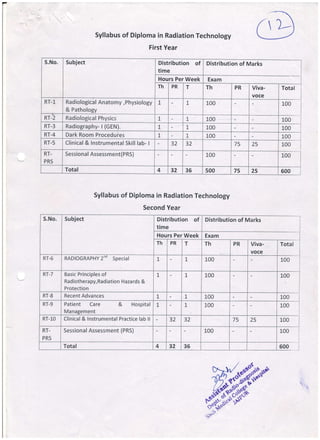
- 1. (l)-- Syllabus of Diploma in Radiation Technology First Year Syllabus of Diploma in Radiation Technology S.No. Subject Distribution of time Distribution of Marks Hours Per Week Exam Th PR T Th PR Viva- voce Total RT-1 Radiological Anatomy,Physiology & Pathology 1 1, 100 100 RT-2 Radiological Physics I t 100 100 RT-3 Radiography- I (cEN). L 1, 100 100 RT-4 Dark Room Procedures t t 1_00 100 RT-5 Clinical & Instrumentalskill lab- | 32 32 75 25 100 RT- PRS Sessional Assessment(PRS) 100 100 Total 4 32 36 500 75 25 600 Second Year S.No. Subject Distribution of time Distribution of Marks Hours Per Week Exam Th PR T Th PR Viva- voce Total RT-6 RADIOGRAPHY2nd Special 1 1 L00 100 RT-7 Basic Principles of Radiotherapy,Radiation Hazards & Protection L 1, 100 L00 RT-8 Recent Advances t t 100 100 RT-9 Patient Care & Hospital Management t I 100 100 RT-10 Clinical & lnstrumental Practice lab ll 32 32 75 25 100 RT- PRS Sessional Assessment (PRS) 100 L00 Total 4 32 36 600
- 2. CODE RT-2 RADIOLOGICAL PHYSICS RATIONALE Every electric current is accompanied by magentic effects & electro magnetism is the branch of physics that deals with the relationship between electricity & Magnetism. X-ray belongs to a group of radiation called electromagnetic radiation. lt is the transport of energy through space as a combination of electric and magnetic field. Any accelerating charge not bound to an atom will emit electromagnetic radiation. CONTENTS Basic Electricity and magnetism and Radiation physics : Units of measurement force, work, energy.Heat and energy. Various method sof transmission of heat. Magnetism, classification of magnets. properties of magnets .magnetic field and line of forces and their measurement, Electro magnetism. ,* :tricity, electrostatic conductor and insulators.elementary electron theory. Units of electric charges potential. Condensers and capacity of condensers. Current, Electricity, Om's Law,various units of current ,Voltage and rectifiers.Heating effect of current, units of point and power consumption,Principal and working of moving coil and moving iron type of meters. Electro Magnetic induction ,Transformers,.their losses,.rating ,induction motors. Direct and Alternating currents, impedance, capacitance, Thermoionic emission , Characteristic curves of diode a nd triode valves, semiconductors. Knowledge of Cathode , anode,rectifier.solid state rectifier,self rectified circuits imbalance of single valve rectifications.half wave and full wave rectifications,transformer and HT cables,HT cable calibration and measurement units of HT.Measurement of out put of x-ray Tube. Apparatus for Radiography,radiotherapy and imaging & its routine maintenance. Mains supply,basic x-ray circuit control,and stablising,Equipment motors,various exposure timers control of scattered radiations fluoroscopy t. rography.mobile equipment.photofluorography.mammographic equipment. REFERENCE BOOKS 1. Radiation physics 2. The Fundamentals of x-ray and Radiation 3. RADIOLOGICAL BOOK FOR TECHNOLOGISTS Satish Bharghav Josaph Selman Bushong & sievert )aw *-qi*ffi*i'#*-
- 3. RADIOLOGICAL ANATOMY,PHYSIOLOGY & PATHOLOGY CODE R T-1 RATIONALE The study of anatomy physiology and pathology is essential because it will help in understanding the basic structure of the organs, their functions and changes due to various diseases affecting the organs of the human body. CONTENTS Gross Radiological surface anatomy of human body. The Human Skeleton bones and joints, formation of bones, growth of skeleton, centers of Ossification, types of bones,type of joints, thoracic contents and general location of organs and vessels, abdominal viscera and location of the major organs, types of cells, composition a nd developme nt,Cell function a nd tissue d iffe rentiation. 2. Anatomy, Physiology and Pathology of Body system-Genes reproductive organs ,embryological development..The nature and appearance of Bacteria.Common Benign Tumors,Malignant Tumors.Dissemination of Malignancy, Primary and Secondary spread.ComposruJh and type of nerve tissue, rnuscular tissue and types.Abnormalities in tissues,ulceration,Sepsis asepsis and anti sepsis.Heart and blood, vessels.structure of heart and function.Major vessels.of the circulatory system: blood circulation , purification.Common terms used for diseases and conditions of this system. 3. Respiratory system. and nasal passages and nasal sinuses, pharynx, Nature and function of respiration.common terms related to diseases and conditions of the system. Lymphatic system. lymphoid tissue and the tonsils.Reticulo endothelial system, liver and spleen. bone morrow.Life cycle of red and white corpuscles of the blood.Alimentary system.Functions of mouth and teeth. 4. Salivary gland,pharynx and oesophagus,stomach, small intestine,.large intestine[colon], liver and biliary tract, and pancreas Functions of alimentary system digestion absorption of food, metabolism, urinary tract- Kidney Ureters and bladder urethra Urinary secretion.Reproductive system male genitalia, female genitalia, mammary glands. Menstruations, pregnancy and lactation. Nerve system and common terms used in this system Main subdivisions organs of sense.Structure and the functions of eye,ear,Surface landmarks and topography in relation to organs of the body for radiograph.- positioning.lnflamation.Pyrexia.Ulcer.bacteria and the specific granulomatous.disorders.endocrine.nutrition and metabolism. Ref. Books:- L.Foundation of Anatomy & physiology -Ross Wilson 2. Atlas of Radiological Anatomy - Weir & Abrahms k%--$".k'* {/ * -,lr9'-*oo -^E a - -fl? -L- r-z nt ."v _ov t9-rs'l,oeP^*" ,1,l" ^ht, #F
- 4. RADIOGRAPHY - | (Gen.) CODE RT- 3 RATIONALE Radiography is a branch of photography in which an image is formed on a film or plate by exposure to X-ray. An opaque object- e.g. Part of human body or a metal casting is placed between the source of the X-rays and the sensitized material; the resulting radiograph shows details of the internal structure which is widely used in medical field for diagnostic purposes. CONTENTS Routine Radiographic Techniques for whole body. (Different views of routine with specialviews of radiography Skull & Neck: Different views of skull bones. Maxilla, mouth, mastoid, Petrous bones, optie foramen, sella tissue neck, nasopharynx, larynx. mandible, zygoma, T.M. Joints. Open mouth & close turcica, internal auditory canal, sphenoid bone, soft Upper Limbs: Fingers individualand as a whole, hand carpal tunnel syndrome, wrist, forearm, elbow, head of r __ ius humerus shoulder joints, acromio clavicular joint, sternoclavicular joint and scapula. Chest and Thorax Bones : Chest PA (Tele radiography), Chest Supine, Lordotic, Oblique Lateral, sternum oblique, lateral and thoracic inlet view & decubitus. Abdomen : Preparation indication and contra indication, acute abdomen, different position of abdomen- upright (standing) sitting, lying, decubitus, supine, and in prone position. Vertebral Column: Atlanto occipital, odontoid, cervical spine, cervico thoracic spine, dorsal spine, thoraco lumbar spine, lumbo sacral spine, sacrum, coccyx, scoliosis, kyphosis, flexion, extension and both oblique views of spines. Hips and Pelvis:Pelvis with Hip joints in different positions. Internal and external rotation, frog positions. S.l. joints. Cephalic tilt and caudaltilt. Lower Limbs: Toes, feet, calcaneum, ankle joints, leg bones. Different view of knee. Patella inter condylar notch and femurs. Owers:Dental radiography, macro and micro radiography, mobile and portable for bed side radiography operation theatre radiography, cine radiography, localization of foreign body, battery operated units, mass miniature radiography and all other emergency radiography. REFERENCE BOOKS: 1. WHO - Manual of radiographic Technique. 2. Radiographic for Technicians 3. Pocket Atlas of Dental Radiology. 4. Clark's positioning in radiography N^ rri wr;h* I Av ^.-(l ^ q rD- ^(>.. ^O-- ^Y E! -AL-rQ -q' ^ Lv r$- |v.r-^{v- 'fet"i"69" tDr' -t'
- 5. - DARK ROOM PROCEDURES CODE RT- 4 RATIONALE Radiography unquestionable begins and ends in the dark room.Where the necessary handling and processing of X-ray film can be carried out safely and efficiently, without the hazard of producing film fog by accidental esposure to light or X-ray. CONTENTS Dark Room Procedures :Photographic Process-Light image.image produced by radiation.light sensitive materia ls,latent. image. Film Material :The structure of X-ray films.resolving power-graininess of film.sensitivity of film.speed of film.contrast of film and types of film. Sensitivity :Cha'racteristic curve and its usefulness. X'- Ray Film Storage :Storage 6f unexposed films. Screens : Construction of intensifying screens. Choice of fluorescent material.intensifying factor detail Sharpness,Speed,screen contact,care of intensifying screens and type of screens. Cassettes :Cassettes design and care of cassettes.Mounting of intensifying screens in the cassettes. Film Processing :Consitutions of the processing solution and replenisher.Factors affecting the developer type of developer and fixer.factors affecting the use of the fixer,silver recovery method. Film Rinsing Washing and Drying :lntermediate rinse. washing and drying of films. Film processing Equipment :Manual and automatic processing. Dark Room Design :Layout and material used The radiographic image :The sharpness, contrast detail definition.viewing conditions. Administration :Trimming, identification of film legends,relevant papers of the patients.records filling,Reptrt d istribution Dark Room Process:Light proof with colour.ventilation and temperature.maintenance.Technical and processing film faults.Fog static pressure and static currents, Artefacts of different types.Darkroom illuminations, orientation of laser cameras. REFERENCE BOOKS 1. WHO-Manual of darkroom Technique. 2, Radiographic physics and darkroom procedure.- Gupta. 3. Radiographic Photography. -CHESNEY D.H. & CHESNEY M.O. ,,#'ffi;ffi[;:14{{i"
- 6. CLINICAL & INSTRUMENTAL SKILT LAB. TRAINING.I CODE RT.5 RATIONALE It is very important for a X-ray trainee to have practical knowledge of various laboratory tests.The student will be able to interpret correctly the test results and correct diagnosis of a disease. Practicals & trair*ng related to theory papers-Radiological Anatomy,Physiology&Pathology,Radiological Physics, Radiography -l (G EN.)Dark Room Procedures .Note : The EssentialTheory should be taught during the Practicals. REFERENCE BOOKS : L WHO- A Guide to X-ray Department ,
- 7. RADIOGRAPHY2nd (Special) CODE RT-6 RATIONALE Radiography is branch of photography in which an image is formed on a film or plate by exposure to X- ray, an opaque object-e.g. Part of human body or a metal casting is placed between the source of the X-rays and the sensitized material; the resulting radiography shows details of the internalstructure which are widely used in medicalfield for diagnosis. CoNTENTS 1.. Special Radiographic Techniques & Applications & uses of contrast media Carotid Angiography, Investig'ation related to the blood Supply of the brain. Ventriculography - Position and techniques Pneumo-Encephalography trolley equipment, preparation of the patient and after care. Angiography:- fourvessel, Selective cath lab procedure Gastro intestinal tract:- Ba. Swallow, Ba. Meal, Ba, Mealfollow through, Ba. Enema. Biliary Tract: Oral Cholecystography, lVC, trans hepatic percutaneous cholangiography, preoperative chola ngiogra phy, T-tu be chola ngiogra phy a nd ERCP. Myelography:- Vertebral Angiography, preparation of patient, contrast media equipment and techniques of oroced ure, Urinary Tract - KUB, IVU ,Retro grade, cystourethrogram; micturating urethrography. " 'stero-Salpingography:- Investigation of uterus and fallopian tubes. Tomography - Principle, equipment with type of rnovement, procedures. Theatre technique - Sterile technique in OT, Cleanliness of mobile unit or C- arm. Others - Dacrocystography, sialography, sinography; angiography (Cerebral and venography) Bronchography, arteriography, mammography, Spleenoportovenography, Lymphangiography, xerography and all other special investigations. Ref. Books:- 1. Clark's positioning of Radiography
- 8. BASIC PRINCIPLES OF RADIOTHERAPY, RADIATION HMARDS & PROTECTION CODE RT -7 RATIONALE X-ray may cause harm. Many somatic dangers of radiation became evident a few months after X-rays were discovered. Small doses of radiation can cause both mutations & neoplasm. No one knows just how much radiation is tolerable. Protection must be provided against any type of radiation to general public as well as radiation workers. The greatest risk from X-rays is for the operator and doctor, who may be exposed repeatedly over the years while they are working. CONTENTS General principiL of radiotherapy, therapeutic ratio, cell cycle, Factors influencing radiation effects on normal tumour cells, Radiotherapy management of various malignancies treatment and side effects of radiations. Knowledge of Linear accelerators, brachytherapy & Teletherpy Machine & their Applications ,Radioactive isotopes & their applications Fundamentals of computers & its application in Radiodiagnosis & Radiotherapy Radiation hazards and its protection for occupational workers and general public, Planning of department of radiology, Radiotherpy.Structure of Atom, Radio Activity naturaland artificialproduction. Interaction of radiation with matter, quantity and quality of radiation and the factors on which it depends. H.V.T. T.V.T Various radiation units - Roentgen, rad, rem, etc, Dosimetry, various radiation measuring instruments, ICRP recommendations, measurement of X-ray and other radiation, rules of AERB , effects of radiation, radiation hazards,, film badge. REFERENCE BOOKS l-.Radiation Physics 2.The Fundamentals of X-ray and Radiation 3. A book of radiologicalTechnologists Satish Bharghav Josaphy Selman Bushong & sivert w *f#*ffil"t#*.
- 9. RECENT ADVANCES CODE RT - 8 RATIONALE Eu9& electric current is accompaniend by magnetic effects & electromagnetism is the branch of physics that deals with the relationship between electricity & Magnetism.X-ray belongs to a group of radiations called electromagnetic radiation.lf the transport of energy through space as a combination of electric and magnetic field.Any accelerating charge not bound to an atom will emit electromagnetic radiation. units ! CoNTENTS Recent Advances in lmaging radiology lmage intensifiers Rapid serial'changers pressure syringe x-ray tube and complete knowledge of x-ray along with all accessories. mobile and portable x-ray units. (ii) (ilr) (iv) (v) (vi) (vii) (viii) (ix) (x) (xi) (xii) Satish Bharghav Josah Selman Rumack Recent advance in imaging technology: - Knowledge of Ultra sonograhy, Color Doppler, different types of tra nsd ucers. CT Scan, gonventional, spiral (Helical), Multislice. Magnetic resonance imaging (MRl) Spectroscopy (MRS) Computerized radiography Digital Radiography DSA P ictu re Archivi ng comm unication syste m ( PACS) Mammography Orthopantogra phy Positron em ission Tomogra phy (PET) Different type of cameras e.g. laser, photography etc. REFERENCE BOOK: 1. Radiation Physics The Fundamentals of X-ray and Radiation Diagnostic Ultrasound Computed Tomography & Magnetic Resonance lmaging of the Whole Body Foundation of Computing Haaga P.K Sinha & P Sinha BPB Publication 2. 3. 4. sw
- 10. Patient Care & Hospital Management CODE RT - 9 CONTENTS Cleaning and care of enamel, stainless steel and glass instruments/cleaning of rubber and polythene goods, care of linen, woolen blankets, mattress and other sheets, bed making, giving bedpan, urinal and removing them. Lifting of patients and first aid procedures. Transferring patients from wheel chairs, trolley or stretcherto the bed and x-ray couch and vice versa. Temperature, pulse, respiration and blood pressure, enema water and soap water enema' Explanation of hospital charts, sterilization and sterile technique of handling the sterile instruments. Injection Technique : Intra Muscular, Intra Venous, setting up of drip, supply of oxygen, dignity of patient. Psychology of the sick. Preparation of the patient for any major investigation. Use of X-ray and radiation hazards. Preparation of the trays for special investigation and care of cancer patients. Maintaining up to date medico legal case (MLC) Radiographic record and verification of patient's marks of identity. Storage and 'istribution of reported films, storage of waste films and used solutions. F[ospital managcment Rules & Regulations: Licensing & registration procedure, Shop & Commercial Establishment act. Municipal bye laws & insurance cove rage. Management Teclrniques : Leadership authority responsibility, Functions of Hospital Management Quality Control & Quality Acceptance Meaning importance of keeping standard, Factors responsible for deviation from standards.lsO and ISO 9OOO tq9006, Tptalquality management. ""rman Relations & Personality Development Motivating the employees,lnter personnel relations, Grievances and their handling, Staff requirement, training and monitoring. Bio lVledical Waste Management: Environmental impact of radiation, Introduction to bio-medicinalwaste, Types of Collection of bio-medical waste, treatment and safe disposal of bio-medical waste bio-medica I waste, REFERENCE BOOK: L WHO - A Guide to X-Ray Department 2. WHO - Manual of Radiographic Technique.
- 11. 3. Radiographic for Technicians. 4. Hand Bok on entrepreneurship Development 5. Environmental lmpact ASsessment O.P. harkut. Mc Graw Hill, NewYark,1977 CLINICAL & INSTRUMENTAL SKILL LAB TRAINING- II CODE RT - 10 RATIONALE It is very important for an X-ray trainee to have practical knowledge of various laboratory tests. The student will bf able to interpret correctly the test results and correct diagnosis of a disease. . PRACTICALS Practical & training related to theory papers - Radiography -ll (Special). Radiotherapy Radiation Hazards & Protection, Physics of Recent Advances, Patient care & Hospital Management. Since the trainee has to work on various medical instruments & equipments, he must have the basii- knowledge and practical training about the different machines so that in case of any trouble during work. He/She will be able to correct and repair the faults. PRACTICALS I ntrod uction to equ ipments Simple usage Indication & Contraindication use Repair & Maintenance of Instruments. Note : The Essential Theory should be taught during the Practicals. REFERENCE BOOKS: WHO - A Guide to X-Ray Department. "ffi .z:11Y - ^0> f,t- 'r' t.Iv gs"-