Call Girls Guntur Just Call 8250077686 Top Class Call Girl Service Available
Small cell lung cancer - oncology
1. Small cell lung cancer
Introduction
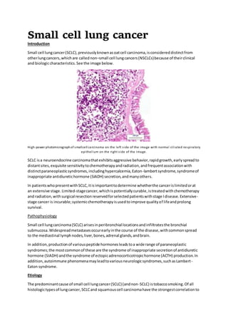
Small cell lungcancer(SCLC),previouslyknownasoatcell carcinoma,isconsidereddistinctfrom
otherlungcancers,whichare callednon–small cell lungcancers(NSCLCs)because of theirclinical
and biologiccharacteristics.See the image below.
High-power photomicrographof smallcell carcinoma on the left side of the image with normal ciliated respiratory
epithelium on the right side of the image.
SCLC isa neuroendocrine carcinomathatexhibitsaggressive behavior,rapidgrowth,earlyspreadto
distantsites,exquisite sensitivitytochemotherapyandradiation,andfrequentassociationwith
distinctparaneoplasticsyndromes,includinghypercalcemia,Eaton-lambertsyndrome,syndromeof
inappropriate antidiuretichormone (SIADH) secretion,andmanyothers.
In patientswhopresentwithSCLC,itisimportanttodetermine whetherthe cancerislimitedorat
an extensive stage.Limited-stagecancer,whichispotentiallycurable,istreatedwithchemotherapy
and radiation,withsurgical resectionreservedforselectedpatientswithstage Idisease.Extensive-
stage cancer is incurable;systemicchemotherapyisusedtoimprove qualityof lifeandprolong
survival.
Pathophysiology
Small cell lungcarcinoma(SCLC) arisesinperibronchial locationsandinfiltratesthe bronchial
submucosa.Widespreadmetastasesoccurearlyinthe course of the disease,withcommonspread
to the mediastinal lymphnodes,liver,bones,adrenal glands,andbrain.
In addition,productionof variouspeptide hormonesleadstoa wide range of paraneoplastic
syndromes;the mostcommonof these are the syndrome of inappropriate secretionof antidiuretic
hormone (SIADH) andthe syndrome of ectopicadrenocorticotropichormone (ACTH) production.In
addition,autoimmune phenomenamayleadtovariousneurologicsyndromes,suchasLambert-
Eaton syndrome.
Etiology
The predominantcause of small cell lungcancer(SCLC) (andnon-SCLC) istobaccosmoking.Of all
histologictypesof lungcancer,SCLCand squamouscell carcinomahave the strongestcorrelationto
2. tobacco. Approximately98%of patientswithSCLChave a smokinghistory.PatientswithSCLCshould
be encouragedtostop smoking,assmokingcessationisassociatedwithimprovedsurvival.
All typesof lungcancer occur withincreasedfrequencyinuraniumminers,butSCLCisthe most
common.The incidence of lungcancerisincreasedfurtherinthese individualsif theyalsosmoke
tobacco.
Exposure toradon,an inertgas that isa productof uraniumdecay, hasalsobeenreportedtocause
SCLC.
Epidemiology
International occurrence
Globally,lungcanceristhe mostfrequentmalignancyinmen(inEurope,lungcancerissecondonly
to prostate cancer) and the fifthmostcommoncancer inwomen.Althoughthe incidenceof lung
cancer has beenfallinginthe US,it isincreasingata staggeringpace indevelopingcountriesdue to
the risingprevalence of tobaccouse.AccordingtoWorldHealthOrganization(WHO) statistics,about
1.69 milliondeathsfromlungcancer occur annuallythroughoutthe world.
Separate worldwide dataforsmall cell carcinomaare not available.The incidence of lungcancer
startedto decline amongmeninthe early1980s and has continuedtodoso overthe past 20 years.
In contrast,the incidence inwomenstartedtoincrease inthe late 1970s anddid notbeginto decline
until the mid-2000s.
Age –and sex relateddemographics
As withotherhistopathologictypesof lungcancer,mostcasesof SCLC occur inindividualsaged60-
80 years.
Overthe past twodecades,the incidence of lungcancerhasgenerallydecreasedinbothmenand
women30 to 54 yearsof age inall races and ethnicgroups.However,the incidencehasdeclined
more steeplyinmen.Asa result,lungcancerratesinyoungerwomenhave become higherthan
those inyoungermen.Innon-HispanicwhitesandHispanicsages44 to 49 years,forexample,the
female-to-male rate ratioforlungcancerincidence rose from0.88 during1995-1999 to 1.17 during
2010-2014.
Thisreversal canbe explainedinpartbyincreasedratesof cigarette smokinginwomenbornsince
1965. However,while the difference insmokingratesinthatage group has narrowed,ratesin
womenhave generallynotexceededthe ratesinmen,sootherfactors maybe playingarole. For
example,womenmaybe more susceptible tothe oncogeniceffectsof smoking
Prognosis
Approximately60-70%of patientswithsmall cell lungcancer(SCLC) have clinicallydisseminatedor
extensive diseaseatpresentation.Extensive-stageSCLCisincurable.Whengivencombination
chemotherapy,patientswithextensive-stagedisease have acomplete responserate of more than
20% anda mediansurvival longerthan7 months;however,only2% are alive at5 years. For
individualswithlimited-stagedisease thatistreatedwithcombinationchemotherapypluschest
radiation,acomplete response rate of 80% and survival of 17 monthshave beenreported;12-15%
of patientsare alive at5 years.
Genome-wide associationstudieshave identifiedsingle-nucleotidepolymorphisms(eg,withinthe
promoterregionof YAP1 onchromosome 11q22) that mayaffectsurvival inpatientswithSCLC.
3. Indicatorsof poor prognosisincludethe following:
Relapseddisease
Weightlossof greaterthan 10% of baseline body
Poorperformance status
Hyponatremia
Patienteducation
Because tobaccosmokingisthe predominantcause of lungcancer,the onlymeansof decreasingthe
incidence of thisdiseaseoverall,aswell asthatof small cell lungcancer(SCLC) specifically,isto
decrease the prevalence of smoking.The evidence isclearthatthe decliningincidence of lungcancer
inmenin the UnitedStateshas coincidedwithadecrease insmokingamongmales.
Clinical Presentation
History
Fewerthan5% of patientswithsmall cell lungcancer(SCLC) are asymptomaticatpresentation.
Commonpresentingsignsandsymptomsof the disease,whichveryoftenoccurinadvanced-stage
disease,include the following:
Shortnessof breath
Cough
Bone pain
Weightloss
Fatigue
Neurologicdysfunction
Most patients withthisdisease presentwithashortdurationof symptoms,usuallyonly8-12weeks
before presentation.The clinical manifestationsof SCLCcan resultfromlocal tumorgrowth,
intrathoracicspread,distantspread,and/orparaneoplasticsyndromes.
Local tumorgrowth
SCLCs are usuallycentrallylocatedandmaycause irritationand/orobstructionof the majorairways.
Commonsymptomsresultingfromlocal tumorgrowthinclude cough,dyspnea,andhemoptysis.
Squamouscell canceralsopresentsasa central lesion,butunlike SCLC,itfrequentlyexhibitscentral
cavitation.
Rapidtumor growthmay leadtoobstructionof majorairways,withdistal collapse leadingto
postobstructive pneumonitis,infection,andfever.
Intrathoracicspread
SCLCs usuallygrowrapidly andmetastasizetomediastinal lymphnodesrelativelyearlyinthe course
of the disease.Atpresentation,patientsmayhave verylarge intrathoracictumors,and
distinguishingthe primarytumorfromlymphnode metastasesmaybe impossible.
Pressure onmediastinalstructurescancause varioussymptoms,includingthe following:
Superiorvenacava(SVC) obstruction
Hoarseness - Due to compressionof the recurrentlaryngeal nerve
Hemi-diaphragmparalysis - Due tophrenicnerve compression
4. Dysphagia- due toesophageal compression
Stridor- Due tocompressionof majorairways
SCLC causesSVCobstructionmore oftenthannon-SCLC(NSCLC).Patientspresentwithswellingof
the face andupperextremities,andcandevelopstridordue tolaryngeal edemaorheadache,
dizziness,andotherneurologicsymptomsdue tocerebral edema.Hoarsenessof recentonsetcanbe
causedby compressionof the leftrecurrentlaryngeal nervebyamediastinal massinvolvingthe
aortopulmonarywindow(i.e.,primarytumororlymphnode metastasis).
Compressionof the phrenicnerve causesparalysisof the ipsilateral hemidiaphragm,contributingto
shortnessof breath.Inaddition,esophageal compressioncanleadtodysphagiaandodynophagia,
and compressionof the mainstembronchi andtrachea cancause severe shortnessof breathand
stridoror wheezing.
Symptomsfromdistantspread
Commonsitesof hematogenousmetastasesinclude the brain,bones,liver,adrenal glands,andbone
marrow.The symptomsdependuponthe site of spread.
Neurologicdysfunctioncanoccur due to brainmetastasesorspinal cord compression.Patientswith
symptomaticbrainmetastasesmayhave raisedintracranial pressure secondarytomasslesionsand
vasogenicedema.Commonsymptomsinclude the following:
Headache- usually worse inthe morning
Blurredvision
Photophobia
Nausea
Vomiting
Slurredspeech
Confusion
Localizingsymptoms-suchasextremityweakness.
Suspectedspinal cordcompressionisanoncologicemergency.Earlyrecognitionof vertebral and
paraspinal metastasesisimportant,because adelayindiagnosisandtreatmentfrequentlyresultsin
permanentlossof neurologicfunction.The initial symptomisusuallybackpain,withorwithout
neurologicdysfunction.Once present,neurologicdysfunctioncanprogressveryrapidly(i.e.,within
hours) to cause quadriplegiaorparaplegia,dependinguponthe locationof the lesion.
Othersymptomsfromdistantmetastasismayinclude painfrombone metastasis,aswell asjaundice
or abdominal/rightupperquadrantpaindue toliver metastasis.
Paraneoplasticsyndromes
Paraneoplasticsyndromesare rare disordersthatare triggeredbyan alteredimmune system
response toa neoplasmorectopicproductionof a hormone orcytokine.Table 1,below,shows
some examplesof the paraneoplasticsyndromesaffectingthe endocrine andneurologicsystemsin
patientswithSCLC.
5. Organ System Syndrome Mechanism Frequency
Endocrine SIADH Antidiuretichormone 15%
Ectopic secretionof
ACTH
ACTH 2-5%
Neurologic Eaton-Lambert
reverse myasthenic
syndrome
3%
Subacute cerebellar
degeneration
Subacute sensory
neuropathy
Limbic
encephalopathy
Anti-Hu,Anti-Yoantibodies
Physical examination
Physical findingsinsmall cell lungcancer(SCLC) dependuponthe extentof local and distantspread
and the organ systeminvolved.
Respiratorysystem
Patientsusuallyexperience shortnessof breath;physical examinationmayreveal use of the
accessorymusclesof respiration(scalene muscles,intercostal muscles) andflaringof the nasal alae.
In addition,byvirtue of acentral tumor location,patientsmaydevelopdistal atelectasisand
postobstructive pneumonia.Withpleural effusion,the examinationrevealsdullnesstopercussion
and decreasedorabsentbreathsoundsonthe side of the effusion.
Cardiovascularsystem
Pericardial effusionsmaybe asymptomaticwhensmall,ortheymayresultintamponade if theyare
large or accumulate overa short period.Patientsare usuallyshortof breathandtheirheartsounds
may be distanton auscultation.Jugularvenouspulsationiselevated,and,paradoxically,itriseswith
inspiration.
Tamponade isan emergencyandrequiresimmediate decompressionof the pericardium.Pulsus
paradoxusisa classicsignof pericardial tamponade.If tamponade issuspected,anechocardiogram
shouldbe performed.The definitive diagnosisisestablishedwithcardiaccatheterization,which
revealsequalizationof pressuresincardiacchambers.Definitivemanagementmayinclude
chemotherapyand/orsurgical creationof apleuropericardial window.
Examinationof the extremitiesmayreveal clubbing,cyanosis,oredema.Inthe presence of superior
venacava (SVC) obstruction,the rightupperextremityisusuallyedematous.
Central nervoussystem
6. Asymptomaticbrainmetastasesoccurin5-10% of patientswithSCLC(see Workup).Patientswith
symptomaticbrainmetastasesmayhave raisedintracranial pressure secondarytomasslesionsand
surroundingbrainedema.The physical findingsdependonthe site of the brainlesions.
Performfunduscopytolookforsignsof raisedintracranial pressure,aswell asa thoroughneurologic
examinationandanevaluationof cerebellarfunction,coordination,andgait.
Gastrointestinal system
The liverisa commonsite of metastaticspread.Physical examinationmayreveal icterus(secondary
to widespreadlivermetastasisorobstructionof biliaryoutflow) and/orhepatomegaly.However,
mostpatientsdonot have any specificfindingrelatedtothe gastrointestinal(GI) tracton
examination.Veryoftenpatientsare asymptomaticbutmayhave mildelevationof liverenzyme
levels.
Lymphaticsystem
Carefullyperformalymphnode examination.Currently,enlargedipsilateral supraclavicularlymph
nodesare includedinlimited-stage disease,butenlargedaxillarylymphnodesupstagethe diagnosis
to extensive-stagedisease.
Complications
Multiple complicationsmaybe noted,dependingonthe site of metastasisorthe metabolicfactor
that the tumor affects.Hypercalcemiacouldinitiallybe asymptomaticbutin late stagescouldleadto
weakness,fatigue,andsleepiness,andinextremecasestosevere constipationandlethargy.
Brain metastasisisoftenasymptomaticbutcouldmanifestasa unilateral eye abnormality,focal
neurologicdeficit,orattimeswitha new-onsetheadache thatwakesthe patientup.Seizuresare a
possible manifestation.
Differential diagnosis
Atypical CarcinoidLungTumor
Large Cell Neuroendocrine Carcinoma
Lung Adenoma
Lung Hamartoma Imaging
Mediastinal Lymphoma
Non-Small Cell LungCancer(NSCLC)
APPROACH CONSIDERATIONS
Lung cancer screening
The USPSTF recommendsannual screeningforlungcancerwithlow-dose computedtomography
(LDCT) in adults55 to 80 yearsof age whohave a 30 pack-yearsmokinghistoryandcurrentlysmoke
or have quitwithinthe past15 years.The USPSTFrecommendsthatscreeningbe discontinuedonce
a personhas notsmokedfor15 years or developsahealthproblemthatsubstantiallylimitslife
expectancyorthe abilityorwillingnesstohave curative lungsurgery.
The ACS recommendsLDCTscreeninginapparentlyhealthypatients55-74years of age whohave at
leasta 30 pack-yearsmokinghistoryandwhocurrentlysmoke orhave quitwithinthe past15 years.
The ACS stressesthatthe decisiontoinitiate lungcancerscreeningshouldbe sharedbetweenthe
7. clinicianandpatientandshouldinvolve discussionof the potentialbenefits,limitations,andharms
associatedwithsuchscreening.
A studyby researchersfromthe National CancerInstitute(NCI)andthe AmericanCancer Society
that reviewednine riskpredictionmodelsdeterminedthatthe followingfourmodelsweremore
accurate than the othersforpredictinglungcancerriskand forselectingpatientswhohadever-
smokedforlungcancer screening:
Bach model
OvarianCancerScreeningTrial Model 2012(PLCO-M2012)
Lung CancerRiskAssessmentTool (LCRAT)
Lung CancerDeath RISKAssessmentTool (LCDRAT)
Althoughthe researchersconcludedthatthatany of those modelscouldbe usedtoselectUS
smokerswhoare at the greatest riskforlungcancer incidence ordeath,all the modelshave
limitations.The Bachmodel doesnotaccountfor race/ethnicity,familyhistoryof lungcancer,or
presence of chronicobstructive pulmonarydisease;the PLCO-M2012model underestimatedlung
cancer riskinpeople of Hispanicdescentbyafactor of 2 to 3, and the LCRAT andLCDRAT models
bothunderestimatedriskinthe "Asian/other"subgroup.
Initial workup
A thoroughhistoryandphysical examinationusuallyprovidescluestothe organsystemsinvolvedin
small cell lungcancer(SCLC),andthese are usedto guide furtherworkup. Investigationsare
performedtodelineate the extentof disease andtoassessorganfunctionbefore therapybegins.In
general,dependingontumorlocalization,biopsiesfromthe primarytumorshouldbe obtainedusing
bronchoscopyorany of the following techniques:
Mediastinoscopy
Endobronchial Ultrasound(EBUS)
EndoscopicUltrasound
Transthoracicneedle aspiration
Thoracoscopy(if necessary)
A metastaticlesion,if easily andsafelyaccessible,maybe the preferredoptionforabiopsy
specimen;thiswillalsoprovidepathological staging.
Stagingworkup
The purpose of a stagingworkupforsmall cell lungcancer(SCLC) isto determine the prognosisand
managementof thisdisease.Patientswithlimited-stage disease are usuallytreatedwithcombined
chemoradiotherapy,whereasthose withextensive-stage diseaseare usuallytreatedwith
chemotherapyalone.Stagingworkupof SCLCisas follows:
Complete historyandphysical examination
Complete bloodcount(CBC) withdifferential
Serumelectrolyteslevel,includingcalcium
Liverfunctiontests(LFTs)
Renal functiontests(RFTs)
Serumlactate dehydrogenase(LDH) level
Serumalkaline phosphate (ALP)level
Chestradiography
8. CT scanningof the chestand abdomenwithintravenouscontrast(includingliverandadrenal
glands)
CT scanning/magneticresonance imaging(MRI) of the brainwithIV contrast
Bone scanning
Bone marrow aspirationandbiopsyif abnormalitiesare presentinthe CBCor peripheral
smear.
Stagingshouldbe adequate beforemakingthe diagnosisof limited-stage SCLC.Anypleural effusion
shouldbe testedcytologicallyformalignantcells,andisolatedliveroradrenal lesionsshouldbe
sampledbyfine-needle aspiration(FNA) before adiagnosisof limited-stage diseaseismade.Some
authoritiessuggestabone marrowexaminationinthe absence of anyotherevidence of spread.
TNMCLASSIFICATION FORSMALLCELL LUNG CANCER
Primary Tumor (T)
TX Primarytumorcannot be assessed, ortumoris provenbythe presence of malignantcellsinsputum
or bronchial washingsbutnotvisualizedbyimagingorbronchoscopy.
TO No evidenceof primarytumor
Tis Carcinomainsitu
Squamouscell carcinomainsitu(SCIS)
Adenocarcinomainsitu (AIS):adenocarcinomawithpure lepidicpattern,≤3 cm in greatest
dimension
T1 Tumor ≤ 3 cm in greatestdimension,surroundedbylungorvisceral pleura,withoutbronchoscopic
evidence of invasionmore proximal thanthe lobarbronchus(i.e.,notinthe mainbronchus)
T1mi Minimallyinvasive adenocarcinoma:adenocarcinoma(≤3 cm in greatestdimension)witha
predominantlylepidicpatternand≤ 5 mm invasioningreatestdimension.
T1a Tumor ≤ 1 cm in greatestdimension.A superficial,spreadingtumorof anysize whose invasive
componentislimitedtothe bronchial wall andmayextendproximaltothe mainbronchusalsois
classifiedasT1a,but those tumorsare uncommon.
T1b Tumor > 1 cm but ≤ 2 cm ingreatestdimension
T1c Tumor > 2 cm but ≤ 3 cm in greatestdimension
T2 Tumor > 3 cm but ≤ 5 cm or havinganyof the followingfeatures:
Involvesthe mainbronchusregardlessof distance tothe carina,butwithoutinvolvementof
the carina
Invadesvisceral pleura(PL1or PL2)
Associatedwithatelectasis orobstructivepneumonitisextendingtothe hilarregion,
involvingpartorall of the lung
T2 tumorswiththese featuresare classifiedasT2a if ≤ 4 cm or if the size cannotbe determinedand
T2b if > 4 cm but ≤ 5 cm
T2a Tumor > 3 cm but ≤ 4 cm in greatestdimension
9. T2b Tumor > 4 cm but ≤ 5 cm ingreatestdimension
T3 Tumor > 5 cm but ≤ 7 cm ingreatestdimensionordirectlyinvadinganyof the following:parietal
pleural (PL3),chestwall (includingsuperiorsulcustumors),phrenicnerve,parietalpericardium;or
separate tumornodule(s)inthe same lobe asthe primary
T4 Tumor > 7 cm or tumorof anysize that invadesone ormore of the following:diaphragm,
mediastinum,heart,greatvessels,trachea,recurrentlaryngeal nerve,esophagus, vertebral body,or
carina; or separate tumornodule(s)inanipsilaterallobe differentfromthatof the primary
Regional lymph nodes(N)
NX Regional lymphnodescannotbe assessed
N0 No regional lymphnode metastasis
N1 Metastasisto ipsilateral peribronchial and/oripsilateral hilarlymphnodesandintrapulmonary
nodes,includinginvolvementbydirectextension
N2 Metastasisinipsilateral mediastinaland/orsubcarinal lymphnode(s)
N3 Metastasisincontralateral mediastinal,contralateral hilar,ipsilateral orcontralateral scalene,or
supraclavicularlymphnode(s)
Distant metastasis (M)
M0 No distantmetastasis
M1 Distantmetastasis
M1a Separate tumornodule(s)inacontralateral lobe tumor;tumorwithpleural orpericardial nodules or
malignantpleural orpericardial effusion.Mostpleural (pericardial) effusionwithlungcancerare a
resultof the tumor.In a few patients,however,multiple miscroscopicexaminationsof pleural
(pericardial) fluidare negative fortumor,andthe fluidisnonbloodyandnotan exudate.If these
elementsandclinical judgmentdictate thatthe effusionisnotrelatedtothe tumor,the effusion
shouldbe excludedasastagingdescriptor.
M1b Single extrathoracicmetastasisinasingle organand involvementof asingle nonregional node
M1c Multiple extrathoracicmetastasesinasingle organorinmultiple organs
ROUTINE LABORATORY STUDIES
A complete bloodcell count(CBC) withdifferential,serumelectrolytelevels,renal functionstudies,
and liverfunctiontests(LFTs) are all partof the routine stagingworkup,andinsome cases,these
studiesmayreveal the sitesof metastasis(eg,elevatedserumcalciumand/oralkalinephosphatase
[ALP] levelswithbone metastasis).These testsare alsoimportanttoassessorganfunctionbefore
startingtherapy.
10. Serumlactate dehydrogenase(LDH) andsodiumlevelsalsoprovide prognosticinformation.
Increaseduricacidlevelsandimpairedrenal functionmayindicate the potential fortumorlysis
syndrome withtherapy.
Complete bloodcount
In 5-10% of patients,small cell lungcancer(SCLC) mayhave spreadto bone marrow at presentation.
Bone marrow examinationisnotroutinelyperformedinSCLCunlessabnormalitiesare identifiedin
the CBC or peripheral smearexamination,raisingthe possibilityof bone marrow spread.These
abnormalitiesmayinclude cytopeniaorthe presence of immature white andredbloodcells(a
leukoerythroblasticbloodpicture),whichraisesthe possibilityof myelophthisicanemia.
Additionally,beforeinstitutinginitial full-dose combinationchemotherapy,the CBCshould
demonstrate the following:
Absolute neutrophilcount(ANC) - Shouldbe greaterthan1000 x 103/µL
Hemoglobinlevel- Shouldbe above 10 g/dL
Plateletcount- Shouldbe more than100 x 103/µL
Serumchemistries
The presence of elevatedserumcalciumandALPlevelsraisesthe suspicionof bone metastasis,and
insuch casesa bone scan shouldbe orderedeveninthe absence of symptoms.Serumelectrolytes
shouldbe obtainedto lookforparaneoplasticsyndromes,suchassyndrome of inappropriate
antidiuretichormone(SIADH) secretion.The presence of hyponatremiaisconsideredanadverse
prognosticindicator.
ElevatedserumLDHindicatesanincreasedtumormassand highcell turnover;thisfindingisalsoan
adverse prognosticindicator.Abnormal liverfunctionfindingsraise the possibilityof hepatic
metastasisandmayrequire adjustmentstoplannedtherapy.
THORACIC IMAGING STUDIES
Radiography
Good posteroanteriorandlateral radiographsare useful inidentifyingthe primarytumor,aswell as
concurrentparenchymal abnormalities.Mediastinalwideningmayindicate mediastinal lymphnode
involvement.
Computedtomography
Computedtomography(CT) scanningof all commonsitesof metastasisshouldbe performedto
stage the disease adequately.EvaluationviaCTscanningof the thorax (lungsandmediastinum) and
commonlyinvolvedabdominal viscera(ie,liver,adrenals) isthe minimumrequirementinstandard
stagingworkupof SCLC. Intravenouscontrastagentsshould be usedwheneverpossible.
Brain andspinal cord imaging
Brain metastasismaybe presentinasmanyas 10-15% of patientsatdiagnosis andmaybe occultin
5% of patients.Consequently,magneticresonance imaging(MRI) of the brainshouldbe orderedin
asymptomaticpatientsaswell asinthose withneurologicsymptoms. Because MRIismore sensitive
than computedtomography(CT) scanningwithcontrastfordetectionof brainmetastasis,MRIis
usedas the first-lineimagingstudyinmanyinstitutions.
11. MRI hasan increasedabilitytodetectdisease inproximitytoneurovascularstructuresandisalso
consideredstandardinthe workupof patientsinwhomspinal cordcompressionissuspected.
AlthoughaCT myelogramcanestablish the diagnosisof vertebral andparaspinal metastases,itis
currentlyrarelyused.MRIis noninvasive andverysensitive inestablishingthe diagnosisinalmostall
cases.
SKELETAL RADIONUCLIDE IMAGING
Bone isa commonsite of metastasisforsmall cell lungcancer(SCLC).A radionuclide bonescan
shouldtherefore be obtainedtoidentifybone metastases.
Bone metastasesfromSCLCusuallycontainbothosteolyticandosteoblasticcomponents,anda
bone scan issuperiortoplainradiographsindetectingosteoblasticlesions.However,becausesome
benignlesionscanalsocause abnormalitiesonbone scans,obtainingplainradiographsof abnormal
areas forradiographiccorrelationisimportant,particularlyinweight-bearingbonesatriskfor
fracture.
Bone scans shouldbe obtainedinall patientswithSCLCat diagnosisorduringfollow-upif new bone-
relatedsymptomsdeveloporif the serumcalciumor alkaline phosphatase level iselevated.
PET SCANNING
Positronemissiontomography(PET) scanning(see the image below) remainsunderevaluationfor
the stagingof small cell lungcancer(SCLC).The AmericanCollege of ChestPhysicians(ACCP)doesnot
recommendPETscanninginthe routine stagingof SCLC, althoughthe National Comprehensive
Cancer Network(NCCN)guidelinesrecommendcombinedPET-CT(computedtomography) scanning
if limited-stage disease ormetastasisissuspected. PET-CTimagingissuperiortoPET scanningalone.
(PET scanningisinferiortoMRI or CT scanningfor the detectionof brainmetastases.)
This coronal PET shows large, focal, hypermetabolic area on the right that is consistent with a large mass in the central
portionof the right upper pulmonarylobe. Multiple other small hypermetabolic areas suggest lymph node metastatic
disease in chest, abdomen, and right subclavicular region.
In small,uncontrolledstudies,PETscanninghasshowngoodaccuracy (83-99%) instagingextensive-
versuslimited-stage SCLC. AlthoughPETscanningmayimprove the accuracy of staging,however,
any lesionidentifiedusingthismodalitythatwouldalterstagingrequirespathologicconfirmation
due to the possibilityof afalse-positive finding. The full roleof PETimaginginthissettingremainsto
be determined.
BRONCOSCOPYAND FINE NEEDLE ASPIRATION
12. Small cell lungcancer(SCLC) isusuallycentrallylocatedandcanbe approachedeasilywitha
bronchoscope.The advantage of endoscopyisdirectvisualizationof the tumor,allowingfordirect
biopsyaswell ascytologicexaminationof bronchial washings.
For tumorsthat cannot be diagnosedwithtransbronchial biopsy,transthoracicpercutaneousfine-
needle aspiration(FNA) carriedoutundercomputedtomography(CT) guidance isareasonable
alternative.
SPUTUMCYTOLOGY
Sputumcytologyisa noninvasivetestand,if positive,canprovide anaccurate diagnosisof central
lungcancers.Althoughsmall cell lungcancer(SCLC) usuallypresentsasalarge,central tumor,tumor
cellsfrequentlyinvolve the submucosal layerof the bronchuswithlittle ornoexophytic
endobronchial extension.Therefore,sputumcytologyisnotas useful fordiagnosingSCLCasit isfor
the diagnosisof squamouscell carcinoma.
THORACENTECIS
In small cell lungcancer(SCLC),the presence of malignantpleural effusionupstagesthe disease to
extensive-stageSCLC.Foradequate staging,pleural effusionsshouldbe aspiratedandexaminedfor
malignantcellsif noothersitesof distantspreadare identified.
HISTOLOGIC FINDINGS
Small cell lungcancer(SCLC) istypicallycentrallylocated,arisinginperibronchial locations.These
tumorsare thoughtto developfromneuroendocrineKulchitskycellsandare composedof sheetsof
small,roundtospindledcellswithdarknuclei,scantcytoplasm, andfine,granular(“saltand
pepper”) nuclearchromatinwithindistinctnucleoli.
Veryhighratesof cell divisionare observed,andnecrosis,sometimesextensive,maybe seen.
Because of the central location,the tumorcellsmayexfoliateintosputumandbronchial washings.
Crushartifact of the relativelyfragile tumorcellsisacommonfindinginsmall biopsies,butthis
feature isnotconsidereddiagnosticinandof itself.
Neurosecretorygranulescanbe identifiedwiththe aidof electronmicroscopy.The neuroendocrine
nature of the neoplasmissuggestedbyitsfrequent associationwithneurologicandendocrine
paraneoplasticsyndromes.
Immunohistochemical stainsforchromogranin,neuron-specificenolase,CD56,and synaptophysin
are usuallypositive,butthese are notan absolute requirementforthe diagnosis.
Approximately5%of SCLCsexhibitfeaturesof mixedsmall cell andnon–smallcell components,
suggestingphenotypicplasticityandlendingsupporttothe cancer stemcell hypothesis.Patients
withmixedSCLC/NSCLChistologyare managedaccordingtothe same guidelinesasthose for
patientswithSCLC.
TYPES OF STAGING
Typesof staging
The AmericanCancerSociety(ACS) uses2typesof staging—clinical andpathologic—forSCLC.
Clinical staginginvolvesphysical examination,biopsyexaminations,andimagingscans;the majority
of patientsare stagedwithclinical staging,andthistype of stagingisusuallyusedtodescribeSCLC
tumor extent.
13. Pathologicstagingisgenerallymore accurate,asit includesclinical stagingandaddspostsurgical
findings.Occasionally,findingsbetweenthe 2stagesmay be different,suchasduringproceduresin
whichcancer isinan areathat is notseenonradiologicstudies.The surgical findingsmaygive the
cancer a more advancedpathologicstage.
Stagingsystems
VALSG 2-Staging system: The stagingsystemmostcommonlyusedforSCLCis the Veterans
AdministrationLungGroup(VALSG) 2-stage system, whichdefineslimited-stageand
extensive-stagedisease. Patientswithdisease confinedtoone hemithorax,withorwithout
involvementof the mediastinal,contralateral hilaroripsilateral supraclavicular,orscalene
lymphnodesare consideredtohave limited-stagedisease,whereasthose withdisease
involvementatany otherlocationare consideredtohave extensive-stagedisease.
TNM system: Almostall solidtumors,includinglungcarcinomas,are stagedusingthe tumor,
node,metastasis(TNM) system, because itprovidesimportantprognosticinformationandis
usedto designmanagementplans.However,olderliterature hasstatedthatthe TNM
systemfailstoprovide importantprognosticinformationinpatientswithSCLCandisuseful
onlyforthe fewpatients(<5%) whomightbe eligible forsurgical resection.
IASLC TNM system:The International Associationof the Studyof LungCancer (IASLC)
developedanewTNMstagingsystemforlungcancer in2007; thisstagingsystemincluded
non-SCLC(NSCLC) andSCLC.
AJCC stagingsystem: Underthe new tumor,node,metastasis(TNM) stagingsystem, from
the AmericanJointCommitteeonCancer(AJCC) ,limited-stage SCLCisdefinedasanyT, any
N,M0; the exceptionisT3-4,owingtomultiple lungnodulesthatextendbeyondasingle
radiationfield.
TREATMENT AND MANAGEMENT
Small cell lungcancer(SCLC) ischaracterizedbyrapidgrowthand earlydissemination.Prompt
initiationof treatmentisimportant.
Treatmentprotocols:
Treatmentprotocolsforsmall cell lungcancer(SCLC) are providedbelow,includingfirst-line therapy,
therapyforlimited-stage disease,andtherapyforextensive-stagedisease.
Treatment recommendationsforlimitedstage SCLC
Stage I-IIIdisease:
Limited-stagedisease istypicallytreatedwithsystemictherapy,withorwithoutradiation
therapy
Chemotherapyandradiationtherapyare typicallygivenconcurrently,butcanalsobe given
sequentiallyforlimited-stage disease inpatientsunable totolerate concurrent
chemoradiation;chemotherapyisgivenfirst,followedbyradiationtherapybecause of the
highrate of responsivenesstochemotherapyforSCLC.
A selectgroupof patientsmaybe eligible forsurgical resection.Clinical stage I - IIA (T1 - 2,
N0, M0) patientswhoare surgical candidatesshouldundergopathological mediastinal
stagingto determine if thereismedastinal lymphnodeinvolvement.
Patientswithpathologicallynegative medastinal lymphnodesshouldgoonto
lobectomywithmediastinal lymphnode dissectionorsampling.
14. Patientsfoundtohave pN0disease atthe time of surgical resectionshouldreceive
adjuvantsystemictherapy(seeoptionsbelow)
Patientsfoundtohave pN1or pN2 disease shouldreceive systeictherapy+/-
mediastinal radiationtherapy.
Concurrentchemotherapyrecommendationswithradiationforlimitedstage include:
Cisplatin60 mg/m2IV onday 1 plusetoposide120 mg/m2 IV on days1-3 every21 - 28d.
Cisplatin75 - 80 mg/m2IV onday 1 plusetoposide 100mg/m2 IV on days1-3 every21 - 28d.
Cisplatin25 mg/m2IV ondays 1 - 3 plusetoposide 100mg/m2 IV ondays 1-3 every21 - 28d.
CarboplatinAUC5-6 IV day 1 plusetoposide 100 mg/m2IV days1-3 every21 - 28d.
Chemotherapyshouldbe givenupto4 - 6 cycles.
Radiotherapyforlimited-stage diseaseshouldstartwithcycle 1 or 2 of chemotherapy.
Chemotherapyrecommendationsforpatientsnotable totolerate concurrentchemotherapyand
radiation:
Patientswithlimited-stage(stagesI–III) disease whoare notable totolerate chemotherapy
and radiationconcurrentlyshouldbe treatedwithchemotherapyasfirst-line therapy
Cisplatin60-80 mg/m2 IV on day1 plusetoposide 80-120 mg/m2 IV on days1-3 every21-
28d or
CarboplatinAUC5-6 IV on day1 plusetoposide 80-100 mg/m2 IV on days1-3 every28d.
First-line chemotherapyforextensive stage disease
Stage IV disease:
The followingtreatmentrecommendationsshouldbe givenfora maximumof 4-6 cycles:
Atezolizumab1200 mg IV onday 1 pluscarboplatinAUC5 onDay 1 plusetoposide 100
mg/m2 IV on days1-3 every21d x 4 cycles;follow withmaintenance atezolizumabevery21d
Cisplatin60-80 mg/m2 IV on day1 plusetoposide 80-120 mg/m2 IV on days1-3 every21-
28d
CarboplatinAUC5-6 IV on day1 plusetoposide 80-100 mg/m2 IV on days1-3 every28d
Cisplatin60 mg/m2IV onday 1 plusirinotecan60mg/m2 IV on days 1, 8, and15 every28d
Cisplatin30 mg/m2IV ondays 1 and8 or 80 mg/m2 IV onday 1 plusirinotecan65 mg/m2IV
on days1 and 8 every21d
CarboplatinAUC5 IV on day1 plusirinotecan50 mg/m2 IV on days1, 8, and 15 every28d
CarboplatinAUC4-5 IV on day1 plusirinotecan150-200 mg/m2 IV on day 1 every21d
Cisplatin25 mg/m2IV ondays 1 - 3 plusetoposide 100mg/m2 IV ondays 1-3 every21 - 28d
Cyclophosphamide 800-1000 mg/m2 IV onday 1 plusdoxorubicin40-50 mg/m2 IV on day1
plusvincristine1-1.4mg/m2 IV on day 1 every21-28d.
Second-line chemotherapyforrelapsedorrefractorydisease
Stage IV disease:
Second-line chemotherapyisgivenforatleast4-6 cyclesbut can be givenuntil disease
progressionastoleratedinsome cases
Patientswhohave relapseddiseasemore than6moaftercompletingfirst-line chemotherapy
can be treatedwiththatoriginal first-line regimen(typicallyaplatinum-baseddoublet)
again,withan expectedresponse rate of 62-100%.
15. SystemictherapyrecommendationsforrelapsedorrefractorySCLCinclude:
Etoposide 50 mg/m2 POdailyfor3wk every4wk
Topotecan2.3 mg/m2PO on days1-5 every21d
Topotecan1.5 mg/m2IV on days1-5 every21d
CarboplatinAUC5 IV on day1 plusirinotecan50 mg/m2 IV on days1, 8, and 15 every28d
CarboplatinAUC4 - 5 IV onday 1 plusirinotecan150-200 mg/m2 IV on day1 every21d
Cisplatin30 mg/m2IV ondays 1, 8, and15 plusirinotecan60mg/m2 IV ondays 1, 8, and15
every28d
Cisplatin60 mg/m2IV onday 1 plusirinotecan60 mg/m2IV on days1, 8, and 15 every28d
Cisplatin30 mg/m2IV ondays 1 and8 or 80 mg/m2 IV on day 1 plusirinotecan65 mg/m2IV
on days1 and 8 every21d
Paclitaxel 80mg/m2 IV weeklyfor6wkevery8wk
Paclitaxel 175mg/m2 IV onday 1 every3wk
Cyclophosphamide 800-1000 mg/m2 IV onday 1 plusdoxorubicin40-50 mg/m2 IV on day1
plusvincristine1-1.4mg/m2 IV on day 1 every21-28d
Pembrolizumab200 mg IV every3 weeksuntil disease progression
Nivolumab240 mg IV every2wkor nivolumab480 mg IV every 4 wkuntil disease
progression
Nivolumab1mg/kgIV plusipilimumab3mg/kgIV every21 days for4 cyclesfollowedby
nivolumabmaintenance (240mg IV every2wkor nivolumab480 mg IV every4 wk
InstitutionReviewBoard(IRB)–approvedclinical trial
Special considerations:
PatientswithmixedSCLC/non-SCLChistologyshouldbe giventhe same treatmentas
patientswithSCLC
Prophylacticcranial irradiationisrecommendedforSCLCpatientswithacomplete orpartial
remission(total of 25 Gy in10 fractionsor 30 Gy in10-15 fractions)
Thoracic radiationtherapyshouldbe consideredforpatientswithextensive stage disease
aftertheycomplete systemictherapy
Dose-denseordose-escalationchemotherapyregimensare notrecommendedoutsideof a
randomizedclinicaltrial
Patientswithbrainmetastasescanreceivechemotherapypriortobrainradiationdue to
highresponse rateswithchemotherapy
A studyevaluatingtreatmentof patientswithstereotacticbodyradiationtherapyconcluded
that itis a promisingalternative tosurgeryforpatientswithstage Inon-SCLC