SlideShare a Scribd company logo
1 of 1
Mujer de 45 años y 10 mesesMujer de 45 años y 10 meses
con tumoración cervical izda.con tumoración cervical izda.
Ecografía cervical:Ecografía cervical: Imagen ovoidea conImagen ovoidea con
buena transmisión del sonido sin aparentebuena transmisión del sonido sin aparente
vascularizaciónvascularización
RM cervical:RM cervical: TSTIR coronal, axial DP-T2TSTIR coronal, axial DP-T2
Fat Sat, postGd axial con Fat Sat.Fat Sat, postGd axial con Fat Sat.
Lesión hiperintensa T2 con tabiquesLesión hiperintensa T2 con tabiques
periféricos posteriores captadoresperiféricos posteriores captadores

More Related Content

Viewers also liked (18)

Manchas En La Piel Y Arrugas
Manchas En La Piel Y ArrugasManchas En La Piel Y Arrugas
Manchas En La Piel Y Arrugas
 
Malformaciones Vasculares
Malformaciones VascularesMalformaciones Vasculares
Malformaciones Vasculares
 
Tumores cutáneos benignos
Tumores cutáneos benignosTumores cutáneos benignos
Tumores cutáneos benignos
 
Alteraciones benignas de la piel
Alteraciones benignas de la pielAlteraciones benignas de la piel
Alteraciones benignas de la piel
 
Hemangiomas en Pediatría
Hemangiomas en PediatríaHemangiomas en Pediatría
Hemangiomas en Pediatría
 
Anomalías vasculares
Anomalías vascularesAnomalías vasculares
Anomalías vasculares
 
NEVO DE OPORTO
NEVO DE OPORTONEVO DE OPORTO
NEVO DE OPORTO
 
Malformaciones vasculares
Malformaciones vascularesMalformaciones vasculares
Malformaciones vasculares
 
Hemangiomas y malformaciones vasculares en paciente pediátrico
Hemangiomas y malformaciones vasculares en paciente pediátricoHemangiomas y malformaciones vasculares en paciente pediátrico
Hemangiomas y malformaciones vasculares en paciente pediátrico
 
Hemangiomas
HemangiomasHemangiomas
Hemangiomas
 
Eje hipotálamo hipófisis-gónada
Eje hipotálamo hipófisis-gónadaEje hipotálamo hipófisis-gónada
Eje hipotálamo hipófisis-gónada
 
Hemangiomas
HemangiomasHemangiomas
Hemangiomas
 
Tumores Benignos de la piel
Tumores Benignos de la pielTumores Benignos de la piel
Tumores Benignos de la piel
 
Hemangioma
HemangiomaHemangioma
Hemangioma
 
Hemangioma
HemangiomaHemangioma
Hemangioma
 
Dermatología - Hemangiomas y Nevos
Dermatología - Hemangiomas y NevosDermatología - Hemangiomas y Nevos
Dermatología - Hemangiomas y Nevos
 
HEMANGIOMA
HEMANGIOMAHEMANGIOMA
HEMANGIOMA
 
Exploracion fisica de piel y tegumentos
Exploracion fisica de piel y tegumentosExploracion fisica de piel y tegumentos
Exploracion fisica de piel y tegumentos
 

More from Ruth Martín Boizas

Congenital Cholesteatoma of the Mastoid Bone on CT& MRI
Congenital Cholesteatoma of the Mastoid Bone on CT& MRICongenital Cholesteatoma of the Mastoid Bone on CT& MRI
Congenital Cholesteatoma of the Mastoid Bone on CT& MRIRuth Martín Boizas
 
Mantle B-Cell Lymphoma: Extradural & Foraminal Relapse
Mantle B-Cell Lymphoma: Extradural & Foraminal RelapseMantle B-Cell Lymphoma: Extradural & Foraminal Relapse
Mantle B-Cell Lymphoma: Extradural & Foraminal RelapseRuth Martín Boizas
 
Intracavernous Arachnoid Cyst MRI
Intracavernous Arachnoid Cyst MRIIntracavernous Arachnoid Cyst MRI
Intracavernous Arachnoid Cyst MRIRuth Martín Boizas
 
Virchow-Robin versus Lacunar Infarct CT & MRI
Virchow-Robin versus Lacunar Infarct CT & MRIVirchow-Robin versus Lacunar Infarct CT & MRI
Virchow-Robin versus Lacunar Infarct CT & MRIRuth Martín Boizas
 
Alzheimer’s Disease Image Findings
Alzheimer’s Disease Image FindingsAlzheimer’s Disease Image Findings
Alzheimer’s Disease Image FindingsRuth Martín Boizas
 
Mild cognitive Impairment & Dementia MRI Protocol
Mild cognitive Impairment & Dementia MRI ProtocolMild cognitive Impairment & Dementia MRI Protocol
Mild cognitive Impairment & Dementia MRI ProtocolRuth Martín Boizas
 
Voxel-Based Morphometry in Alzheimer's Disease 1
Voxel-Based Morphometry in Alzheimer's Disease 1Voxel-Based Morphometry in Alzheimer's Disease 1
Voxel-Based Morphometry in Alzheimer's Disease 1Ruth Martín Boizas
 
Giant Fusiform Medial Cerebral Artery Aneurysm
Giant Fusiform Medial Cerebral Artery AneurysmGiant Fusiform Medial Cerebral Artery Aneurysm
Giant Fusiform Medial Cerebral Artery AneurysmRuth Martín Boizas
 
Multiple System Atrophy Cerebellar type
Multiple System Atrophy Cerebellar typeMultiple System Atrophy Cerebellar type
Multiple System Atrophy Cerebellar typeRuth Martín Boizas
 
Spinal tuberculous leptomeningitis & subdural empyema & lately intraspinal tu...
Spinal tuberculous leptomeningitis & subdural empyema & lately intraspinal tu...Spinal tuberculous leptomeningitis & subdural empyema & lately intraspinal tu...
Spinal tuberculous leptomeningitis & subdural empyema & lately intraspinal tu...Ruth Martín Boizas
 

More from Ruth Martín Boizas (20)

Right varicocele. Ultrasound.
Right varicocele. Ultrasound.Right varicocele. Ultrasound.
Right varicocele. Ultrasound.
 
2nd meningiomas meet up
2nd meningiomas  meet up2nd meningiomas  meet up
2nd meningiomas meet up
 
Type 3 Frontal Cell
Type 3 Frontal CellType 3 Frontal Cell
Type 3 Frontal Cell
 
Hypoglossal Nerve Schwannoma
Hypoglossal Nerve SchwannomaHypoglossal Nerve Schwannoma
Hypoglossal Nerve Schwannoma
 
Congenital Cholesteatoma of the Mastoid Bone on CT& MRI
Congenital Cholesteatoma of the Mastoid Bone on CT& MRICongenital Cholesteatoma of the Mastoid Bone on CT& MRI
Congenital Cholesteatoma of the Mastoid Bone on CT& MRI
 
Mantle B-Cell Lymphoma: Extradural & Foraminal Relapse
Mantle B-Cell Lymphoma: Extradural & Foraminal RelapseMantle B-Cell Lymphoma: Extradural & Foraminal Relapse
Mantle B-Cell Lymphoma: Extradural & Foraminal Relapse
 
Epilepsy in Preterm Neonate MRI
Epilepsy in Preterm Neonate MRIEpilepsy in Preterm Neonate MRI
Epilepsy in Preterm Neonate MRI
 
Intracavernous Arachnoid Cyst MRI
Intracavernous Arachnoid Cyst MRIIntracavernous Arachnoid Cyst MRI
Intracavernous Arachnoid Cyst MRI
 
Choroidal Angioma on Image
Choroidal Angioma on ImageChoroidal Angioma on Image
Choroidal Angioma on Image
 
Virchow-Robin versus Lacunar Infarct CT & MRI
Virchow-Robin versus Lacunar Infarct CT & MRIVirchow-Robin versus Lacunar Infarct CT & MRI
Virchow-Robin versus Lacunar Infarct CT & MRI
 
Alzheimer’s Disease Image Findings
Alzheimer’s Disease Image FindingsAlzheimer’s Disease Image Findings
Alzheimer’s Disease Image Findings
 
Mild cognitive Impairment & Dementia MRI Protocol
Mild cognitive Impairment & Dementia MRI ProtocolMild cognitive Impairment & Dementia MRI Protocol
Mild cognitive Impairment & Dementia MRI Protocol
 
Voxel-Based Morphometry in Alzheimer's Disease 1
Voxel-Based Morphometry in Alzheimer's Disease 1Voxel-Based Morphometry in Alzheimer's Disease 1
Voxel-Based Morphometry in Alzheimer's Disease 1
 
Ecchordosis Physaliphora
Ecchordosis PhysaliphoraEcchordosis Physaliphora
Ecchordosis Physaliphora
 
Petrous Apex Mucocele
Petrous Apex MucocelePetrous Apex Mucocele
Petrous Apex Mucocele
 
Giant Fusiform Medial Cerebral Artery Aneurysm
Giant Fusiform Medial Cerebral Artery AneurysmGiant Fusiform Medial Cerebral Artery Aneurysm
Giant Fusiform Medial Cerebral Artery Aneurysm
 
Multiple System Atrophy Cerebellar type
Multiple System Atrophy Cerebellar typeMultiple System Atrophy Cerebellar type
Multiple System Atrophy Cerebellar type
 
Pulmonary Embolism CT
Pulmonary Embolism CTPulmonary Embolism CT
Pulmonary Embolism CT
 
Surgicel MRI 2
Surgicel MRI 2Surgicel MRI 2
Surgicel MRI 2
 
Spinal tuberculous leptomeningitis & subdural empyema & lately intraspinal tu...
Spinal tuberculous leptomeningitis & subdural empyema & lately intraspinal tu...Spinal tuberculous leptomeningitis & subdural empyema & lately intraspinal tu...
Spinal tuberculous leptomeningitis & subdural empyema & lately intraspinal tu...
 

Linfangioma

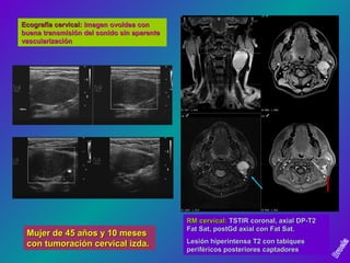
  • 1. Mujer de 45 años y 10 mesesMujer de 45 años y 10 meses con tumoración cervical izda.con tumoración cervical izda. Ecografía cervical:Ecografía cervical: Imagen ovoidea conImagen ovoidea con buena transmisión del sonido sin aparentebuena transmisión del sonido sin aparente vascularizaciónvascularización RM cervical:RM cervical: TSTIR coronal, axial DP-T2TSTIR coronal, axial DP-T2 Fat Sat, postGd axial con Fat Sat.Fat Sat, postGd axial con Fat Sat. Lesión hiperintensa T2 con tabiquesLesión hiperintensa T2 con tabiques periféricos posteriores captadoresperiféricos posteriores captadores