1. Special Clinical Advisors
Alan Altman, MD
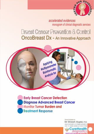
accelerated evidences
monogram of clinical diagnostic services
Clinical Compilation by
Dr Dinesh Gupta, PhD
www.CureHealth.in || info@CureHealth.in
+91 11 2646 2636 || 4105 5709
Early Breast Cancer DetectionEarly Breast Cancer Detection
Diagnose Advanced Breast CancerDiagnose Advanced Breast Cancer
Monitor Tumor Burden andMonitor Tumor Burden and
OncoBreast Dx - An Innovative Approach
Applying
Applying
Multiparameter
Multiparameter
Bioinformatic
Bioinformatic
Analysis for
Analysis for
CureHealth
IMMACULATE LAB MEDICINE SOLUTIONSIMMACULATE LAB MEDICINE SOLUTIONS
diagnostics
Exr co
e
f
l
e
l
r
en
t
c
n
e
e
C
2. CureHealth Diagnostics
(national clinical reference lab)
New Delhi - 110065. India
www.curehealth.in
withbestcomplimentsof
pushing boundaries
Indian healthcare is largely in the hands of
private sector. The prevalence of any disease
is so much over-burdening our system that
we practically have no mind set for preventive
care standards which is the need of the larger
section of our rising middle class nuclear families.
3. ABOUTUS..
Years ago, we made a tryst with our destiny..
that, we would dedicate ourselves to the cause of women’s health and in
particular, women’s cancer, in the Indian healthcare domain.
that, to make our appeal more effective and impacting in our mission, we
had to partner ourselves with key international players.
that, we needed clear deliverables that could make a difference to end-
user beneficiaries in terms of improved health standards utilising our niche
technologies...
Hence, we focussed and catalysed the transformation of cervical cancer
screening & diagnostics in India with the introduction of HPV Tests, LBC Tests
and HPV E6/E7mRNA more recently. The impact on women’s healthcare is
there to see for everyone in the past one and half decade.
This has been a tremendously encouraging journey and the great
cooperation of the entire Indian gynaecological and gyn oncological
fraternity that we ever remain indebted to.
We now take on with BREAST CANCER SCREENING AND EARLY DETECTION, as
the other major cause of women’s agony and anxiety, that has yet not been
sufficiently addressed. We wish to bring in a paradigm shift from the
subjective disease assessment in practice currently, to the more objective
and clinically meaningful disease evaluation to be able to distinctly bring
down the incidence over a next decade.
We are sure, with your continued support and encouragement, it is there for
everyone to see, yet again! Our Vision and Mission is pretty much clear.
pushing boundaries
Vision:
CureHealthwasestablishedwithavisiontopromote
packagedpreventive careintheareaoflower genital
cancers and sexually transmittable infectionsbyenhancing
awarenessamongcommonpublic,byworking with the
clinical fraternityandmakingstate-of-the-modern-care
technologies ataffordable costsformany.
“Our Mission is to incorporate advanced molecular
and cellular diagnostic procedures to improved patient
care at affordable prices by many. By being the partner
reference laboratory of choice for the clinicians to allow
accurate disease prognosis, we aim to be the most
responsive and personalized patient-oriented service
provider laboratory in India”
Mission
4. ABOUTOURTECHNOLOGYPARTNERS..
reast cancer is not a single biological entity with a distinct
Betiology and natural history. It is a genetically heterogenous
family of diseases. By quantifying multiple breast cancer
biomarkers that have established clinical significance, we deliver a
definitive and unequivocal breast cancer assay result that is
meaningfully relevant in personalised decision taking..
Our assay quantitatively measures proteomics, genomics, & cell
morphometric features, provided by none other assay procedure
today..
We have developed a first-ever biometric model to analyze the data
derived from our assay..
The results we have achieved during 2013 and 2014 conform to the
independent immunohistochemistry results..
Our Assay data has been successfully validated on stringency levels
and replicated by a reputable, independent medical research
institution in the United States..
pushing boundaries
Our Core Technology Partners
USA
Simple Technology, Simple Solutions
5. ABBREVIATIONSANDACRONYMS
ADH - Atypical Ductal Hyperplasia
AGOI - Association of Gynaecological Oncologists of India
ALND - Axillary Lymph Node Dissection
AOGIN - Asia-Oceania Research Organization in Genital
Infection and Neoplasia
BSE - Self Breast Examination
CBE - Clinical Breast Examination
CDC - Centre for Disease Control, Atlanta, USA
CT - Clinical Trials
DCIS - Ductal Carcinoma in-situ
DES - DiEthylStilbestrol
DNA - Deoxyribose Nucleic Acid
ER+/- - Estrogen Receptor Positive or Negative
FISH - Fluorescence in situ Hybridization
FNA - Fine Needle Aspiration
HBOC - Hereditary Breast & Ovarian Cancer
HDL-C - High Density Lipoprotein-Cholesterol
HR HPV - High Risk types of human papillomavirus
FFPE - Formalin-xed Parafn Embedded sections
HER2 - Human Epidermal Growth Factor Receptor 2
HRT - Hormone Replacement Therapy
HMECs - Human Mammary Epithelial Cells
IACR - Indian Association for Cancer Research
IARC - International Agency for Research on Cancer
IDC - Inltrating Ductal Carcinoma
IHC - ImmunoHistoChemistry
ILC - Inltrating Lobular Carcinoma
IMS - Indian Menopause Society
LCIS - Lobular Carcinoma in situ
MRI - Magnetic Resonance Imaging
mRNA - Messenger Ribose Nucleic Acid
PCR - Polymerase Chain Reaction
PR+/- - Progesterone Receptor Positive or Negative
RT - RadioTherapy
SLNB - Sentinel Lymph Node Biopsy
TAT - Turn-around-Time (for test result reporting)
TNBC - Triple Negative Breast Cancer
WHI - Women’s Health Initiative
WHO - World Health Organization
pushing boundariesPage 5
6. Breast Cancer Prevention & Control
Dr Dinesh Gupta,PhD
Laboratory Director
CureHealth Diagnostics
(National Clinical Reference Laboratory)
New Delhi. India
www.curehealth.in
Our POLICY STATEMENT to the
Prevention & Control of Women’s
Preventive Health in India...
We will promote the most contemporary
preventive health care in the area of women’s
cancers and sexually transmittable infections by
enhancing awareness among common public,
by working with the clinical fraternity and
making state-of-the-modern-care technologies
available at affordable costs for many in India.
By being the partner reference laboratory of
choice for the clinicians to allow accurate
disease prognosis, we aim to be the most
responsive and personalized patient-oriented
service provider laboratory in India.
Page 6
OurPolicyStatement
accelerated evidences
monogram of clinical diagnostic services
OncoBreast Dx - An Innovative Approach
7. Section Head Page
01 Foreword 08
02 The Breast Physiology 10
03 The Factors for Breast Cancer 12
04 Symptoms of Breast Cancer 15
05 Types of Breast Cancer 17
06 Breast Cancer Physio-Pathology 19
07 ER/PR & HER2/nue Status 20
08 ASCO Recommendation 23
09 OncoBreast Dx Assay 24
10 OncoBreast Dx Results 26
11 Head-to-Head Comparison 31
11 Specimen Types 33
12 Diagnostic Work Up & Assay Info 34
13 Patient Counselling 35
Page 7
Pager
Pager
8. Foreword
REAST CANCER is the foremost leading cause of women deaths in India. The
BGlobocan 2012 (IARC) statistics estimates annual incidence at 144,937 new cases,
of which nearly 87% occur in women less than 65 years of age. Going forward, it
estimates over 20% rise in the incidence; to increase to 174,706 victims by 2020. More
younger women are going to be a target of this disease, much against a belief that the
disease burden is higher among older women. What does India need to do to avert this?
SCREENING for early disease detection may set in steady decline in the incidence rate. But
an etiologic heterogeneity of breast cancer has not allowed a comprehensive and
convincing screening strategy like what HR HPV has been able to establish for cervical
cancer. While clinical experts almost everywhere agree on a pathological diagnosis of
invasive breast cancer, there is lot needed to improve diagnosing atypia, for instance,
atypical ductal hyperplasia (ADH) or ductal carcinoma in-situ (DCIS). The availability of
OncoBreast Dx as a new-gen breast cancer screen test therefore promises to ll up this
clinical deciency in India.
THE current methodologies to identify women at risk of breast cancer include regular self
examination (BSE) or clinical breast examination (CBE) by physician, mammography and
ultrasonography to ascertain primary disease. Pathologically, the disease detection is
practised by hormone tests based on diverse qualitative technologies such as ISH or FISH.
More advanced genetic tests are available using realtime PCR, gene microarray or even
gene sequencing but are expensive and clinically less relevant.
LARGER section of this monogram therefore discusses the merits of OncoBreast Dx, a new-
gen multi-parameter, single platform molecular technology that not only promises to
provide a more comprehensive pathological diagnosis, monitor tumour burden and guide
a treatment response as well. What more, the technique also allows screening for early
detection of breast cancer when the tumour could take several years to be detect by
even ultrasound.
WE are a strong nation of economically polarized population. For a larger section of our
society, affordability is challenging. We believe if the more affordable class of patients nd
means to save themselves, the most ill-affordable class of patient will benet from the
tertiary care healthcare facilities more importantly, in the public sector, reducing the
Dr Dinesh Gupta,PhD
Lab Director, CureHealth Diagnostics
· Executive Member, IACR 2009-12:
· Executive Member, ISCCP
· Life Member, AOGIN
· Life Member, AGOI
· Life Member, IMS
Page 8
9. Foreword
cancer burden on the national exchequer!
TARGETED therapies in breast cancer have considerably evolved in last three decades at
international level. The HER2 gene was identied around 1984-85 by a pioneering work by
Dennis Slaman and others, showing HER2 overexpression with poor prognoses. A targeted
therapy Trastuzumab -a synthetic monoclonal antibody- was introduced in the early 1990s
and soon became a standard of chemotherapy by late 90s. By 2000, it was applied for the
treatment of almost every metastatic HER2 positive advanced breast cancers. More
recently, we begin to see a duel targeted therapy with tratuzumab and lapatinib, taking
our understanding of HER2 positive breast cancers to a denitive level of treatment and
almost doubling the overall 5-year survival.
BREAST cancer prevention has received lot less attention but holds a major promise for the
pathologists because they play a very important role not only in its early and accurate
detection but also offering high-risk women appropriate monitoring, personalized care and
preventative therapeutic options guiding the most appropriate treatment recourse. A
comprehensive breast cancer multiple biomarker proling is poised to bring a paradigm
shift in molecular characterization of tumours based on which we will rationally make
therapeutic decisions.
WE wish to offer a novel approach in the targeted detection of early breast cancer,
assisting chemotherapy for only a selected few, saving large number of other patients who
may not benet from it. This would mean a more cost-effective approach. Going forward,
we would encourage using tailored treatment based on multiple specic breast cancer
biomarkers, and the way we look at breast cancer prevention and control.
This is also our mission statement on Women’s Health at CureHealth!!
Page 9
10. THE BREAST PHYSIOLOGY
Page 10
he Breast: The normal female breast is composed of lobules and ducts
Tembeded in to fatty connective tissue called stroma. Lobules are the milk-
producing glands and are supported by a networking of tiny tubules that
carry the milk from the lobules to the nipple. There are about 18 to 20 lobes in
each female breast, and each lobe contains smaller lobules. The entire tissue is
also supported with a net work of blood vessels for the transport of gases and
micro-nutrients as well as lymphatic system for providing necessary immunity and
defending the breast tissue from cancer.
The lymphatic system consisting of lymph nodes which are small bean-shaped
organs and lymph ducts meant for transporting a watery uid that carry immune
cells to ght any opportunistic infection. The lymphatic system gets critically
engaged in cancer spread by a process called metastasis in the advanced
stages of cancer. While the blood vessels provide oxygen and micronutrients to
the breast tissue, the lymphatic system take away the waste products so
produced as a provision of immune defence mechanism. The lymph nodes form
the clusters or nodes near the breast under arm area (axillary nodes), or above
the collarbone (clavicular nodes) and in the chest (mammary nodes).
Cancer Development: The lining of the milk ducts are primarily the site of initial
cellular changes that lead to cancer development and are identiable as
ductal carcinoma. Occasionally, the terminal lobules become a site for cancer
development and get identied as lobular carcinoma.
During the lifetime, women undergo a variety of normal breast changes as a
result of changes in the female hormone levels after the onset of puberty and all
along the menstrual phase. There are also various benign, noncancerous tissue
growths or lumps. Breast tissue is generally prone to developing cancer. Though
the clear etiology of breast cancer is yet unknown, certain inherited and/or
acquired mutations in oncogenes and tumour suppressor genes seem to affect
the normal cell cycle, inducing tumerigenesis. The female hormone, estrogen
however has a direct consequence on breast cancer development. The breast
cells divide rapidly upon a trigger by estrogen and growth factor changes. The
Lobes
Lobules
Tubules
Nipple
Lymph Node
Muscles
11. Page 11
rapidly dividing metaplastic cells are at higher risk of acquiring mutations during
their multiplication phases and that may contribute to tumorigenesis.
Most cancers are mostly lifestyle diseases and breast cancer is no exception.
The way we live – sustained stressful life, delaying marriage, postponing
childbirth, shorter breast feeding, irrational use of oral contraceptives, higher
propensity for high-fat, fast foods, smoking or excessive alcohol consumption
contribute major external factors and disturbed female hormone expressions
e.g. Estrogen and Progesterone contribute to direct internal or intrinsic factors.
While clinically breast cancers are monitored for hormone expressions for
effective treatment and cure, it is imperative that certain lifestyle related factors
are controlled by the patient herself to benet from medical interventions.
Ducts
Tumor
THE BREAST PHYSIOLOGY.2
12. THE FACTORS FOR BREAST CANCER
Page 12
reast cancer is a most important malignancy risk for any woman and
Bbecomes progressively bigger with advancing age. No woman should
consider herself too young to need a screening. Typically, the following risk
factors increase a breast cancer probability for a woman:
Family or Own History of Breast Cancer: Women who have had breast cancer
history in a close family, especially at a younger age have increased risk of
getting breast cancer. If a woman herself had a past history of breast cancer
or other breast problem she has increased risk of recurrence (other breast).
Genetic Alterations: The BrCa1 and BrCa2 are human tumour suppressor
genes that are engaged in repair of damaged DNA. Errors in these genes due
to unknown factors increase the risk of breast cancer. These genes get
mutated in some hereditary breast cancers (as well as ovarian cancers and
termed as HBOC). These genes otherwise maintain genomic stability, prevent
DNA damage and cell cycle arrest and regulate transcription process in
healthy cells. Silencing or inactivation of one copy of these genes results in
mutations (accumulation of multiple genomic damages) that induces
structural changes in the genome, increasing risk of breast cancer. There are 5
to 10% of hereditary breast cancers those result from alterations in these
genes. In some infrequent cases there are other cancer causing genes e.g.
TP53, PTEN, CHECK2 STK11 or ATM involved. Due to limited clinical utility of
genetic testing and in particular BrCa1 and BrCa2, benets for a particular
patient are still not fully understood. Some recent large-scale genomic
analyses have also uncovered dozens of common genetic variants (known as
polygenic risk score) that are associated with breast cancer but these
variants are found to be contributing only a tiny amount to a person's overall
(1)
risk of developing the disease . More common genetic variations known as
single nucleotide polymorphisms or SNPs also contribute to cancer
susceptibility, but the individual contributions are too small to predict breast
(1)
cancer risk .
1. Celine Vachon, Fergus Couch.(2015) Mayo Clinic-led researchers combine genetic variants to
improve identication of women with breast cancer. J Natl Can Inst. March 2015.
Normal
Breast
Breast
Cancer
13. Page 13
Breast Changes: Women are genetically predisposed for breast changes.
Shorter breast feeding durations also induce breast changes. Fibroadenomas
or cysts (breast lumps) are non-cancerous tumors but may hide cancer.
Having a diagnosis of atypical hyperplasia or lobular carcinoma in situ (LCIS)
may increase a woman’s risk for developing cancer.
Women with dense breasts, who use menopausal hormone therapy have
(1)
been detected at a particularly high risk of breast cancer
Longer Exposure to Estrogen: Some breast cancers are sensitive to female
hormones e.g. estrogen or progesterone. Longer a woman is exposed to
hormone in any form (as a drug, or epidermal patch), the more likely is a risk
to develop cancer. Women who begin menstruation at an early age as well
as those reaching late menopause, those on HRT for longer periods are at an
increased probability to develop breast cancer.
A synthetic form of estrogen known as DES is not in use anymore but some
elder women in their 50s and 60s now, who may have taken it during their
pregnancy 20-30 years back to prevent certain complications are at a
relatively higher risk. There is also a moderate risk for their daughters who were
exposed to DES before birth.
Late Childbearing: Women who have their rst child after about 30 or those
who never had children have a greater chance of developing breast
cancer.
Dietary Factors: While diets with high animal fat (especially HDL-C) increases
the risk of breast cancer, a regular dietary consumption of fruits and
vegetables decrease the lifetime risk considerably. Being over-weight,
especially after menopause.
Smoking and Alcoholic Beverages: Smoking and excessive consumption of
alcoholic beverages increases a risk of breast cancer, particularly the types
THE FACTORS FOR BREAST CANCER.2
1. Mammographic Breast Density and Breast Cancer Risk. Interactions of percent density, absolute
dense and non-dense areas with breast cancer risk factors. Lusine Yaghjyan et al. Breast Cancer Res
and Treat. 2015 Vol 150 (1) 181-189).
14. Page 14
affected by hormonal changes (e.g. ER+). Smoking and alcohol cause a slow
DNA damage in the healthy cells.
Radiation Therapy (RT): Younger women who were exposed to radiation
therapy for diseases such as Hodgkin’s Lymphoma or previous breast cancer
treatment are at an increased risk for recurring breast cancer.
Diabetes: Recent studies show diabetes is associated with more advanced
stage breast cancer. Breast cancer patients with diabetes were signicantly
more likely to present with advanced stage breast cancer than those without
diabetes. The results also show lower mammogram rates in women with
diabetes, which could account for later stage disease. Women with diabetes
also had a higher risk of lymph node metastases and larger tumours than
(1)
women without diabetes .
Sometimes, occurrence of a lump among the breast of adolescent girls leads to
an excisional biopsy to conrm. However, this procedure can result in pain,
scarring and breast deformity. Breast cancer is rare in adolescents, and the vast
majority of teenage breast lumps turn out to be benign masses that are related
to hormones and often go away over time.
Breast Cancer, like any other cancers is a lifestyle disease to a larger extent and
it goes together with the choices we make for ourselves. Hence by making the
right choices, we can lower the relative risk of breast cancer even if there are
uncontrollable risks for a particular woman, such as genetic predisposition.
1. Lorraine Lipscombe. Study by the Institute for Clinical Evaluative Sciences (ICES) and Women's
College Hospital. Published on March 25, 2015 at 4:52 AM. Source: Women's College Hospital. G1 06,
2075 Bayview Avenue, Toronto, Ontario M4N 3M5
Electron micrograph of a
single breast cancer cell. By
Dr Ananya Mandal,MD
THE FACTORS FOR BREAST CANCER.3
15. THE SYMPTOMS OF BREAST CANCER
Page 15
ike most other cancers, breast cancer has no denite signs or symptoms until
Lit gets in to invasive form. Early breast cancer usually does not cause pain
either. Hence it is strongly recommended for women to get themselves
screened regularly. Breast self-examination (BSE) can guide a woman to the
possible identication of early signs of breast cancer. In addition, all women
above 40 years of age should also have a mammogram and take OncoBreast
Dx at least once in ve years. OncoBreast Dx correctly helps detect breast
cancer earlier and improves your chances of beating it. No sooner a woman is
able to nd certain abnormal changes, she must ensure a clinical examination
by her doctor. Certain early identiable symptoms may include:
• A lump or thickening in or near the breast or in the underarm area. Swelling
or lump or discomfort in the armpit area.
• A change in the size or shape of one or both the breasts.
• Abnormal discharge of uid (other than milk) or blood stain from nipple or
tenderness. At times, the nipple may get pulled back (inverted) into the
breast, may develop rash, look scaly or red or aky.
• Ridges or pitting of the breast skin (the skin looks like the skin of an orange).
• Any abnormal change in the way the skin of the breast, areola, or nipple
looks or feels.
Mammograms may miss one in every four to three breast cancers with clinical
(1)
sensitivity ranging between 50 to70% with younger women and those with
(2)
dense breast mass observing yet lower sensitivity . An interval breast cancers
(cancer detected after a normal mammogram within the next one scheduled)
have also been detected with adverse prognosis when compared to women
with screen-detected breast cancers. Interval cancers are more likely to be
invasive, of a higher grade and stage and with a greater predominance of HER2
1. Pisano, E.D., et al. (2005) Diagnostic Performance of Digital versus Film Mammography for Breast-
Cancer Screening. The New England Journal of Medicine, 353, 1773-1783.
2. Berg, W.A., et al. (2012) Detection of Breast Cancer with Addition of Annual Screening Ultrasound or
a Single Screening MRI to Mammography in Women with Elevated Breast Cancer Risk. The Journal of
the American Medicine Association, 307, 1394-1404.
16. Page 16
and triple negative molecular subtypes. This heterogeneous group of tumours
may be biologically more aggressive and account disproportionately to overall
(1)
breast cancer mortality .
There is risk for every one in three women being diagnosed with a breast cancer
that may become life-threatening. Better technology to identify women who
may have a greater risk of life threatening breast cancers is now available.
CureHealth Laboratory in association with a molecular diagnostic technology
development company, IncellDx from San Francisco have introduced an early
breast cancer detection test in India, known as OncoBreast Dx which is sensitive
and specic to detect hormonal changes associated with tumour development
and is capable of detecting cancerous lump many years before even an
ultrasonography could detect it. The subsequent sections of this monogram
discusses the advantages of OncoBreast 3Dx over the conventional
investigational lab procedures.
Breast Tissue Density: Very low mammographic breast density has been
associated with poor prognosis of breast cancer. Disease free survivals as well as
overall life expectancy are signicantly shorter in women with very low-density
breasts in comparison to women with high density breast tissue. Breast tissue
density is categorised as low when the proportion of glandular tissue is below
25%, and as very low when the proportion of glandular tissue is below 10%.
The incidence of breast cancer has also been reported in men but it is about 1%
of breast cancer women develop. Women’s breast cells are highly responsive to
hormonal changes than men when fully developed post puberty.
THE SYMPTOMS OF BREAST CANCER.2
Breast Self Examination (BSE)
Image courtesy breastcancer.org
1. Meshkat B. et al. (2014) A comparison of clinical–pathological characteristics between symptomatic
and interval breast cancer. The Breast.
17. THE TYPES OF BREAST CANCERS
Page 17
reast cancers are identied as early, curable stages to metatstatic one
Bspreading from the tissue of origin to distal, unrelated parts of the body
primarily through the circulation and/ or through lymphatic system. Breast
cancer is a heterogenous form of disease. Most breast cancers begin at the
lining of the epithelial cells of the ducts or lobules. Hence they are often referred
to as the ductal or lobular carcinoma. Occasionally there are glandular
secretary cells involved in causing the adenocarcinoma. Based on molecular
characterization breast cancer subtypes are associated with distinct biological
features and clinical outcomes. They contribute to insights into cancer initiation
(1,2,3)
and progression, and guide our clinical decisions . Hence, greater accuracy
of molecular testing technologies is critical.
The following sections describe how a multi-parameter, single platform
molecular technology offered by CureHealth improves clinical outcome for
early breast cancer patients. However, the broad category of breast cancers is
outlined below.
1. Ductal Carcinoma in situ (DCIS) is the most common non-invasive breast
cancer type that begins inside the ducts and has not spread elsewhere.
Nearly 20 to 25% new are DCIS type and are almost entirely curable. A
mammogram is often able to detect it if the tumor has sufciently grown. The
multiparameter, quantitative test introduced by CureHealth, the OncoBreast
3Dx provides high specicity to detect DCIS years before they may be picked
up in mammography. It allows accurate identication of cells undergoing
malignant transformation.
2. Lobular carcinoma in situ (LCIS) is less common type wherein the abnormal
cells cluster in the lobules of the milk-producing glands. Women with LCIS may
have a 7- to 1o-fold increased risk of developing invasive cancer in one or
both the breasts. These tumours are generally good prognostic kind, being
1. Parker JS, Mullins M, Cheang MCU, Leung S, Voduc D, Vickery T, et al. Supervised risk predictor of
breast cancer based on intrinsic subtypes. J Clin Oncol. 2009;27:1160.
2. Prat A, Perou CM. Mammary development meets cancer genomics. Nat Med.2009;15:842–4.
3. Russnes HG, Navin N, Hicks J, Borresen-Dale A-L. Insight into the heterogeneity of breast cancer
through next-generation sequencing. J Clin Invest. 2011;121:3810
18. Page 18
low on histological grade, hormone receptor positive, HER2, p53 and basal
marker negative, thus usually provide a good response to hormone therapy.
3. Inltrating Ductal (IDC) or Lobular Carcinoma (ILC): IDC is most common type
of invasive ductal carcinoma that spreads through the wall of the duct into
metastasize. Similarly ILC becomes threatening when it begins to metastasize
originating from the milk producing glands. For every 8 IDC 1 or 2 LDC may get
detected.
4. Inammatory Breast Cancer: Though it is rare, inammatory breast cancers are
difcult to diagnose due to its scattered occurrence in the breast tissue
without a clear lump formation. More likely this may be missed by
mammography as well posing a serious risk to a correct prognosis. It may
make breast skin look red appearing like a peel of an orange and feel
warmer.
5. Triple Negative Breast Cancer (TNBC): In TNBC the cells lack estrogen and
progesterone receptors (ER-, PR-), and also do not have HER2 protein (HER2-)
on their surfaces. They constitute to nearly 15-20% of all breast cancers and
represent a more aggressive form with greater risk to metastasize, often not
responding to hormone therapy. Generally HRT combining estrogen and
progesterone is used for post-menopausal women to prevent osteoporosis
(weakening of bone density). Use of estrogen alone may also seen sufce for
some women but it may have a risk of uterine cancer which is prevented by
combining it with progesterone. Women who have already undergone
hsyterectomy, estrogen alone may be sufcient.
On the basis of the hormone levels and HER2 expression, breast cancers can
be sub-classied as: 1. (ER+ and HER2-) constituting to about 75%; 2. (ER- and
HER2- also known as TNBC including PR-) constituting to 15%; (ER+ and HER+)
about 7.5%; and (ER- and HER+) remaining about 7.5%.
A woman's level of testosterone also goes down as she gets older, though it's
not tied directly to menopause. Testosterone does not seem to have risk for
breast cancer.
THE TYPES OF BREAST CANCERS.2
19. BREAST CANCER PHYSIO-PATHOLOGY
Page 19
Tumor
Physio-
Pathology
Factors
Tumor Size
Lymph Node involvement
Metastasis
Histopathology
ER Status
PR Status
HER2/nue Status
Other Tests
“Personalised”
Treatment &
Management
ntil today, breast cancer physio-pathology revolved around IHC tests for
Uhormonal (ER/PR) and reexed FISH testing for HER2 expression in cases
where the HER2 level is equivocal. These tests have a great deal of
subjectivity and grading pattern for reporting. Several studies demonstrate
(1)
this may be inaccurate in 20 to 25% cases .
We know this approach is not sufcient for the vastly heterogenous nature of the
breast cancer as a disease and “personalised” care is more than necessary for
individual patient if we need to arrive at the most accurate prognosis and
therapy management. Often it leads to the additional gene expression tests
which are not only prohibitive even to the most affordable class of public as well
as clinically decient to be able to guide available options.
To address these deciencies and provide an alternative cost-affordable
approach, we bring an intact cell based diagnostic assay, OncoBreast Dx, that
is objective, with an unparallel ability to simultaneously and quantitatively
detect oncoproteins, mRNA expression, and complete cell cycle analysis on the
same sample and on a single technology platform - a true multi-parameter test
with highly reproducible results for therapy monitoring.
1. Antonio C. Wolff et al. Recommendations for human epidermal growth factor receptor 2 testing in
breast cancer: Am Soc of Clin Oncol/ CAP clinical practice guideline update. J Clin Oncol. 2013.
31(31),3997-4013 and Antonio C et al. J Clin Oncol. 2010. 28(16),2784-2795.
20. ER/PR & HER2/nue STATUS
Page 20
Popularly recognised as Triple Markers for breast Cancers. As their names imply
ER & PR are the nuclear receptors and Her2/neu is an epidermal cell surface
receptor. Together they regulate hormone and growth factor signaling
mechanism in breast cells. They are involved in cell proliferation and survival and
are thus critical in transforming normal cells to cancer cells. Most breast cancers
express ER and PR. The ER binds its ligand, estrogen (or estradiol), and the PR
binds progesterone. The ligand-receptor complex is then translocated to the
nucleus of the cell where the receptor binds specic DNA sequences in the
promoters of specic genes. Once bound to DNA, the receptor associates with
transcriptional co-regulators and consequently control the level of gene
transcription.
Biologically, estrogen sets in a normal mitotic activity in breast cells that leads to
the development of milk ducts. Estrogen also regulates the expression of the PR.
Progesterone stimulates formation of the milk glands. When the hormone
signaling is dysregulated due to over expression of these receptors, it leads to
(1)
uncontrolled cell growth and tumorigenesis . The mainstay of treatment of ER+
breast cancer is therefore to block the estrogen receptor signaling with drugs
such as tamoxifen or toremifene; to degrade the estrogen receptor by using
drugs like fulvestrant in the metastatic setting; or to prevent the production of
estrogens using aromatase inhibitors. This would suggest that blocking the
estrogen pathway, getting rid of the estrogen receptor, once one has breast
(2)
cancer, is an important arm in combating or eradicating breast cancer .
The other main breast cancer marker Her2/neu is a different kind of cell
receptor. It is a cellular membrane-bound receptor tyrosine kinase and is also
normally involved in the signal transduction pathways leading to cell
proliferation and survival. Like the hormone receptors, Her2/neu is involved in
normal breast growth and development by stimulating lobular-alveolar
development of mammary glands. Her2/neu expression is increased in 20 to 30%
ER Status
PR Status
HER2/nue Status
1. Dickson RB, Lippman ME. 1988. Control of human breast cancer by estrogen, growth factors, and
oncogenes. Cancer Treat Res; 40:119-65.
2. http://www.cancernetwork.com/podcasts/soy-breast-cancer-
connection#sthash.dydR2K4m.cbjcFTCg.dpuf
21. Page 21
of breast cancers, and may be 100 times than that of normal cells. This over-
expression of Her2/neu can disrupt the normal balance of ErB/Her2/neu
favouring more potent heterodimers. This increases proliferative and survival
(2)
signaling, potentially leading to the formation of more aggressive tumour cells .
This information therefore translates into clinically improved patient care.
The HER2/nue gene is located on human chromosome#17 and it encodes a
trans-membrane glycoprotein that functions as an epidermal growth factor cell
surface receptor. This gene has been found to be over-expressed most
commonly in invasive ductal carcinomas. Its function follows a cascade of
downstream signaling events that are important for cell growth and maintain
the transformed state. Its role as a potential target for breast cancer treatment
has recently been identied as critically important. HER-2/neu status provides
important information regarding sensitivity to certain forms of conventional
systemic therapy, particularly anthracyclines. A monoclonal antibody directed
against the HER-2/neu protein has also been developed as therapeutic agent
called as Herceptin or Trastuzumab. IHC and FISH have been seen as the most
commonly used assays for evaluation of HER-2/neu in routine clinical practice.
However, these methods suffer inherent qualitative and subjective
interpretational disadvantages. The antibodies used in IHC test vary in their
sensitivity, specicity and are based on subjective grading of results. FISH test
scores over IHC by being more specic and may be semi-quantifying but is
tedious, time consuming and expensive.
Breast cancer is a very heterogeneous disease. Several specic pathological
events are predictable by analysing the quantitative over- expression of
molecular biomarkers such as ER, PR and Her2/neu as quantitatively determined
by OncoBreast Dx Test. These expression patterns along with patient age,
tumour size, nodal involvement, tumour grade, margin status etc are used to
provide a molecular classication of breast carcinoma that potentially has
prognostic and predictive outcomes. This information is also used to determine
ER/PR & HER2/nue STATUS.2
Most women with early-stage breast
cancer now have sentinel node
biopsy (SNB) as opposed to axillary
(1)
lymph node dissection (ALND)
1. http://www.cancernetwork.com/breast-cancer/most-early-breast-cancer-patients-avoid-full-lymph-
node-removal#sthash.4j7yJfaV.dpuf
2. Yarden Y. 2001. Biology of HER2 and its importance in breast cancer. Oncology; 61 Suppl 2(1-13).
22. Page 22
the likelihood for cancer recurrence and/or guide subsequent treatment
(1,2)
options . Increase in ER-positive cells in normal lobules adjacent to tumours is
associated with increased risk for invasive breast cancer. Similarly, Her2/neu
expression in patients with benign breast lesions correlates to nearly a two-fold
(3)
increased risk of developing breast cancer . With respect to lobular neoplasia,
(4)
Her2/neu is elevated in 25% of lobular carcinoma in situ . In ductal carcinoma in
situ Her2/neu is associated with DCIS of a higher grade. Therefore, ER
andHer2/neu are important molecular markers for precursor and pre-invasive
(5)
stage management of breast cancer .
The vast nature of available data as well as emerging knowledge on HER2/nue
therefore is convincing and most promising one in breast cancer therapy ever
since its discovery about three decades ago. The OncoBreast Dx combines the
knowledge of genomics with proteomics not being offered by any other test.
Moreover, the concordance between IHC and FISH results has been found to
be better in the clear unequivocally negative or strong positive IHC breast
cancer cases but they show poorer concordance (10 to 30%) in the cases of
weakly staining IHC (borderline cases) taken for subsequent gene amplication
(6)
by FISH . Pathologically, innumerable breast biopsies in routine practice
produce results range from benign to atypical hyperplasia or carcinoma in situ
to invasive cancer with certain degree of concurrence between pathological
experts or between any two technology platforms such as IHC or FISH or gene
1. Menard S, Fortis S, et al. 2001. HER2 as a prognostic factor in breast cancer. Oncol; 61 Suppl 2(67-72.
2. Tang P, Skinner KA, Hicks DG. 2009. Molecular classication of breast carcinomas by IHC analysis:
are we ready? Diagn Mol Pathol; 18(3):125-32.
3. Stark A, Hulka BS, et al. 2000. HER-2/neu amplication in benign breast disease and the risk of
subsequent breast cancer. J Clin Oncol; 18(2):267-74.
4. Mohsin SK, O’Connell P, et al. 2005. Biomarker prole and genetic abnormalities in lobular
carcinoma in situ. Breast Cancer Res Treat; 90(3):249-56.
5. Nofech-Mozes S, Spayne J, et al. Prognostic and predictive molecular markers in DCIS: a review.
Adv Anat Pathol 2005;12(5):256-64.
6. Joann G Elmore (JAMA) http://jama.jamanetwork.com/multimediaPlayer.aspx?mediaid=9604184.
and http://www.darkdaily.com/jama-report-highlights-inaccuracies-in-pathologists-breast-cancer-
diagnoses-0325
ER/PR & HER2/nue STATUS.3
23. Page 23
array. These critical tissue diagnoses in the area of breast cancer directly
determine most suited management strategies. However such diagnoses do not
explicitly show inter relationship between cellular morphometric changes and
the disease architecture. OncoBreast Dx also lls up the a knowledge gap at a
time when medicine is becoming more evidence-based and personalized.
ASCO RECOMMENDATIONS
SCO Recommendations (2008): The ER and PR status be determined on
Aall invasive breast cancers including recurrences. A testing algorithm
that provides accurate, reproducible assay performance is proposed.
Elements to reliably reduce assay variation are specied. It is recommended
that ER and PR assay be considered positive if there are at least 1% positive
tumor nuclei in the sample on testing in the presence of expected reactivity of
internal and external controls. The absence of benet from endocrine therapy
for women with ER-negative invasive breast cancers has been conrmed in
large RCTs.
24. Page 24
®
OncoBreast Dx Testing:
Our proprietary technology...
otential to limit the current diagnostic inaccuracies inherent using
Pstandard approaches that lead to frequent mismanagement.
ollows multiple regulatory pathways that characterise breast
Fcancer heterogeneity.
ncompassing view of a cancer cell by quantifying multiple
Ebiomarkers on a ow cytometry platform.
OncoBreast Dx Assay
Our combinatory assay on breast tumors has a potential to fulll that
early step missing in current breast cancer detection.
25. ONCOBREAST DX ASSAY
Page 25
Breast Cancer detection complexities look up to some of the “latest
• and greatest” technologies to arrive at a precise prognoses &
decision-to-treat.
A priori knowledge teaches us the solution to most questions will
• involve a multi-factorial approach to nd the best solutions.
We integrate a cellular morphometric characteristics of breast cancer
• development with molecular technology and apply it to the
quantitative estimation of multi-parameters in OncoBrest Dx™ to
arrive at a precision diagnostics.
IncellDx’s patented technology, isothermal in-situ hybridization of
• target markers in intact cells for cytometric quantication, has the
ability to quantify DNA content, proteomic and mRNA targets in single
cells.
In the world of precision breast cancer diagnostics, we developed a
• OncoBrest Dx™ Test that integrates a priori knowledge while building
a platform for additional clinical knowledge.
Gene copy number analysis
Gene Sequencing
One of the greatest
challenges in
developing a
clinically useful
breast cancer assay
is to combine ALL of
these methods IN
ONE to provide a
comprehensive
assessment of
tumor environment
Gene Expression Analysis
Protein analysis
26. ONCOBREAST DX RESULTS
Page 26
OncoBreast Dx assay has been the most advanced development to provide
clinically meaningful, reproducible and fully quantiable value of HER-2/nue
gene as well as its mRNA over-expression simultaneously. It claries a subjectivity
of IHC or FISH tests thus assumes a pivotal role in deciding not only a breast
cancer type but also guiding personalised therapy that may be the most
appropriate for a particular patient.
Fig A: The y-axis exhibits the protein amount of HER2 quantitatively while the x-
axis displays mRNA expression of HER2 from 3 separate cell populations. Vast
majority of localised (<10mm) HER2+ breast carcinomas exhibit a high grade,
diffuse, and extensive in situ component, which may explain the risk of
recurrence among these tumours.
Fig B: Two cell populations as seen in the wild type status of HER2 expression. The
ability to correlate protein and mRNA on a cell by cell basis allows precise
denition of the inter-relationship seen in biological systems.
Even the most advanced gene tests produce clueless results, not clarifying:
•in what cells the genes are expressed? tumour site-specic involvement..
•how does this relate to their protein expression levels?
•what was the proliferative state of the cell with this expression?
A B
Quantitative Detection of HER2/nue mRNA & HER2/neu Protein together
27. Page 27
A Triple-Negative (TN) breast cancers, also called as Basal-like (BL), account for
nearly 15 to 20% of the disease and are negative for ER, PR and HER2. These are
more aggressive than the other types of breast cancers and are less likely to be
detected on mammograms. They grow faster, are more metastatic in nature
and likely to recur more often too. They offer poor prognoses than the ER/PR
positive ones.
Though the available treatment options currently are limited, TN breast cancers
can be successfully treated if they are detected accurately and early.
OncoBreast Dx is the only test that is capable of quantifying hormone
expressions on a single technology platform ensuring reproducibility and clinical
relevance.
Clinical Status
Method ER+/PR+, Her2- Triple Nega ve ER-, Her 2+
IHC 22 3 2
OncoBreast Dx 21 3 2
Her 2 Status
Method Her 2++ Intermediate Borderline
IHC 2 5 2 cases
2 2 Both posi ve
OncoBreast Dx
Since OncoBreast 3Dx
combines HER2 mRNA
expression and its expression
product (HER2 protein) we have
from normal tissue, expanded
HMECS along with known
tumour lines. The combination of
the two makes OncoBreast 3Dx
highly predictable.
ONCOBREAST DX RESULTS.2
28. Page 28
ER-
ER+
FISH for HER2 gene- Copy
Number Analysis..
•In the cells with extra
inserts, what else is
happening??
•Is this cell resting or
actively proliferating??
•What proteins associated
with these cells??
•Where are these cells in
the EMT??
In multiparameter
space driver mutation
is the cell with the
growth advantage.
Diploid Cell,
intact genome
Although HER2 is known
to form heterodimers
with other members of
the family, at a very
high expression levels, it
may lead to
spontaneous
homodimerisation with
its ligand which is
important for cell
growth and
maintenance of the
transformed state.
This may be clinically
important in HER2- over
expressing tumours.
IHC for HER2 protein-
There is weak membrane
staining around some of
the tumour cells. Cases
such as this are subject to
considerable differences
in interpretation. Only a
minority of cases with this
level of HER2 staining
show HER2 gene
(1)
amplication .
Insertion of
DNA with an
elevated
proliferative
rate
1. Stuart J. Schnitt, M.D, Breast Cancer in the 21st Century: Neu Opportunities and Neu Challenges.
Department of Pathology, Beth Israel Deaconess Medical Center and Harvard Medical School,
Boston, Massachusetts. The United States and Canadian Academy of Pathology, Inc. 2001. 14(3),
213-18.
ONCOBREAST DX RESULTS.3
29. Page 29
he integration of assay readout designed to handle heterogeneity
T(cytometry) with a preparative technology that maintains proteins, mRNA
expression and full cell cycle analysis on a cell-by-cell basis (IncellDx
patented technology).
The OncoBreast Dx™ addresses:
• mRNA expression with oligoprobe cocktails designed around the gene(s) in
question.
• Proteins that dene clinical subsets based on receptor expression as well as
metastatic potential and “stem-like” properties.
• Complete cell cycle analysis (genomic integrity and proliferation) rather than
genes associated with cell cycle.
Reference Documents
1. Chargin,A, Patterson,B, Shults, K and Chen, L. “Multi-Parametric Analysis of Breast Tissue Utilizing
Available Cytometric Systems” , Poster 176, 25th Annual CSU Biotechnology Symposium, Jan 3, 2013.
2. Shults,K. “An Intergrated Approach to the Proteomic and Genomic Analysis of Breast Cancer Using a
Cytometric Readout” GLIIFCA 22 September 29,2013 Detroit , Breaking Disciplines Session.
3. “A Truly Integrated Approach Applied to Breast Cancer Diagnostics: An Emerging Solution to Current
Inaccuracies”, Amanda Chargin, Bruce Patterson and Keith Shults AMP Abstract # 3077 and Poster,
Nov 2013.
ONCOBREAST DX RESULTS.4
30. Page 30
ER/PR/HER2 Pathology HER2 mRNA HER2 Protein DNA Index % E-Cadherin + ER/PR
C001045467 ER+/PR-/HER2+ 403 187 1 77% 1288.16
C001045468 ER+/PR+/HER2+ 826 191 1 50% 877,18
C001051701 ER+/PR+/HER2+ 548 114 1 15% 722.64
C001046055 ER-/PR-/HER2+ 1374 1422 2 58% 187.97
C001053362 ER-/PR-/HER2+ 1605 160 1 0% 205.1
C001054450 ER-/PR+/HER2+ 968 131 1 8% 410.81
C001053591 ER-/PR+/HER2+ 1178 164 2 4% 1178.17
HER2 mRNA HER2 Protein DNA Index % E-Cadherin + ER/PR
5 Normal Breast Cases 545 98 1 9.90% 983
5 Normal Breast Tissues Compared to 7 HER2+ Breast Tumors
The use of the FNA mimic allows us to obtain the analytical performance of
HER2 mRNA and HER2 proteins simultaneously. The use of multiple proteins can
break homogenous populations into separate functional components, giving us
a better prognosis.
OncoBreast Dx provides an early stage, pre-surgical clinically useful single
platform diagnostic tool that answers the following questions:
Is the tissue mass cancerous?
If so, is the cancer conned or local? Or is it metastatic?
Beyond the Metastatic Signature,
it helps directing therapy to each patient -“Precision Medicine”
it conrms continuing efcacy of any therapy -“Companion Diagnostics”
it provides a diagnostic template that applies equally to most epithelial
cancers with clinical usefulness - “Wider Applicability”.
Reference Documents
1. Hamed Jafarian “Breast Cancer Data Statistical Analysis” , MS-PS Final May 2014.
2. Shults, K. “Why Derivation of the Denominator is More Important in Biology than Math Class” ;
Presentation to Surgical Oncology Group MD Anderson June 2014.
ONCOBREAST DX RESULTS.5
31. HEAD-TO-HEAD COMPARISON
Comparative Analysis of Current Approach to Breast Cancer Diagnosis versus
OncoBreast Dx as a Molecular Quantitative Intact Cell-based Assay Procedure.
Page 31
IHC/FISH based Assays OncoBreast Dx Assay
Subjective slide-based Tests. IHC targets
Protein and FISH targets Gene.
(mRNA is further targeted by either realtime
PCR or gene microarray techniques).
Objective, intact cell-based assay based on
the sensitive Flow Cytometry platform
capable of quantifying veritable breast
cancer biomarkers with accuracy.
IHC detects HER2 overexpression at the
protein level, and that be affected by
conditions of the testing procedures e.g.
time and duration to xation, processing,
denaturation, heating, antigen retrieval, the
staining procedure used, and the
interpretation of staining. Although there are
antigen retrieval techniques in use, these
may result in false-positive IHC results.
FISH measures HER2 DNA. Some xatives,
chemicals or heat, may interfere with the
FISH assay. However, occasionally an
internal control is used to distinguish
between a FISH-negative and a non-
(1)
informative result .
OncoBreast Dx detects HER2nue gene and
its protein expression simultaneously, and is
unaffected by conditions of the testing
procedures. The assay procedure is highly
reproducible and the interpretation is
objective. No false-positive results.
No interfering radicles that may inuence
assay result. In built internal control with
each assay procedure.
Ideally follows ASCO clinical guidelines to
guide treatment approach for HER2 positive
tumours as well as rst-line or second-line of
(2)
therapy or metastatic tumours .
Saves costs on repeat testing or inter-lab
variable results.
1. Wolff AC, et al. Am Soc Clin Oncol/CAP guideline recommendations for human epidermal growth
factor receptor 2 testing in breast cancer. Arch Pathol Lab Med. (2007b);131-18
2. Giordano SH, et al: Systemic therapy for patients with advanced human epidermal growth factor
receptor 2-positive breast cancer: Am Soc Clin Onco Clinical Practice Guideline. J Clin Oncol 2014.
Difcult to establish a universal quality
control. Variability of specimen and their
aging adds uncertainties to quality control
measures. Methods of prociency testing
between labs differ from country to country.
For ER or PR no gold standard available.
A universal quality control is run with each
assay procedure regardless specimen type
(except for FFPE specimens). External
commercial cell culture preparations
available too.
32. HEAD-TO-HEAD COMPARISON.2
Page 32
1. Oestrogen receptor status of breast carcinoma: Allred/H score conversion table. 2008. S Shousha.
Dept of Histopathology, Charing Cross Hospital, Imperial College & Imperial College Healthcare NHS
Trust, London, UK Correspondence. Journal compilation. Blackwell Publishing Ltd, Histopathology
(2008) 53, 345–367.
2. Comparison of HER2 Status by Fluorescence in Situ Hybridization and Immunohistochemistry to
Predict Benet From Dose Escalation of Adjuvant Doxorubicin-Based Therapy in Node-Positive Breast
Cancer Patients. Lynn G. Dressler et al. J Clin Oncol (2005) 23:4287-4297.
3. Her2/neu testing in gastric cancer: evaluating the risk of sampling errors. Full Text Annals of Oncology,
11/13/2012.
4. HER2 Tests: How do we choose? Bob Carlson, MHA Senior Contributing Editor. Biotechnology
Healthcare, Sept/Oct 2008.
IHC/FISH based Assays OncoBreast Dx Assay
Different IHC centres of ER follow different
grading system e.g. McCarty’s H scoring or
Allred quick score; usually difcult to
(1)
interprete .
One standard universal interpretation of
results easier for consultants to refer to, in
order to arrive at prognostic decision. Cells
expressing ER are quantied by software.
In screening patients with esophageal
cancers for HER2 status, the relative
efciency of (IHC and FISH has been found
to be debatable.
Well suited for other GI cancers (HER2). In
screening patients with esophageal or GI
cancers for HER2 status, the efciency of the
(3)
assay is accurate and clinically relevant .
Can be performed on FFPE tissue blocks
besides other biological specimens.
Can not be performed on FFPE blocks.
OncoBreast Dx is an intact cell based assay
that needs well preserved sample
containing viable cells.
Moderate level of concordance among IHC
& FISH, even addition of PCR does not
resolve discordance. None of the three
(2)
methods is convincingly superior .
OncoBreast Dx Assay resolves inter-assay
disparities and provides objective results.
Helps identify ER/PR +/- and/or HER2nue +/-
subtypes.
A false HER2-positive comes with a price: 52 weeks of chemotherapy and
(4)
trastuzumab exceeds $50,000 plus the expense of relieving the side effects .
Variable time usually spread of couple of
days. Fastest TAT may take several days from
specimen collection to reporting.
Total hands-on time 4 to 5 hours giving
fastest TAT of 24 hours from sample
collection to report with specimen
adequacy feature. Ideal as a batch test.
33. SPECIMEN TYPES
Page 33
he following specimen types may be suitable for OncoBreast Dx
Ttesting.
1. Nipple Discharge: If a woman observes some nipple discharge, min 0.5 ml of
the uid may be collected for detecting hormone receptors or HER2nue
expression if it may contain cancerous cells. The discharge may be red or red-
brown, suggesting that it may contain blood and cancerous cells of interest.
2. Ductal Lavage and Nipple Aspiration: Ductal lavage may be collected to
screen women who are asymptomatic and are at high risk for breast cancer.
The specimen may be collected at an outpatient site. A local anaesthetic
cream is applied to numb the nipple area and 0.5 ml of uid is collected from
the ducts up to the nipple surface by gentle suction. A catheter is inserted into
a duct opening. Saline is slowly infused into the catheter to gently rinse the
duct and collect cells.
3. Fine Needle Aspiration (FNA): A small amount of ne needle aspirate from a
suspicious area may be collected. The needle can be guided into the area of
the breast change with some palpitation. A specimen may be collected by
ultrasono- guided FNA under local anaesthesia. It is important to locate the
site of disease to be able to collect some cancer cells.
4. Core Needle Biopsy: This procedure collects a small cylindrical breast tissue
guided by either ultrasono or mammography or even MRI under local
anaesthesia. Multiple core biopsies may be taken.
5. Lymph Node and Sentinel Lymph Node Biopsy: To determine if the cancer has
spread to one or more axillary lymph nodes in the case of metastatic breast
cancer a lymph node biopsy is taken to be able to provide appropriate
adjuvant therapy before or after the surgery. A good sample may contain
cancer cells at least 200 cells from 0.2 mm area. Usually axillary lymph node
dissection (ALND) leads to lymphedema after the surgery. Therefore sentinel
lymph node biopsy (SLNB) may be collected for OncoBreast Dx test.
34. DIAGNOSTIC WORK UP
Page 34
General Health Assessment: History
Menopausal
Physical Examination
Full Blood Count
LFT/ KFT/ Alk phos/ Ca
Primary Tumour Assessment: Physical Examination
Size 2 - 5cm, stages I & II) Mammography
USG
MRI
Histopath
Local Lymph Node Involvement: Physical Examination
(Size 5cm+, stage III a, b or c) USG
USG-guided biopsy
Metastatic Disease: Physical Examination
(Size 5cm++, stage IV) Other tests are not
recommended routinely unless
locally advanced or when
symptoms suggestive of
metastasis are present.
Clinical Indica ons
OncoBreastDxOncoBreast3Dx
DIAGNOSTIC WORK-UP FOR EARLY BREAST CANCERDIAGNOSTIC WORK-UP FOR EARLY BREAST CANCER
(BeingIntroducedShortly)
Test Code Test Name Parameters included
BRAC01 OncoBreast Dx Hormone Status (ER; PR), HER2/nue, HER2
Protein.
Total 4 quantitative parameters.
BRAC02 OncoBreast 3Dx
(Being introduced soon)
Hormone Status (ER, PR), HER2/nue mRNA,
HER2/nue Protein, E-Cadherin, Ploidy, Cell
Cycle, DAPI.
Total up to 19 quantitative parameters
Assay Information for Ordering Services:
35. PATIENT COUNSELLING
Page 34
Encouraging Women for Screening:
A Hospital to have a sound call-recall system before women are
enrolled for screening breast cancer. Have the ofce staff remind
patient of her upcoming appointment.
Be a good counsellor for a patient to let her know of her risk of
breast cancer, the benets of testing procedures, costs etc and
address fears.
Guide her to the lab where she can get the test done, as well as
provide her with clinical signicance of the tests advised.
If the Screening Test Suggests Breast Cancer:
Talk with your patient about the cost-benets of further tests e.g.
OncoBreast Dx or OncoBreast 3Dx and how it helps better prognosis
and disease management.
Answer questions about your patients' concerns.
Remind your patient of her upcoming appointments.
Schedule a follow-up appointment, based on the results of the tests.
If the Breast Cancer is Conformed:
Know the next step(s) and why they are needed.
Refer your patient promptly to a Breast Cancer specialist.
Keep track of her progress so she continues to see the cancer
specialist(s) and gets all necessary therapy.
Work with a patient navigator, if possible.
TM
based on the CDC’s Vital Signs Nov 2012
As per Globocan 2012 estimates:
India has 144,937 new breast cancer cases detected
annually and is likely to go up to 174,706 by the year
2020. Nearly 85% of the new cases are going to be
women under the age of 65.
Ref: Globocan 2012 (IARC) (20.10.2014)
36. BreastCancerPrevention&Control
OncoBreast Dx - An Innovative Approach
Helping you get your self esteem
Te : +91-11-2646 2636, 4105 5709
Email: info@curehealth.in
Web: www.curehealth.in
www.curehealth.in
1 in 8
di
CureHealth
IMMACULATE LAB MEDICINE SOLUTIONSIMMACULATE LAB MEDICINE SOLUTIONS
diagnostics
An ISO 9001:2008 Certified Laboratory
empoweredYou