2. Immunology and Skin in Health and Disease
Jillian M. Richmond and John E. Harris
Department of Medicine, Division of Dermatology, University of Massachusetts Medical School, Worcester,
Massachusetts 01605
Correspondence: john.harris@umassmed.edu
The skin is a complex organ that, in addition to providing a strong barrier against external
insults, serves as an arena for a wide variety of inflammatory processes, including immunity
against infections, tumor immunity, autoimmunity, and allergy. Avariety of cells collaborate
to mount functional immune responses, which are initiated by resident populations and
evolve through the recruitment of additional cell populations to the skin. Inflammatory
responses are quite diverse, resulting in a wide range of signs and symptoms that depend
on the initiating signals, characteristics of the infiltrating cell populations, and cytokines that
are produced (cytokines are secreted protein that allows for cell–cell communication;
usually refers to communication between immune–immune cells or stromal–immune
cells). In this work, we will review the skin architecture and resident and recruited cell
populations and discuss how these populations contribute to inflammation using human
diseases and treatments when possible to illustrate their importancewithin aclinical context.
Tissues at the interface of the host and the
environment, including the skin, gut, and
other mucosal surfaces, present the first line of
defense against pathogens. The barrier function
of the skin is of critical importance, which is
evident when this barrier is disrupted following
injury, or in atopic dermatitis, ichthyosis, or
irritant contact dermatitis. Once the barrier is
disrupted, the rapid but nonspecific innate im-
mune response is recruited in defense, a process
that relies on detection of both self and foreign
“danger signals” as the initial alarm. Next, the
slower, but specific adaptive immune response
may be required for definitive clearance of a
pathogen.
In addition to providing protection against
invading pathogens, the skin is also an arena
where sterile inflammation, including tumor
immunity, allergy, and autoimmune responses,
may participate in disease. Tumor immunity
is defective in organ transplant patients who
are immunosuppressed, but is co-opted dur-
ing imiquimod treatment of various skin can-
cers and warts. Allergic responses, which likely
evolved to protect against parasitic invasion,
may cause disease when directed against innoc-
uous foreign materials, as in allergiccontact der-
matitis. Autoimmunity, possibly caused byanti-
pathogen or antitumor immunity misdirected
against self, causes a wide range of pathology
in the skin, including vitiligo, lupus, psoriasis,
and other diseases.
Appropriate functioning of the skin in these
roles requires close communication and collab-
Editors: Anthony E. Oro and Fiona M. Watt
Additional Perspectives on The Skin and Its Diseases available at www.perspectivesinmedicine.org
Copyright # 2014 Cold Spring Harbor Laboratory Press; all rights reserved; doi: 10.1101/cshperspect.a015339
Cite this article as Cold Spring Harb Perspect Med 2014;4:a015339
1
www.perspectivesinmedicine.org
Spring Harbor Laboratory Press
at COLUMBIA UNIVERSITY on December 9, 2014 - Published by Cold
http://perspectivesinmedicine.cshlp.org/
Downloaded from
3. oration among a number of various cell types,
including stromal cells (keratinocytes, fibro-
blasts, endothelial cells, and adipocytes) as well
as those derived from the bone marrow (den-
dritic cells, macrophages, natural killer cells,
mast cells, T cells, and others). Of the bone mar-
row–derived cells that can be found in the
skin, some are resident cell populations that
migrate to the skin where they terminally dif-
ferentiate and primarily reside there, some re-
circulate continuously and perform a surveil-
lance role, some are recruited to fight infection
for the short term, and others are recruited and
maintained as memory cells to protect against
future reinvasion. Bone marrow–derived cells
can be further subdivided into innate and adap-
tive immune populations (see Fig. 1). Innate
cells form rapid, but nonspecific, responses
to infection. They generally recognize non-
self-molecular patterns on pathogens or path-
ogen-associated molecular patterns (PAMPs),
through receptors called pattern-recognition
receptors (PRRs are intracellular or cell surface
receptors activated by DAMPs to induce in-
flammation). Adaptive immune populations
form slower, but pathogen-specific, responses
to infection through specialized and unique
antigen-specific receptors formed via genetic
rearrangement (an antigen is any molecule ca-
pable of inducing an antibody response or T-
cell response; it can be protein, lipid, or carbo-
hydrate). These receptors permit immune cells
to mount a more targeted attack on invaders,
and these cells can then become long-lived and
capable of a rapid, specific response against
reinvasion of a pathogen, known as a memory
response.
Initiationofanadaptiveimmuneresponseis
performed by antigen-presenting cells (APCs),
which efficiently take up (phagocytose) pro-
teins, process them into recognizable peptides
(antigens), and present them to T cells on sur-
Innate immune cells
Skin-resident cells
Recruited immune cells
Innate immune cells
Granulocytes
Neutrophil Eosinophil
NK cell Monocyte T cell
Adaptive immune cells
B cell
Adaptive immune cells
T cell
(usually memory,
NKT, or γδ)
Nonhematopoietic origin
Keratinocyte
Fibroblast
Endothelial cell
Langerhans cell
Dermal dendritic cell Mast cell
Macrophage
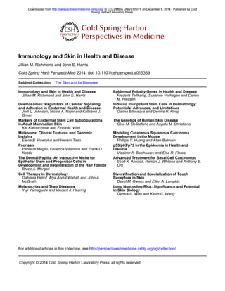
Figure 1. Immune populations in the skin. (Top) Langerhans cells, dermal dendritic cells, macrophages, other
innate cells (mast cells, NK cells, NKT cells, gd-T cells), and memory T cells comprise the skin-resident immune
system under the steady state. Langerhans cells, dermal dendritic cells, macrophages, and gd-T cells first sense
infection or injury and initiate a rapid, innate response that includes the recruitment of effector cells. Innate
effectors (NK populations) provide a rapid, antigen-nonspecific response, whereas memory T cells provide a
rapid, antigen-specific memory response to previously encountered pathogens. Stromal cell populations, such
as keratinocytes, fibroblasts, and endothelial cells, also participate in immune responses by sensing tissue
damage and producing inflammatory cytokines. (Bottom) On activation of the skin-resident immune system,
additional immune cells are recruited to help contain and fight infection and/or to remove cellular debris to aid
in the healing process. These include additional innate cells like neutrophils and eosinophils, as well as adaptive
populations like naı̈ve or central memory T cells and B cells.
J.M. Richmond and J.E. Harris
2 Cite this article as Cold Spring Harb Perspect Med 2014;4:a015339
www.perspectivesinmedicine.org
Spring Harbor Laboratory Press
at COLUMBIA UNIVERSITY on December 9, 2014 - Published by Cold
http://perspectivesinmedicine.cshlp.org/
Downloaded from
4. face human leukocyte antigen (HLA) Iand HLA
II molecules (human leukocyte antigen class I
and II; typically, class I presents intracellular an-
tigens and class II presents extracellular anti-
gens). All nucleated cells in the body express
HLA I, providing an important mechanism for
detection of viral infections as well as malignant
transformation through direct communication
with immune cells. HLA I presents antigens that
are produced within the cell and permits their
recognition by antigen-specific T cells through
theirT-cellreceptor,makingthecellssusceptible
to cytotoxic T-cell-mediated killing. Typically,
if those antigens are from normal self-proteins,
they will be spared, a process called immunolog-
ical tolerance. However, if those antigens are de-
rived from intracellularpathogens (like viruses),
or if the antigen is an abnormal self-protein
generated following malignant transformation
of the cell, then that cell may be killed to pre-
vent further injury to the host. Unlike HLA I,
HLA II is typically only expressed on APCs,
and is capable of presenting antigens derived
from extracellular proteins acquired through
phagocytosis (cell eating; the process by which
antigen-presenting cells and other phagocytic
cells take up pathogens or other debris). Den-
dritic cells (DCs) are recognized as professional
APCs because their main function is to take up
antigens, thereby linking innate and adaptive
immune responses. If DCs phagocytose a path-
ogen in the presence of a PAMP that alerts them
to danger, they then produce proinflammatory
mediators to recruit innate immune cells to ini-
tiate the defense, and then typically migrate out
of the skin through lymphatic channels into
draining lymph nodes. There they position
themselves to directly encounter T cells, and
each T cell will briefly probe the DC to deter-
mine whether it is presenting an antigen that the
T cell recognizes. If so, the T cell will become
activated and initiate a search for the location
of the infection.
In this article, we will describe how all of the
cell populations within the skin contribute to
immune responses, and the general principles
and tools required for effective immunity. We
will use specific examples of common skin dis-
eases and treatments when possible to illustrate
how the immune system works within a clinical
context.
NORMAL SKIN IMMUNE SYSTEM
Tissue Architecture and Composition
Normal skin architecture includes the epider-
mis, dermis, and subcutaneous fat (see Fig. 2).
The resident cell populationsthat make up these
strata can be broadly divided into immune and
nonimmune cells. Nonimmune cell popula-
tions are important for the structure and func-
tion of the skin, but also contribute to skin im-
munity, as they first provide a general barrier
to invasion by foreign materials. They also
directly participate in inflammation, shaping
the immune response as it develops within the
tissue.
Epidermis
The epidermis is comprised primarily of kera-
tinocytes, which are tightly connected to one
another and act similarly to a brick wall to limit
access to the internal environment. As a conse-
quence, these tight connections limit move-
ment within this layer and therefore other cell
types that reside there, which include both stro-
mal and bone marrow–derived cells, are pri-
marily fixed in position. However, when dam-
age occurs and immune cells sense danger, they
release proinflammatory mediators to recruit
innate effector cells, and then exit to draining
lymph nodes where they encounter T cells and
other members of the adaptive immune system.
Cellular Components of the Epidermis
† Keratinocytes maintain tight junctions and
form the stratum corneum, which is critical
for the barrier function of the epidermis (see
Box 1). They may also contribute to inflam-
mation, as they can express HLA II and se-
crete cytokines.
† Melanocytes are pigment-producing cells in
the skin. The melanin pigment they produce
and distribute to keratinocytes helpsto shield
DNA from ultraviolet radiation, which can
Skin Immunology
Cite this article as Cold Spring Harb Perspect Med 2014;4:a015339 3
www.perspectivesinmedicine.org
Spring Harbor Laboratory Press
at COLUMBIA UNIVERSITY on December 9, 2014 - Published by Cold
http://perspectivesinmedicine.cshlp.org/
Downloaded from
5. induce DNA damage. Melanocytes are also
capable of expressing MHC II, and are the
targets of autoimmunity in vitiligo.
† Merkel cells are specialized cellsthat commu-
nicate with cutaneous neurons in skin sen-
sation. A clear contribution of Merkel cells
to cutaneous immunity has not yet been de-
scribed.
Bone Marrow–Derived Cells of the
Epidermis
† Langerhans cells (LHCs) are dendritic cells
(DCs) that spend the majority of their time
in the epidermis. LHCs are tightly connected
to keratinocytes through dendritic processes
that radiate in all directions, allowing them to
probe throughout the entire epidermis. The
exact role of LHCs is not entirely clear, al-
though they may promote tolerance to envi-
ronmental antigens, including commensal
bacteriaandfungi,andhelptopolarizeTcells
into a particular inflammatory response as
described below. Although keratinocytes
andmelanocytesmayalsobecapableofacting
as APCs, the implication of this function is
not yet clear.
† Memory T cells may reside within the epi-
dermis for very long periods of time. Where-
as neutrophils and T cells can infiltrate the
epidermis under some circumstances (atopic
and contact dermatitis, cutaneous T-cell
Epidermis
Dermis
Blood vessel
Subcutaneous fat
Adipocyte
Nerve bundle
Extracellular matrix
protein
Merkel cell
Figure 2. Location of immune cells within normal skin architecture. Distinct populations of immune cells
inhabit local niches within the skin. The epidermis contains Langerhans cells to provide immune surveillance.
Memory T cells are also retained in the epidermis, presumably for early detection and control of re-encountered
pathogens. Keratinocytes may sense pathogens or other damage-associated signals and communicate this to the
immune system through cytokines. The dermis contains dermal dendritic cells, T cells, and fibroblasts. In this
example, we depict a T cell being recruited out of the blood and into the dermis and potentially the epidermis,
depending on the site of infection/injury. Recruitment of cells to peripheral tissues requires production of
chemoattractants. Endothelial cells, which form the walls of the vasculature, may present chemokines and/or
adhesion molecules to immune cells to direct their transmigration into the skin.
BOX 1. SKIN BARRIER DYSFUNCTION
Filaggrin is a protein that multimerizes, binds to keratin fibers, and strengthens connective tissues,
helping to form a tight barrier against exposure to environmental agents. It is essential to barrier
function within the epidermis. In atopic dermatitis and ichthyosis, filaggrin mutations cause barrier
dysfunction, resulting in chronic inflammation and opportunistic infection.
J.M. Richmond and J.E. Harris
4 Cite this article as Cold Spring Harb Perspect Med 2014;4:a015339
www.perspectivesinmedicine.org
Spring Harbor Laboratory Press
at COLUMBIA UNIVERSITY on December 9, 2014 - Published by Cold
http://perspectivesinmedicine.cshlp.org/
Downloaded from
6. lymphoma, psoriasis, and vitiligo), they are
typically excluded.
Dermis
The dermis is primarily comprised of extracel-
lular matrix proteins that give the skin structure
and elasticity. Unlike the epidermis, it permits
the free migration of cell populations. The der-
mis and epidermis are separated by the base-
ment membrane, a thin, tight sheet of extracel-
lular matrix proteins that regulates movement
of cells and proteins in between these two layers.
Cellular Components of the Dermis
† Fibroblasts produce structural proteins,
which in addition to providing a supporting
scaffold, serve as highway systems for migra-
tory immune cells. As in the lymph node,
these highways ensure that immune cells
frequentlycontacteachother,whichisimpor-
tant in communication during immune re-
sponses.Likekeratinocytes,fibroblastsareca-
pable of producing cytokines.
† Endothelial cells form the innermost layer of
the blood vessels in the skin, and regulate the
passage of immune cells into the skin
through the production of adhesion mole-
cules, cytokines, and chemokines (a cytokine
that acts as a chemoattractant to induce cell
migration) (see also Box 2).
† Neurons form nerve bundles in the skin,
which allow for sensation. Recently, it has
been shown that memory T cells can interact
with neurons to regulate innate immunity
(Rosas-Ballina et al. 2011). Neuroimmunol-
ogy is a relatively new field, and the relation-
ship between skin neurons and immune cells
is not yet fully appreciated.
Bone Marrow–Derived Cells of the Dermis
Innate populations of the dermis:
† Dermal DCs (dDCs) and plasmacytoid DCs
(pDCs), two distinct populations of den-
dritic cells, are located in the dermis. They
have fewer dendrites but increased motility
compared with LHCs, and are capable of mi-
grating on collagen paths to monitor the der-
mis. Each DC population has been character-
ized by the production of different cytokines,
and may initiate distinct inflammatory re-
sponses following activation. For example,
pDCs have been reported to initiate antifun-
gal immunity (Ramirez-Ortiz et al. 2011),
whereas dDCs initiate antiviral immunity
(Kaplan 2010). These roles are not likely to
be exclusive, such that different DC popula-
tions may contribute to different immune
responses in multiple ways.
† Macrophages are skin-resident immune cells
with high phagocytic capacity and motility.
Although they are less likely than DCs to pre-
sent antigen to T cells, due to relative cell
numbers, they are capable of activating im-
mune responses through PRRs and cytokine
secretion. They also “clean up” debris from
dead or dying cells, invading pathogens,
or environmental insults, including tattoo
BOX 2. LFA-1 AND LEUKOCYTE ADHESION
LFA-1 is expressed by endothelial cells and promotes adhesion of leukocytes, permitting their mi-
gration into peripheral tissues. A defect in the adhesion molecule CD18, a component of LFA-1,
results in leukocyte adhesion deficiency (LAD). LAD is characterized by chronic bacterial skin
infections owing to impaired neutrophil chemotaxis. In addition, the CD11a component of LFA-1
was the target of the drug efalizumab for psoriasis, which presumably functioned by inhibiting
immune cell migration into the skin. Predictable side effects were infections of multiple organs,
including a potentially lethal JC virus infection within the brain, which led to its withdrawal from the
market in 2009.
Skin Immunology
Cite this article as Cold Spring Harb Perspect Med 2014;4:a015339 5
www.perspectivesinmedicine.org
Spring Harbor Laboratory Press
at COLUMBIA UNIVERSITY on December 9, 2014 - Published by Cold
http://perspectivesinmedicine.cshlp.org/
Downloaded from
7. ink. For example, “melanophages” represent
macrophages that have phagocytosed mela-
nocyte fragments and melanin released into
the dermis following epidermal inflamma-
tion.
† Monocytes are a type of immature macro-
phages that are usually found in the circula-
tion. They can be recruited to the skin to
maintain homeostasis or in response to in-
fection/injury, where they receive cues to dif-
ferentiate into macrophages or myeloid DCs,
a DC population that is not well understood.
† Granulocytesincludeneutrophils(alsocalled
polymorphonuclear cells or PMNs), eosino-
phils, basophils, and mast cells. These innate
immune cell populations can be recruited to
the skin following activation of a tissue-resi-
dent cell and subsequent release of chemo-
kines and activation of the endothelium.
Granulocytes are named for their cytoplas-
mic granules that are filled with proteases,
vasoactive peptides, and antimicrobial pep-
tides, which are released during degranula-
tion. Neutrophils are typically the first cell
typerecruited tothe skin following activation
of dendritic cells and/or macrophages in re-
sponse to PAMPs encountered during infec-
tion, and are capable of efficiently phagocy-
tosing and killing pathogens (see Box 3).
Neutrophils have also recently been shown
to make “sticky” extracellular traps (NETs)
by expelling the DNA from their nucleus, ef-
fectively trapping pathogens like flies caught
in a spider web. Eosinophils and basophils
contribute to antiparasitic and allergic re-
sponses, although we are just beginning to
understand their unique roles in immunity.
Eosinophils degranulate when their IgE re-
ceptors become cross-linked, and they release
proteases, chemokines, and vasoactive pro-
teins. Basophils, which also degranulate in
this manner, can act as potential APCs.
Mast cells are typicallyskin-resident granulo-
cytes that mediate immune responses against
parasites following binding and cross-linking
of IgE antibodies bound to parasitic invad-
ers, followed by degranulation of histamines
and other proinflammatory proteins. They
have also been implicated in the pathogenesis
of allergic responses when IgE antibodies are
produced against innocuous environmental
antigens, like dust mite proteins and animal
dander.
† Natural killer (NK) cells are classified as in-
nate cells because of their pattern-recogni-
tion functions, but may also confer memory,
like adaptive populations. NK cells detect the
level of self-HLA I expressed on the surface
of cells, and are activated when expression
levels are too low, which occurs when either
malignant or virally infected cells impair the
expression of HLA I to escape immune de-
tection by T cells. NK cells can also perform
antibody-dependent cellular cytotoxicity
(ADCC) by binding IgG-coated pathogens
and cells, releasing granules containing per-
forin and granzyme.
Adaptive populations of the dermis:
† Most of the skin-resident adaptive cells are
T lymphocytes (T cells) of different subsets.
Thereareapproximately20billionTcellspre-
sent in the skin, nearly twice the number of T
cells in the blood (Clark 2010) (a lymphocyte
is a type of adaptive immune cell including T
and B cells, capable of genetically rearranging
antigen-specific receptors). CD8þ
cytotoxic
T cells (CTLs) are effector cells that recognize
a specific antigen presented on a cell surface
by HLA I, and subsequently kill that cell.
BOX 3. CHRONIC GRANULOMATOUS DISEASE
Neutrophils kill pathogens through the use of a “respiratory burst,” which depends on NADPH
oxidase to generate oxygen free radicals. This gene is defective in chronic granulomatous disease
(CGD), and patients with CGD develop chronic skin bacterial and fungal infections.
J.M. Richmond and J.E. Harris
6 Cite this article as Cold Spring Harb Perspect Med 2014;4:a015339
www.perspectivesinmedicine.org
Spring Harbor Laboratory Press
at COLUMBIA UNIVERSITY on December 9, 2014 - Published by Cold
http://perspectivesinmedicine.cshlp.org/
Downloaded from
8. CD4þ
helper T cells (TH cells) “help” effector
cellpopulations like CTLsand Bcellsthrough
the production of cytokines and promatura-
tion signals to DCs, which both license the
effector cells to carry on an attack, and steer
the response in a particular direction, either
antiviral, antitumor, antibacterial, or anti-
parasitic, as described in more detail below.
This TH-cell help provides an important
check to the development of an immune re-
sponse, essentially licensing other antigen-
specific T and B cells before allowing the re-
sponse to develop. T-regulatory cells (Tregs)
are typically CD4þ
and express FoxP3, a crit-
ical transcription factor for their develop-
ment (see Box 4). Their role is to suppress
immune responses as a major contributor
to peripheral tolerance, helping to prevent
autoimmunity and to resolve inflammation
once a threat has been controlled. As men-
tioned above, any of these cell populations
may be temporary, migrating through the
skin for surveillance, or may remain long
term as skin-resident memory T cells (TRM)
to prevent reinfection.
† gd-T cells, which have a limited repertoire of
T-cell receptors (TCRs), and natural killer T
cells (NKTs), which are classified by their
expression of both NK cell receptors and
the universal T-cell marker CD3, are other
T-cell populations of the dermis. Both pop-
ulations respond to lipid antigens.
† Once activated, B lymphocytes (B cells) ma-
ture into antibody-producing plasma cells
that usually reside in the lymph nodes and
bone marrow, producing antibodies that be-
come passively delivered to the skin through
the circulation. These antibodies may medi-
ate either infectious or autoimmune re-
sponses, depending on their specificity.
Whereas B cells and plasma cells can occa-
sionally be found in the skin (syphilitic infec-
tion, lupus, and other diseases), the reason
for this localization to the skin is unknown.
Subcutaneous Fat
The subcutaneous fat layer is mainly comprised
of adipocytes, but also contains nerves, blood,
and lymphatic vessels. Adipocytes are fat cells
that sequester potentially inflammatory fatty
acids in the form of lipids. They are also capable
of producing proinflammatory cytokines.
Soluble Proteins of the Skin Immune System
† There are several families of proteins that are
importantforskinimmunity.Complementis
a family of soluble plasma proteins that can
bind to pathogens, self-catalyze to form solu-
ble chemoattractants and membrane-bound
opsonins that promote phagocytosis, and
membrane attack complexes that can punc-
ture bacteria (see Box 5). Complement fixa-
tion to the bacterium can occur by three
different pathways: classical, alternative, or
lectin. Classical activation of complement is
catalyzed by antibodies bound to the patho-
gen, alternative activation occurs through di-
rect binding of complement to the pathogen,
and lectin activation is catalyzed by mannose
binding lectin(MBL)boundtothepathogen.
Phagocytes express complement receptors
that recognize complement bound to an in-
vading organism, which facilitates phagocy-
BOX 4. IPEX SYNDROME
Immunodysregulation polyendocrinopathy enteropathy X-linked syndrome (IPEX) is a genetic dis-
ruption of the FoxP3 transcription factor, which is important for the generation of Tregs. Tregs are
critical for the maintenance of peripheral tolerance to self-tissues and thus in the prevention of
autoimmunity. Without this, patients develop multiple autoimmune diseases, including enteropathy;
insulin-dependent diabetes mellitus, thyroid disease, and other endocrinopathies; and skin disease
including eczema, psoriasiform dermatitis, urticaria, and alopecia universalis, among others.
Skin Immunology
Cite this article as Cold Spring Harb Perspect Med 2014;4:a015339 7
www.perspectivesinmedicine.org
Spring Harbor Laboratory Press
at COLUMBIA UNIVERSITY on December 9, 2014 - Published by Cold
http://perspectivesinmedicine.cshlp.org/
Downloaded from
9. tosis of the invader regardless of howcomple-
ment was first fixed to the surface (a form of
opsonization, which is the process of coating
with proteins that facilitate phagocytosis).
† Antimicrobial peptides are another family of
proteins that contribute to skin immunity.
Examples include b-defensins, psoriasin,
and lysozyme, which have varying but direct
antimicrobial activity against bacteria and
fungi. Keratinocytes are one of the main
sources of antimicrobial peptides, which are
induced by TLR ligation and/or cytokine
production.
† Interferons (IFN) belong to a family of cyto-
kines that are proinflammatory and also di-
rectly“interfere”withviralreplicationthrough
reducing protein translation and increasing
p53 levels, which promotes apoptosis.
† Antibodies bind with high specificity and af-
finity to both foreign proteins during an in-
fectious immune response, or self-proteins in
autoimmunity. They can injure cells by pro-
moting phagocytosis through opsonization,
complement-mediated lysis, activation of
signaling cascades, and through neutraliza-
tion of surface proteins.
INITIATION AND EVOLUTION OF AN
IMMUNE RESPONSE IN THE SKIN
DCs regularly take up proteins within the skin,
but must distinguish whether they are presented
in a dangerous context like infection or malig-
nancy, or a safe context like normal cell turn-
over during tissue homeostasis. A number of
molecular patterns alert immune cells to dan-
ger. These patterns are typically present only
when normal cellular processes have been dis-
rupted, as seen in infection, malignancy, or
necrotic cell death. These patterns are broad-
ly referred to as danger-associated molecular
patterns (DAMPs; can be pathogen-derived or
altered-self signals), microbe-associated molec-
ular patterns (MAMPs), viral-associated molec-
ular patterns (V
AMPs), or pathogen-associated
molecular patterns (PAMPs), depending on
from where they are derived. Examples include
lipopolysaccharide (LPS), flagellin, and pepti-
doglycan from bacteria, double-stranded RNA
from viruses, or mislocalized (extranuclear)
double-stranded DNA during necrotic cell
death. The receptors that recognize these pat-
terns include Toll-like receptors (TLRs) and
NOD-like receptors (NLRs), and are expressed
by DCs, other immune cells, and sometimes
stromal cells. There are both surface and intra-
cellular PRRs to allow for both extra- and intra-
cellular recognition of pathogens. Activation of
these receptors by DAMPs promotes antigen
processing and presentation, up-regulation of
costimulatory receptors for T-cell activation,
and secretion of proinflammatory cytokines in-
cluding IL-6, IL-1b, TNF-a, and others. There-
fore, it is these “danger signals” that initiate a
proinflammatory response.
On activation, tissue-resident cells secrete
proinflammatory cytokines that promote the
recruitment of innate effector cells, including
neutrophils, monocytes, and NK cells. These
first responders can begin the attack using the
mechanisms described above. However, an ef-
fective immune response usually requires the
BOX 5. COMPLEMENT ABNORMALITIES
Genetic defects in complement can result in too much or too little complement activity. Loss-of-
function mutations ultimately result in recurrent infections because of decreased opsonization and/
or decreased lytic activity. Common infections resulting from complement deficiency include recur-
rent meningococcal infection (Neisseria subspecies), Streptococcus pneumoniae infection, or other
encapsulated bacterial infections. Hereditary or autoimmune angioedema results from too much
complement activity caused bya loss of the C1 esterase inhibitor. As a result, complement activation
occurs spontaneously, which induces production of bradykinin. Bradykinin is a vasoactive peptide
that results in blood vessel dilation, thereby causing the edema and other symptoms of the disease.
J.M. Richmond and J.E. Harris
8 Cite this article as Cold Spring Harb Perspect Med 2014;4:a015339
www.perspectivesinmedicine.org
Spring Harbor Laboratory Press
at COLUMBIA UNIVERSITY on December 9, 2014 - Published by Cold
http://perspectivesinmedicine.cshlp.org/
Downloaded from
10. subsequent involvement of the adaptive im-
mune system, and so DCs must interact with a
multitude of T cells to identify the correct cells
with the capacity torecognizethe specificinvad-
ing pathogen. Because very few T cells are capa-
ble of responding to any particular challenge,
DCs must migrate to a draining lymph node,
the equivalent of Grand Central Station in
New York, to most efficientlysurvey the millions
of potential responders and find those that can
participate in any particular response.
Following the successful activation of an an-
tigen-specific T cell, that cell will proliferate, exit
the lymph node into the blood stream, and
search for the location in the body that contains
its target. The DC will help to direct the T cell to
the skin through a mechanism that is not fully
understood, called“imprinting”;however,itap-
pears that vitamins play a role. Vitamin D is
produced primarily within the skin following
exposure to UV light, and therefore skin DCs
contain large amounts of this vitamin. When T
cells are activated by DCs containing vitamin D,
the T cells express skin-homing receptors, in-
cluding CCR4, CCR10, and CLA. A similar T-
cell educational program occurs via intestinal
DCs that contain vitamin A, which is acquired
through the diet, inducing a6b4 protein, a gut-
homing receptor. Once the T cells are in the
bloodstream and express skin-homing recep-
tors, the initial cytokines and adhesion mole-
cules on endothelial cells at the site of inflam-
mation help to guide those cells to the correct
site. Although activated B cells, which produce
antibodies, may also be recruited to the skin
during inflammation, they more often remain
within the lymph nodes or bone marrow, secret-
ing the antibody into the blood where it is pas-
sively carried to the skin and contributes to the
immune response.
As described above, T-cell subsets have spe-
cialized functions, including those that are di-
rectly cytotoxic, others that suppress responses,
and still others that oversee and help to shape
the response through the secretion of cytokines.
TH subsets are usuallydesignated with a number
to delineate which types of cytokines they pro-
duce. For example, TH1 cells produce interfer-
on-g (IFN-g) and tumor necrosis factor (TNF);
TH17 cells produce IL-17, IL-21, and IL-22; and
TH2 cells produce IL-4, IL-5, and IL-13. These
different cytokine patterns are usually associat-
ed with recruitment of slightly different types
of immune effector populations. For example,
TH2 responses recruit basophils, eosinophils,
and mast cells to coordinate an antiparasitic
response, whereas TH1 responses result in re-
cruitment of CTLs for an antiviral or antitumor
response, and TH17 promotes an antibacterial
or antifungal response through the recruitment
of neutrophils and production of cytokines and
antimicrobial peptides. TH1 and TH17 respons-
es may also promote autoimmunity, whereas
TH2 responses may mediate allergy (see also
the section on Diseased Skin). For efficiency
and efficacy, immune responses often polarize
toward a single specific pathway; however,
mixed responses may also occur, creating a sig-
nificant level of complexity that has yet to be
fully understood for all such responses in vivo.
The subpopulation of DC that initiates the
immune response and the signals present dur-
ing the initiation can influence the nature of that
response. For example, LHC may suppress im-
mune responses, acting as tolerogenic media-
tors because they regularly sample foreign pro-
teins and organisms present on the intact skin
surface, which are primarily nonthreatening.
When LHC-deficient mice are exposed to acon-
tact allergen, the response is exacerbated, sup-
porting this suppressive role of LHCs in the
skin. In contrast, dDC-deficient mice elicit a
dampened response, suggesting this dDC pop-
ulation is proinflammatory in this context. This
role is intuitive, because exposure of dermal
DCs to a foreign antigen would require disrup-
tion of the epidermis, which is a potentially
dangerous event. Recent studies also support a
nuanced proinflammatory role for these two
populations. Whereas activated LHCs seem to
promote an antibacterial/antifungal immune
response through the production of TH17-spe-
cific cytokines, dDC promote antiviral respons-
es through the production of IFNs and other
TH1-specific cytokines. This may be because of
the fact that viruses often enter the skin systemi-
cally or through epidermal disruption and
therefore primarily enter the dermis, whereas
Skin Immunology
Cite this article as Cold Spring Harb Perspect Med 2014;4:a015339 9
www.perspectivesinmedicine.org
Spring Harbor Laboratory Press
at COLUMBIA UNIVERSITY on December 9, 2014 - Published by Cold
http://perspectivesinmedicine.cshlp.org/
Downloaded from
11. bacteria most often enter through the epider-
mis. Little is known about the mechanism by
which DCs distinguish these stimuli, although
it is likely mediated through PRRs.
Turning off Inflammation and Initiating
Wound Healing
Animportantpartofnormalimmuneresponses
isturning off inflammation once the infection is
cleared or injury is healed. Skin-resident Treg
populations have been shown to be activated
by epidermal LHC (Seneschal et al. 2012), and
playan important role in dampening inflamma-
tion.ThereareseveraldifferentTreg populations
that are usually subdivided into central and pe-
ripheralsubtypes.WhereascentralTregsdevelop
in the thymus, peripheral Tregs are generated
during immune responses in lymphoid organs
and/or tissues, where asymmetric division of
effectorcellsallowsforgenerationofasmallpop-
ulation of Tregs. Examples of these include TH3
and TR1 cells, which produce the anti-inflam-
matorycytokine IL-10.Both centralandperiph-
eral Tregs are capable of down-modulating in-
flammation via cytokine production, uptake,
and removal of the T-cell growth factor IL-2,
and through DC interactions. Interleukin-10
(IL-10) is an anti-inflammatory cytokine that
will down-regulate the expression of other cyto-
kines, MHC II, and costimulatory molecules.
Tregs express the high-affinity IL-2 receptor,
CD25, and will therefore preferentially bind
and “sop up” IL-2 in the milieu, thereby remov-
ing the T-cell growth and survival signal. Tregs
are also capable of directly interacting with DCs
and inducing down-regulation of costimulatory
molecules through cell–cell interactions. Tregs
also produce transforming growth factor-b
(TGF-b), which can both inhibit immune cell
proliferation and stimulate fibroblast produc-
tion of extracellular matrix proteins (see Box
6). In both the skin and the gut, Tregs have
been shown to play an important role in main-
taining tolerance to normal bacterial flora. Ithas
recently been postulated that Treg sensing of flo-
ra is also important for normal wound healing
(Chen et al. 2013).
DISEASED SKIN
Proper Immune Responses in Disease—
Infectious Immunity
Infectious Immunity: Part I. Examples of
Bacterial Immunity
Staphylococcus aureus is a prevalent human skin
commensal and pathogen that can cause super-
ficial skin infections (impetigo and exacerba-
tion of atopic dermatitis), infection of the hair
follicle (folliculitis and furunculosis), as well as
deep infections (ecthyma and abscesses). Like
most other bacterial infections of the skin, these
conditions are characterized clinically by ten-
der, red, inflamed pustules and abscesses that
form as a result of cytokine expression, neutro-
phil recruitment, and an epidermal response,
including keratinocyte proliferation and pro-
duction of antimicrobial peptides. Twenty per-
cent of the population is colonized with S. au-
reus, and infections like those described above
are quite common.
Methicillin-resistant S. aureus (MRSA) is a
particularly recalcitrant infection due both to
its antibiotic resistance as well as multiple viru-
lence factors (a component of a pathogen that
permits infection and survival; can be protein,
lipid, carbohydrate, or nucleic acid) (reviewed
by Foster 2005). S. aureus is a Gram-positive
coccus, which has an outer cell wall. Innate im-
BOX 6. IMIQUIMOD TREATMENT OF WARTS
Imiquimod is a synthetic TLR-7/8 agonist that can be used as adjuvant therapy. In the case of HPV,
imiquimod can induce production of type I IFN, IL-6, and TNF to overcome the lack of inflammatory
mediator production and actively suppress Treg function. An additional approach to treating warts is
intradermal injection of Candida antigens, which are natural TLR ligands.
J.M. Richmond and J.E. Harris
10 Cite this article as Cold Spring Harb Perspect Med 2014;4:a015339
www.perspectivesinmedicine.org
Spring Harbor Laboratory Press
at COLUMBIA UNIVERSITY on December 9, 2014 - Published by Cold
http://perspectivesinmedicine.cshlp.org/
Downloaded from
12. mune receptors and PRRs important in the de-
tection of Gram-positive bacteria include Toll-
like receptor 2 (TLR-2), and mannose-binding
lectin (MBL), which are capable of detecting
the sugars and lipids that comprise bacterial
cell walls. As described in detail above, ligation
of PRRs associated with skin-resident immune
cells induces activation and maturation of DCs,
keratinocyte proliferation and antimicrobial
peptide production, and the production of cy-
tokines and chemokines to promote the recruit-
ment of neutrophils and macrophagesto the site
of infection. Once in the skin, neutrophils and
macrophages phagocytose and destroy the bac-
teria. Asmentioned above,phagocytosisisaided
byopsonizationwithantibodiesandthebinding
of complement. Once bacteria are taken into the
phagosome, it fuses with the lysosome resulting
inacidification,activationofproteases,andpro-
duction of reactive oxygen species, all of which
are capable of breaking down the pathogens.
This process also results in the generation of
antigens, which can be presented on HLA II to
activate the adaptive immune response. During
this process, some macrophages and DCs will
migrate to the draining lymph node to activate
Tand B cells via the HLA II–antigen complexes
for initiation of the adaptive immune response.
Activation of T cells and TH17 skewing seems
to be particularly important for antibacterial
immunity. This is attributable to the functions
of TH17-associated cytokines: IL-17, 22, and 23
can promote thickening of the epidermis, pro-
duction of antimicrobial peptides, and recruit-
ment of neutrophils, which further enhances
bacterial clearance.
S. aureus has developed several mechanisms
to avoid the host immune response. It can avoid
phagocytosis through the virulence factors Pro-
tein A (interferes with IgG binding to its FcgR),
ClfA (promotes coating with fibrinogen, which
outcompetes the binding of opsonins includ-
ing complement), and capsule formation (pro-
motes biofilm formation via polysaccharide
intercellular adhesion), which inhibits comple-
ment and antibody binding. It can also prevent
phagolysosomal fusion via the virulence fac-
tor SarA, thereby evading death and surviving
within the immune cells themselves. Inside the
host phagocytes, S. aureus can also scavenge free
radicals via superoxide dismutases. The unique
structure of its wall is resistant to degradation by
lysozyme. These factors may in part explain why
itis sodifficult to induce goodadaptiveimmune
responses to S. aureus, as there could be less ef-
ficient antigen generation and presentation.
S. aureus produces the chemotaxis inhibi-
tory protein of staphylococci (CHIPS), which
can bind to and inhibit chemoattractant recep-
tors on the surface of neutrophils. It also pro-
duces Eap, a protein that disrupts LFA-1 and
ICAM-1 interactions that are required for neu-
trophils to adhere to endothelial cells and trans-
migrate into the skin (see also Box 2). The bac-
teria are also capable of preventing complement
fixation through production of a Staphylococcus
complement inhibitor (SCIN), Efb, or through
Staphylokinase, an enzyme that promotes deg-
radation of complement, antibodies, and clots.
As mentioned above, complement is important
not only for phagocytosis of bacteria, but also
for neutrophil chemotaxis. By inhibiting com-
plement fixation, therefore, the bacteria are not
only avoiding complement-mediated lysis and
opsonization, but also potentially reducing the
number of neutrophils that are recruited to the
skin by the complement proteins C3a and C5a.
S. aureus is capable of expressing nucleases,
which can cut neutrophilic NETs comprised of
DNA discussed above, in addition to lipases and
proteases, which supports their dissemination.
Another way that S. aureus can avoid the
host immune response is by invading epithelial
and endothelial cells and living inside them in
a semi-dormant state. Intracellular pathogens
usually require cell-mediated immunity to clear
infection. However, S. aureus has also developed
mechanisms to evade T cell and other leukocyte
responses, namely through production of tox-
ins and superantigens. The a-toxin is capable of
inserting into host membranes and multimeriz-
ing to form a pore, much like the way comple-
ment can form a pore in bacterial cell walls.
Other toxins include the Panton–Valentine leu-
kocidin (PVL), which can lyse leukocytes, and
the leukocidin D/E, leukocidin M/F, and the g-
hemolysin (Hlg), which can lyse erythrocytes
and leukocytes. Recurrent furunculosis and
Skin Immunology
Cite this article as Cold Spring Harb Perspect Med 2014;4:a015339 11
www.perspectivesinmedicine.org
Spring Harbor Laboratory Press
at COLUMBIA UNIVERSITY on December 9, 2014 - Published by Cold
http://perspectivesinmedicine.cshlp.org/
Downloaded from
13. other conditions have been associated with PVL
expression by S. aureus. S. aureus also produces
one of the best-characterized superantigens,
which nonspecifically activates T cells via strong
binding of both the MHC II and the T-cell re-
ceptor. Toxic shock syndrome toxin-1 (TSST-1,
which can initiate tampon-associated toxic
shock syndrome) prevents the generation of
normal T-cell responses against the bacterium
and also affects the ability to generate antibody
responses by B cells, which often need T cell help
for appropriate class switching and activation.
In light of all of the ways in which S. aureus
can evade immune responses, it is easy to see
why drug-resistant strains pose a threat. Immu-
nocompromised patients are especially vulner-
able (see Box 3), making it of utmost impor-
tance to understand skin immunity to bacteria.
Infectious Immunity: Part II. Examples
of Viral Immunity
Antiviral immunity largely depends on NK and
CTL responses. Both NKs and CTLs kill their
targets viaperforin/granzyme-inducedapopto-
sis. The inflammatory response to most viral
infections of the skin consists of minimal in-
flammationandsymptomscausedbythetarget-
ed destruction of virally infected cells and there-
foreusuallylackredness,swelling,orpusthatare
characteristic of bacterial or fungal infections.
TH1-type cytokines are important for antiviral
immunity because they drive CTL responses
and antiviral mechanisms through production
of IFN-g. We will discuss human papilloma vi-
rus (HPV) as an example of antiviral immunity
in the skin. HPV is the most common STD; it is
estimated that 50% of sexually active men and
womenget infected.The majorityofpeoplewho
contractHPVcleartheinfectionwithin2 yr,and
not everyone that has HPV will develop cancer,
asthisisstraindependent. Inthissection,wewill
highlight concepts of antiviral immunity and
ways in which HPV can subvert the host im-
mune response.
More than 70 different strains of HPVexist.
HPV belongs to the papillomaviridae family of
nonenveloped DNA viruses. HPV productively
infects keratinocytes, and takes over the cellular
machinery to manufacture progeny viruses. The
HPVreplication cycle is tied to keratinocyte dif-
ferentiation, in which HPV early proteins (“E”
1–7) are produced in undifferentiated keratino-
cytes and late proteins (“L” 1 and 2), which are
involved in capsid formation, are produced in
more superficial cells to promote sloughing of
virus. Some HPV strains can be transmitted sex-
ually and cause genital warts, whereas some can
cause warts on other parts of the skin. Different
HPV strains are adapted to infect specific ana-
tomic locations. For example, strain 1 usually
infects the soles of the feet, strain 2 the palms
of the hands, strains 6 and 11 are associated with
genital warts, and strains 16 and 18 can cause
cervical cancer. Recently, two vaccines were de-
veloped against HPV: Gardasil (types 6, 11, 16,
and 18) and Cervarix (types 16 and 18) in hopes
of preventing most cervical cancers (90% con-
tain HPV DNA).
HPV produces proteins E6 and E7 that in-
hibit host p53 and Rb, respectively, which nor-
mally are involved in sensing DNA damage
and repair mechanismsto halt cell-cycle progres-
sion. This is accomplished via ubiquitin-medi-
ated proteasomal degradation. P53 and Rb are
known as the “guardians of the genome” be-
cause the DNA damage-sensing mechanism is
important for cells to maintain the health of the
genome and to avoid improper growth. Because
viruses need to use the host cell machinery to
replicate, overriding these proteins allows them
to replicate more efficiently. A byproduct of this
is the hyperproliferation of keratinocytes that
characterizes a wart. HPV can also avoid IFN-
mediated antiviral responses through E7, which
can bind to and inhibit the promoters of type-I
IFN-related genes. Treatment with IFN-acan be
used for genital warts, although patients with
higher E7 levels tend to respond less well than
patients with low E7. E6 can down-regulate IL-
18 expression, which is important for the gen-
eration of TH1 and CTL responses.
Productive antiviral immunity depends on
NK cell responses, CTL responses, and antibody
production. NK cells look for the presence or
absence of self HLA I on the surface of cells,
whereas CTLs respond to specific HLA I–pep-
tide complexes. To avoid detection by CTLs,
J.M. Richmond and J.E. Harris
12 Cite this article as Cold Spring Harb Perspect Med 2014;4:a015339
www.perspectivesinmedicine.org
Spring Harbor Laboratory Press
at COLUMBIA UNIVERSITY on December 9, 2014 - Published by Cold
http://perspectivesinmedicine.cshlp.org/
Downloaded from
14. many viruses have developed the ability to
down-regulate HLA I. For example, the E5 pro-
teinofHPViscapableofdown-regulatingHLAI
expression via inhibition of tapasin and HLA I
promoter binding by transcription factors. Yet
even though the chance of activating a CTL is
decreased by E5, an NK cell could still kill an
HPV-infected target cell. NK cells receive acti-
vating signals through killer activating receptors
(KARs),whereasinhibitorysignalsaretransmit-
ted through killer inhibitory receptors (KIRs),
which detect presence of HLA I on the cell’s
surface. Therefore, it is the balance of positive
and negative signalsthat the NK cell receivesthat
will determine whether or not it will kill the
target cell. However, not many NK cells are re-
cruited to sites of HPV infection, as the virus
has devised other mechanisms to subvert in-
flammatory responses. Part of this is because
of the sequestration of the virus inside primarily
differentiated keratinocytes, which are located
high in the epidermis. HPV induces the recruit-
ment of CD4þ
CD25þ
FoxP3þ
Tregs via CCL17
andCCL22productionbyLHCandmacrophag-
es within the wart. Consistent with their func-
tion discussed above, these Tregs can dampen
inflammation and promote viral survival. In ad-
dition, Tregsspecificfor HPVantigens havebeen
found in cervical cancer patients (see Box 6).
Antibody responses toviruses like HPV help
prevent initial infection or spread of infection
via neutralization of binding of the virus to the
host cells. Yet the availability of HPV antigens
during infection is often low owing to the fact
that, unlike other viruses, HPV does not induce
lysis of its target cells to allow for release of vi-
rions thereby producing free antigens. Addi-
tionally, the L proteins are most immunogenic
but are primarily produced in superficial kera-
tinocytes, thereby allowing the virus to avoid
detection by most LHC, which reside closer to
the basement membrane. Therefore, antibody
responses to HPV are often delayed and inade-
quate. Some LHC and other APCs are able to
take up HPV antigens either through phagocy-
tosis of cellular debris or possibly through exo-
somes, which are nanometer-sized structures
secreted by the cell. However, HPV infection is
not a highly inflammatory process, and there-
fore lacks stimulation of APCsto mature, secrete
proinflammatory cytokines or promote effector
T-cell activation. Several HPV proteins mimic
host proteins and therefore can be tolerogenic.
(Subversion of host immunity by HPV is nicely
summarized by Tindle [2002].)
Infectious immunity: Part III. Examples
of fungal and yeast immunity
Antifungalimmunityreliesheavilyontheinnate
immune system, and protective immunity de-
pends on both TH1 and TH17 responses as well
as antibody production. Like S. aureus, Candida
albicans colonizes much of the population,
but is primarily pathogenic in immunocompro-
mised patients, where disease incidence is 24
cases per 100,000. Treatments for Candida are
antifungal drugs, such as miconazole and flu-
conazole, which inhibit formation of fungal cell
membranes. Several antifungal vaccines have
made it to phase I clinical trials, howeverachiev-
ing both efficacy and safety has proven difficult.
PRRs that are important for recognition of
Candida include TLRs 2, 4, and 6, as well as
lectin-likereceptorsincludingdectins,galectins,
DC-SIGNandthemannosereceptor.Allofthese
PRRs recognize sugar and lipid components of
the fungal cell wall, such as b-glucan, zymosan,
and chitin. Complement receptor 3 (CR3) is
important for recognizing opsonized Candida
and other pathogens. TLR-9, an intracellular
PRR, is important for recognition of fungal
DNA, which is hypomethylated compared with
mammalian DNA (recognition of Candida is
summarized by Netea et al. 2008). Skin-resident
macrophages and DCs use these PRRs to detect
Candida, and initiate the recruitment of neutro-
phils and monocytes via cytokine and chemo-
kine production. Recruited populations of neu-
trophils and monocytes phagocytose Candida
and kill it via generation of reactive oxygen spe-
cies. Extracellular killing is also induced, al-
thoughthe mechanismfor this is not yet known.
As described above, skin-resident LHCs
have been reported to initiate TH17 cell respons-
es. Interestingly, ligation of different PRRs
generates different TH profiles: ligation of the
mannose receptor usually results in IFN-g pro-
Skin Immunology
Cite this article as Cold Spring Harb Perspect Med 2014;4:a015339 13
www.perspectivesinmedicine.org
Spring Harbor Laboratory Press
at COLUMBIA UNIVERSITY on December 9, 2014 - Published by Cold
http://perspectivesinmedicine.cshlp.org/
Downloaded from
15. duction and TH1 responses, chitin recognition
and fungal DNA recognition results in IL-4 and
IL-13 production and TH2 responses, and hy-
phae recognition by dectin 1 results in IL-17 pro-
duction and TH17 responses. Treg responses can
also be generated through tolerogenic DC pop-
ulations and TRIF-dependent signaling down-
stream of some PRRs. Most of these associations
have been determined using PRR knockout
mouse models of Candidiasis. However, little
is known about how these mixed T-cell respons-
es ultimately influence the host’s ability to clear
the pathogen, although it seems that early gen-
eration of Treg responses allows Candida to sub-
vert host immunity. It is possible that confusing
the immune system by inducing these mixed T-
cell responses also dampens host immunity ow-
ing to a lack of positive-feedback loops. One
possible mechanism for this differential induc-
tion of immune responses would be the mor-
phological changes that occur when Candida
transitions from yeast to hyphae, which results
in differences in bioavailability of PAMPs from
the cell wall. Understanding this modulation of
DCs by different fungal epitopes is important
for the design of effective antifungal vaccines.
Infectious Immunity: Part IV. Examples
of Parasite Immunity
Immunityagainstparasitesismediatedbyamix-
ture of innate and adaptive immune responses
that rely on IgE, granulocytes, and TH2 cells.
Parasites present challenges to the immune sys-
tem because of several factors, including their
size and complexity, migration to different tis-
sues within the body, and ability to subvert the
host immune response. These immune evasion
mechanisms have made it notoriously difficult
todevelopvaccinesagainstparasites.Wewilldis-
cuss skin immune responses to the hookworm
as an example of antiendoparasite immunity
(see Box 7 for a note on ectoparasites). Hook-
worms are nematodes that belong to the fam-
ily Ancylostomatidae, and infect approximately
20% of theworldpopulation. The larvae burrow
into the skin, often through the feet that come
into contact with contaminated soil or water.
As they mature, they travel through the blood
to the lungs and eventually the intestines, where
they feed off of blood and reproduce. Hook-
worm eggs are secreted in feces, and embryos
develop and hatch in the soil or water where
they consume bacteria until they develop into
the infective worm stage, L3, thus completing
the life cycle (reviewed by Loukas and Prociv
2001).
On entering the skin, L3 hookworm larvae
shed their outer cuticle and begin expressing
enzymes that permit their movement through
tissues. Cuticle or sheath antigens can be taken
up by APCs and presented to T and B cells.
Initially following skin invasion, zoonotic spe-
cies elicit inflammatory responses that cause
a creeping eruption or ground itch, whereas
anthropophilic species can do so silently. Anti-
body responses have been detected to the cuticle
as well as the exsheathing fluid, and it has been
suggested that this allows the worm to misdirect
the immune response, similar to countermea-
sure decoys to distract a heat-seeking missile.
Antibody responses to fluid and cuticle proteins
may be used clinically as a diagnostic tool for
detecting infection. Although T-cell responses
seem to be weak and their exact specificities are
unknown, TH2-cell responses against hook-
worms result in production of IL-4 and IL-13,
which induce B-cell class switching to IgE. In-
terleukin-5 (IL-5), a major eosinophil survival
and activation factor, is also produced by TH2
BOX 7. ARTHROPOD BITES AND IMMUNITY TO VECTOR-BORNE DISEASES
Many insects have specialized salivary proteins, which allow them to consume a blood meal without
inducing coagulation, and some have developed proteins that inhibit immune responses. Research is
being conducted to examine the effects of an insect bite on cytokine and chemokine production in
the skin.
J.M. Richmond and J.E. Harris
14 Cite this article as Cold Spring Harb Perspect Med 2014;4:a015339
www.perspectivesinmedicine.org
Spring Harbor Laboratory Press
at COLUMBIA UNIVERSITY on December 9, 2014 - Published by Cold
http://perspectivesinmedicine.cshlp.org/
Downloaded from
16. cells. Eosinophils are recruited to sites of infec-
tion and are major players in antiparasite im-
munity. They bind IgE-opsonized larvae via the
Fc1R, causing them to degranulate. Enzymes,
and the reactive oxygen species that they release,
degrade larvae. Once the larvae matures into an
adult worm in the intestine, immune responses
seem to become elevated most likely in response
to the greater availability of worm antigens.
Coinciding with this, peripheral eosinophilia
is often seen in patients with hookworm infec-
tions. Tissue-resident mast cells also degranu-
late on encountering a larvae or worm, which
may result in the recruitment of more eosino-
phils. Their mechanism of activation is slightly
different from that of eosinophils in that they
may have IgE preloaded in their Fc1Rs. IL-9
seems to be important for mast-cell activation
and production of proteases.
In addition to misdirecting antibody re-
sponses to their cuticle, hookworms have de-
veloped several other mechanisms to subvert
the host immune response. They produce a neu-
trophil inhibitory factor protein (NIF), which
interferes with neutrophil recognition of opson-
ized parasites; C-type lectins that mimic those
of the host, which interfere with immune re-
cognition and coagulation; metalloproteinases
and peptides that interfere with coagulation and
permit feeding; cysteine proteases that cleave
Igs and the low-affinity IgE receptor; and aspar-
tic proteases, which help them digest hemoglo-
bin but also may be involved in cleaving Igs and
complement. They also produce protease in-
hibitors and antioxidants that help them with-
stand the effects of degranulation byeosinophils
and mast cells. Hookworms also produce ace-
tylcholinesterases, which are thought to both
inhibit gut peristalsis and interfere with im-
mune function.
Improper Immune Responses in Disease
Improper Immune Responses: Part I. Allergy
Allergy is an improper immune response to an
otherwise innocuous antigen. The incidence of
allergic disease is rising in developed countries,
and treatments cost $400 million annually in the
United States alone (see Box 8). Allergic contact
dermatitis occurs following chemical or envi-
ronmental exposure, resulting in generation of
neoantigens via haptenization of self-molecules
(reviewed by Kaplan et al. 2012). Haptensthem-
selves can cause oxidative stress in keratino-
cytes, resulting in release of reactive oxygen
species and danger signals, such as ATP
. Neo-
antigens produced in response to hapten ex-
posure, such as byproducts of hyaluronic acid
degradation, can activate TLRs, resulting in pro-
duction of proinflammatory cytokines by skin-
resident immune cells. Studies in mice have indi-
cated that recognition of neoantigens is mediated
by TLR-2 and/or TLR-4 in IL-12-dependent and
-independent mechanisms. It is also thought that
NLRs and the inflammasome are capable of
recognizing haptens and neoantigens, although
many ligand-receptor partners have yet to be de-
termined. Repeated exposure induces sensitiza-
tion and subsequent type IV hypersensitivity
(see Box 9). This response is characterized by an
influx of T cells to the skin, resulting in tissue
damage and an inflammatory eruption.
Tissue penetration of haptens is crucial for
induction of allergic contact dermatitis. Close
to 3000 compounds have been discovered that
are capable of inducing contact dermatitis.
Small molecular weight compounds can diffuse
through the epidermis, although disruption of
the barrier function of keratinocytes is thought
to speed up the sensitization process (see Box
1). Examples of compounds that can elicit al-
BOX 8. HYGIENE HYPOTHESIS
The hygiene hypothesis, which was first proposed by Strachan in 1989, states that exposure to
pathogens in childhood helps to develop normal, balanced immune responses. Growing up in
ultraclean environments, therefore, results in the failure to appropriately modulate Th2 responses
in childhood, thereby creating a predisposition to allergic immune responses.
Skin Immunology
Cite this article as Cold Spring Harb Perspect Med 2014;4:a015339 15
www.perspectivesinmedicine.org
Spring Harbor Laboratory Press
at COLUMBIA UNIVERSITY on December 9, 2014 - Published by Cold
http://perspectivesinmedicine.cshlp.org/
Downloaded from
17. lergic contact dermatitis include nickel, which is
the most common contact allergen and can
bind directly to TLR-4 (see Box 10 and Schmidt
et al. 2010), and the dust mite allergens Der p 2
and Der f 2, which are homologs of the en-
dogenous TLR-4-binding protein MD2. Dust
mites, or Dermatophagoides subspecies, con-
sume sloughed keratinocytes, and are often
found in clothing, bedding, and on the skin.
Dust mites often cause allergic responses attrib-
utable to the type of immune responses they
elicit: IgG and IgE responses confer immunity
but also hypersensitivity reactions. Tissue-resi-
dent mast cells also play a role in skin allergy,
caused by their ability to degranulate after their
surface-loaded IgE is cross-linked on an aller-
gen encounter. Whereas systemic and topical
steroids are sometimes used to manage acute
symptoms, the preferred treatment for contact
dermatitis is avoidance of the allergen.
Improper Immune Responses: Part II.
Autoimmunity
Autoimmunity results when the immune sys-
tem targets self-tissues, resulting in destruction,
and, potentially, organ failure. There are check-
points to prevent the immune system from tar-
geting self-tissues; and, therefore, autoimmune
disease is believed to require multiple hits that
deactivate those checkpoints (similar to the
multihit hypothesis of cancer development).
These mechanisms include central tolerance
through deletion of autoreactive T and B cells
in the thymus (see Box 11), peripheral tolerance
through the action of CD25þ
Tregs (see Box
4), production of anti-inflammatory cytokines,
such as IL-10 and TGF-b, and down-modula-
tion of proinflammatory cytokine production
by ligation of certain PRRs on innate and tis-
sue-resident immune cells, such asthe phospha-
tidyl serine receptor on macrophages, which
aids in uptake of apoptotic cells in a physiologic
context. Because autoimmunity is a destructive
process and does not appear to be beneficial for
survival, these responses likely evolved as over-
zealous anti-infectious or antitumor responses.
For example, Japanese have a very low incidence
of psoriasis compared with U.S. or European
populations, whereas their risk for tuberculosis
is much higher, suggesting that their genetic
makeup puts them at low risk for psoriasis but
BOX 9. HYPERSENSITIVITY REACTIONS
† Type I hypersensitivity—immediate hypersensitivity (15–30 min, but can be delayed up to 10–
12 h); IgE-mediated, involves mast cells, basophils, and eosinophils (e.g., asthma).
† Type II hypersensitivity—cytotoxic hypersensitivity (min-h); IgG and IgM antibody plus comple-
ment-mediated (e.g., blood group incompatibility).
† Type III hypersensitivity—immune-complex hypersensitivity (3–10 h); circulating IgG com-
plexed with antigen deposits on basement membranes (e.g., arthus reaction/serum sickness).
† Type IV hypersensitivity—delayed type hypersensitivity (peaks at 48 h); memory T-cell mediated
(e.g., contact dermatitis).
BOX 10. NICKEL ALLERGY
One of the most common triggers of contact hypersensitivity is nickel, which is present in jewelry,
orthopedicmaterials,andcoins.Schmidtetal.(2010)showedthatnickelis aninorganicligandfor the
TLR-4–MD2 complex. On binding to histidine residues in human TLR-4, nickel can induce cross-
linking of the receptor that induces a proinflammatory signal via the MyD88 adapter protein. This
leads to subsequent cytokine production and elicitation of contact hypersensitivity.
J.M. Richmond and J.E. Harris
16 Cite this article as Cold Spring Harb Perspect Med 2014;4:a015339
www.perspectivesinmedicine.org
Spring Harbor Laboratory Press
at COLUMBIA UNIVERSITY on December 9, 2014 - Published by Cold
http://perspectivesinmedicine.cshlp.org/
Downloaded from
18. high risk for tuberculosis. IFIT1 is a component
of interferon signaling, and is a risk allele for
type I diabetes, but the high-risk allele for dia-
betes is protective against coxsackie viral infec-
tion, suggesting that this allele evolved to pro-
tect from infection. Native Americans were
devastated by tuberculosis infection on arrival
of Europeans—the descendants of those who
survived now have a very high risk for rheuma-
toid arthritis, suggesting that tuberculosis sur-
vivors possessed aggressive immune responses
that now promote autoimmunity. These exam-
ples support the hypothesis that autoimmu-
nity developed as a consequence of evolving
potent anti-infectious responses. Therefore, we
will categorize autoimmune responses accord-
ingly. In addition to improving our understand-
ing of pathogenesis, an advantage to thinking
about autoimmunity in this way is that treat-
ments used to interfere with autoimmunity are
likely to increase the risk for infections con-
trolled by that response.
In this section, we will discuss both T-cell-
driven autoimmune disease and B-cell-driven
autoantibody-mediated disease in the context
of improper antiviral/tumor, or antibacterial/
fungal immune responses.
Antibacterial-Like Autoimmunity
Psoriasis, which afflicts 2%–3% of the world
population, is a highly inflammatory disease
of the skin that presents with pruritus, pain,
erythema, occasional pustules, and thickened
scales. Histologically, it is characterized by epi-
dermal acanthosis, vascular proliferation, and a
significant inflammatory infiltrate consisting of
neutrophils,Tcells,DCs,andotherpopulations.
Keratinocytes produce significant levels of an-
timicrobial peptides, and the cytokines IL-23,
IL-17, IL-6, and IL-22 appear to play a promi-
nent role in pathogenesis. These characteristics
mirror those seen in an antibacterial response
and, in fact, lesions of psoriasis rarely become
superinfected, unlike in atopic dermatitis. This
resistance to infection is probably caused by this
overzealous antibacterial-like response.
Treatment for psoriasis includes local, gene-
ral immunosuppressive medications like topical
steroids and calcineurin inhibitors, systemic ge-
neral immunosuppressants like cyclosporine A
and methotrexate, as well as newer, more target-
ed systemic biologic medications like TNF-a
inhibitors, p40 (a component of IL-23) inhibi-
tors, and IL-17 inhibitors. Although these treat-
ments are likely to have improved safety profiles
over more general immunosuppressants, they
still have rare, but predictable, side effects based
on their ability to interfere with the antibacteri-
al response, including an increased incidence of
bacterial and fungal infections. A more detailed
discussion concerning psoriasis and its treat-
ments is addressed in the literature.
Antiviral-Like Autoimmunity
Vitiligo is an autoimmune disease of the skin
that results in the loss of melanocytes from the
epidermis, which can be quite psychologically
devastating for patients. It affects 0.5% of the
population worldwide, without preference for
race or gender, and targets all areas of the skin,
with a preference for the face and genitals great-
er than hands and feet greater than trunk and
proximal extremities. The patchy depigmenta-
tion istypicallysymmetrical except in the case of
BOX 11. APECED SYNDROME
Autoimmune polyendocrinopathy-candidiasis-ectodermal dystrophy (APECED) syndrome results
from a genetic disruption of the transcription factor autoimmune regulator (AIRE). AIRE is expressed
by specialized cells in the thymus and induces the expression of tissue-specific self-antigens from
peripheral tissues to allow for negative selection of autoreactive T cells as they develop. Without
AIRE, patients are unable to delete autoreactive T cells, resulting in development of multiple auto-
immune diseases including autoimmune skin diseases, such as alopecia, vitiligo, nail dystrophy, and
enamel hypoplasia.
Skin Immunology
Cite this article as Cold Spring Harb Perspect Med 2014;4:a015339 17
www.perspectivesinmedicine.org
Spring Harbor Laboratory Press
at COLUMBIA UNIVERSITY on December 9, 2014 - Published by Cold
http://perspectivesinmedicine.cshlp.org/
Downloaded from
19. segmental vitiligo, in which unilateral depig-
mentation typically remains localized and lim-
ited by the midline. Segmental vitiligo affects
5% of all vitiligo patients but is responsible
for a larger component of childhood vitiligo,
affecting 20%. Patients with vitiligo are typi-
callyasymptomatic,withonly 20%complain-
ing of mild itching in lesional skin.
On physical exam, there is typically no ery-
thema or scale, signs seen only in the rare in-
flammatory subtype. Histological examination
may reveal a subtle infiltrate made up of CD4þ
and CD8þ
T cells in the absence of neutrophils
or epidermal proliferation. Often, lesional skin
contains few T cells. Because keratinocytes re-
tain the pigment that they acquire from mela-
nocytes as they differentiate through the layers
of the epidermis until they are sloughed off,
melanocyte destruction does not result in visi-
ble depigmentation until complete turnover of
the overlying keratinocytes, a process that re-
quires 14–48 d. During this time, destructive
T cells likely migrate laterally through the skin
to perpetuate disease, and therefore are not
present once the depigmented epidermis be-
comes clinicallyvisible.Thus, biopsy recommen-
dations to “catch” an immune infiltrate include
selecting perilesional skin up to 1 cm beyond
the visible border, a slightly hyperpigmented
region beyond the border, or better, an elliptical
biopsy radiating outward from the lesion, in-
cluding the border itself and up to 1 cm beyond.
Vitiligo is mediated by CTLs and IFN-g
production, reflecting a TH1 immune response
within the skin. Unlike some autoimmune dis-
eases where it has been difficult to determine
which antigens the autoreactive T cells are re-
sponding to, the TCR specificities in vitiligo
have been determined for a number of peptides,
including MART-1/Melan-A (melanoma anti-
genrecognizedbyTcells),tyrosinase(anenzyme
required for melanin production), and gp100
(also known as premelanosome protein, atrans-
membrane protein expressed on the surface of
pigment-producing cells in the skin and eye).
This clinical, histological, and mechanistic pic-
tureissimilartowhatisseeninresponsetoaviral
infection or antitumor response, in contrast to
psoriasis. Consequently, attempts to treat vitili-
gousinganti-TNF-ainhibitorshavebeendisap-
pointing, and no other systemic therapies for
vitiligo are widely available. Typical treatments
for vitiligo include topical steroids and calci-
neurin inhibitors, as well as narrow-band UVB
light therapy. New approaches to treatment will
likely benefit from targeting IFN-g and other
components of the TH1 inflammatory pathway.
Antibody-Mediated Autoimmunity
The skin is affected bya numberof autoimmune
diseases that are clearly mediated by antibodies.
These diseases can be subdivided according to
whether they are tissue specific, or tissue non-
specific. Tissue-specific diseases are relative-
ly straightforward, as the antibodies interfere
with protein–protein binding, and the symp-
toms develop from a lack of protein func-
tion. Tissue-specific antibody-mediated auto-
immune diseases of the skin include pemphigus
vulgaris, pemphigus foliaceus, bullous pemphi-
goid, epidermolysis bullosa acquisita, linear
IgA, and others. Pemphigus vulgaris is mediated
by antibody production against desmoglein 3,
which is predominantly expressed in the basal
epidermis and acts as a glue to hold keratino-
cytes together. The result is separation of the
epidermis just superior to the basal keratinocyte
layer, which then accumulates fluid and forms a
bullaonthesurfaceoftheskin.Incontrast,pem-
phigus foliaceus is mediated by desmoglein 1
antibodies, forming bullae in the superficial lay-
ers of the epidermis and mucosae, because
of predominant expression of desmoglein 1 at
that location. Because these diseases are primar-
ily antibody mediated, they require little contri-
bution from immune cells during the effector
phase apart from the plasma cells that produce
the antibodies. As mentioned above, despite be-
ing a B-cell-driven autoimmune disease of the
skin, B cells are not typically found within the
skin, but reside within the lymph nodes and
bone marrow, secreting antibody into the blood
stream, which is then delivered passively to the
skin and sterically disrupts protein–protein
binding. Consequently, there is little inflamma-
tion present within lesional skin, and topical
immunosuppression has limited efficacy. Treat-
J.M. Richmond and J.E. Harris
18 Cite this article as Cold Spring Harb Perspect Med 2014;4:a015339
www.perspectivesinmedicine.org
Spring Harbor Laboratory Press
at COLUMBIA UNIVERSITY on December 9, 2014 - Published by Cold
http://perspectivesinmedicine.cshlp.org/
Downloaded from
20. ment is aimed at systemic general immunosup-
pression, including prednisone, mycophenylate
mofetil, azathioprine, cyclophosphamide, and/
or cyclosporine, which ultimately results in de-
creased antibody production. Recent studies
suggest that rituximab, an antibody that targets
B cells but not plasma cells, may be an effective
treatment, so it is not yet clear exactly how it
modulates this plasma cell-driven disease.
Tissue nonspecific antibody-mediated au-
toimmune diseases are best characterized by
the connective tissue diseases. Lupus erythema-
tosus is probably the best understood of these
diseases, yet mechanisms of pathogenesis are far
from clear. Autoantibodies in lupus appear to
target self-proteins that are not limited to a spe-
cific tissue, unlike the tissue-specific diseases.
Common targets include DNA, RNA, and their
associated proteins. It is likely that necrotic cell
death initiates the inflammatory response fol-
lowing release of nuclearcontents, a processthat
is prevented during programmed cell death,
or apoptosis. Sun exposure may contribute to
pathogenesis through damage of keratinocytes
and subsequent release of nuclear contents.
Antibody–protein complexes form within the
vasculature and precipitate onto small vessel en-
dothelium in specific organs, including the skin,
kidney, joints, and others. Therefore, inflamma-
tion mayappear in anyof these organs, resulting
in a wide variety in clinical presentation and
symptoms, evenwithin the same individual. Re-
cent data suggest that nuclearcomponents act as
self-DAMPs (see above) and initiate immune
responses through TLRs and NLRs within the
tissues. More research will be required to under-
standthedetailedpathogenesisofthesecomplex
systemic diseases.
Malignancy
Proper immune responses to combat malig-
nancy are similar to antiviral responses, often
involving NK cells and CTLs, which are capable
of killing altered-self cells. Often, tumors down-
regulate HLA I expression and avoid CTL-me-
diate killing, yet they then become susceptible
to NK cells. They are also often stressed because
of hyperproliferation, and can present altered
self-peptides on HLA I that can be recognized
by CTLs. For in-depth discussions of malignan-
cies of the skin, please refer to the chapters cover-
ing melanoma and cutaneous T-cell lymphoma.
CONCLUDING REMARKS
Skin structural, stromal, and hematopoietic
cells are crucial for protective immunity to a
multitude of pathogens. Genetic susceptibility
or subversion of the host immune response can
lead to chronic, continued infections. Similarly,
genetic susceptibility in addition to environ-
mental triggers can lead to development of con-
ditions like allergy and autoimmunity. Under-
standing the relationships between the cells and
proteins that confer protective immunity in the
skin will ultimately shed light on new potential
treatments for a myriad of diseases.
ACKNOWLEDGMENTS
J.E.H. is supported by National Institute of Ar-
thritis and Musculoskeletal and Skin Diseases,
part of the National Institutes of Health, under
Award Number AR061437 and research grants
from the Charles H. Hood Foundation, Vitiligo
Research Foundation, and Dermatology Foun-
dation. Conflicts of interest: J.E.H. has received
grant support from Combe Inc., Abbvie, and
Sanofi-Genzyme.
REFERENCES
Chen L, Guo S, Ranzer MJ, Dipietro LA. 2013. Toll-like
receptor 4 has an essential role in early skin wound heal-
ing. J Invest Dermatol 133: 258–267.
Clark RA. 2010. Skin-resident T cells: The ups and downs of
on site immunity. J Invest Dermatol 130: 362–370.
Foster TJ. 2005. Immune evasion by staphylococci. Nat Rev
Microbiol 3: 948–958.
Kaplan DH. 2010. In vivo function of Langerhans
cells and dermal dendritic cells. Trends Immunol 31:
446–451.
Kaplan DH, Igyarto BZ, Gaspari AA. 2012. Early immune
events in the induction of allergic contact dermatitis. Nat
Rev Immunol 12: 114–124.
Loukas A, Prociv P. 2001. Immune responses in hookworm
infections. Clin Microbiol Rev 14: 689–703.
Netea MG, Brown GD, Kullberg BJ, Gow NA. 2008. An
integrated model of the recognition of Candida albicans
Skin Immunology
Cite this article as Cold Spring Harb Perspect Med 2014;4:a015339 19
www.perspectivesinmedicine.org
Spring Harbor Laboratory Press
at COLUMBIA UNIVERSITY on December 9, 2014 - Published by Cold
http://perspectivesinmedicine.cshlp.org/
Downloaded from
21. by the innate immune system. Nat Rev Microbiol 6:
67–78.
Ramirez-Ortiz ZG, Lee CK, Wang JP, Boon L, Specht CA,
Levitz SM. 2011. A nonredundant role for plasmacytoid
dendritic cells in host defense against the human fungal
pathogen Aspergillus fumigatus. Cell Host Microbe 9:
415–424.
Rosas-Ballina M, Olofsson PS, Ochani M, Valdes-Ferrer SI,
Levine YA, Reardon C, Tusche MW, Pavlov V
A, Andersson
U, Chavan S, et al. 2011. Acetylcholine-synthesizing T
cells relay neural signals in a vagus nerve circuit. Science
334: 98–101.
Schmidt M, Raghavan B, Muller V, Vogl T, Fejer G,
Tchaptchet S, Keck S, Kalis C, Nielsen PJ, Galanos C, et
al. 2010. Crucial role for human Toll-like receptor 4 in the
development of contact allergy to nickel. Nat Immunol
11: 814–819.
Seneschal J, Clark RA, Gehad A, Baecher-Allan CM, Kupper
TS. 2012. Human epidermal Langerhans cells maintain
immune homeostasis in skin by activating skin-resident
regulatory T cells. Immunity 36: 873–884.
Tindle RW. 2002. Immune evasion in human papillo-
mavirus-associated cervical cancer. Nat Rev Cancer 2:
59–65.
J.M. Richmond and J.E. Harris
20 Cite this article as Cold Spring Harb Perspect Med 2014;4:a015339
www.perspectivesinmedicine.org
Spring Harbor Laboratory Press
at COLUMBIA UNIVERSITY on December 9, 2014 - Published by Cold
http://perspectivesinmedicine.cshlp.org/
Downloaded from