Biosensors And Bioelectronics Presentation by Sijung Hu

Excellent presentation by Sijung Hu, Loughborough University, United Kingdom. He talks about - "Opto-physiological modeling to drive an effective physiological monitoring: from contact to noncontact, from point to imaging" at the 2nd International Webinar on Biosensors And Bioelectronics Date: July 12-13, 2021 Visit here for more details: https://conferencemind.com/conference/biosensorsandbioelectronics Follow us:- https://www.facebook.com/Conference-Mind-103557674347276/ https://twitter.com/ConferenceMind https://www.instagram.com/conferencemind/ https://www.linkedin.com/company/conferencemind-conferences/ https://www.youtube.com/channel/UC2KH-I3EBpPSMkJEZQKIU0A https://in.pinterest.com/academic0532/_created/ https://www.flickr.com/photos/190611570@N06/

Recommended
Recommended
More Related Content
What's hot
What's hot (20)
Similar to Biosensors And Bioelectronics Presentation by Sijung Hu
Similar to Biosensors And Bioelectronics Presentation by Sijung Hu (20)
More from ConferenceMind
More from ConferenceMind (7)
Recently uploaded
Recently uploaded (19)
Biosensors And Bioelectronics Presentation by Sijung Hu
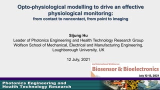
- 1. Opto-physiological modelling to drive an effective physiological monitoring: from contact to noncontact, from point to imaging Sijung Hu Leader of Photonics Engineering and Health Technology Research Group Wolfson School of Mechanical, Electrical and Manufacturing Engineering, Loughborough University, UK 12 July, 2021
- 2. Content of presentation • Introductions • Limitations with photoplethysmography (PPG) • A new model for light trans-illuminating tissue (LTT) • Opto-Physiological Monitoring (OPM) • Commercial opportunities for OPM
- 3. Highlights
- 4. Wolfson School of Mechanical, Electrical, and Manufacturing Engineering
- 5. The LU Photonics Engineering & Health Technology conducts research into: 1. The use of light for sensing and characterising dynamic systems of industrial and biological origin. 2. The research is mainly specialised in Photonics based Biomedical Engineering, i.e., photoplethysmography, Tissue optics engineering simulation, light scattering etc., and has the following generic expertise: Optics/Tissue Optics, (µ)electronics , software design and systems integration at component and systems levels, combined with a team approach to problem solving, allows for a clear and rapid progression from fundamental research ideas to industrial prototypes. The majority of the Group's projects are pursued to the level of engineering implementations and further commercial exploitation. 3. Five academic members including associated members, one visiting clinical professor, two visiting academics, 23 graduated PhDs (since 2006) and eight on-going PhDs arranging from opto-physiological monitoring (point and imaging) to in vitro diagnosis (IVD) optical detector system for point-of-care testing (POC) and dynamic breathy pressure sensing.
- 6. Remarkable achievements 2008: “Opto-physiological monitoring” a milestone of optoelectronic research worldwide www.pages.drexel.edu/~kmg462/currentresearch.html 2010: Multiplexed POCT Instrument in Future Pavilion, World Expo 2010, Shanghai. 2012: LU non-contact vital sign monitoring, as highlighted in NASA/TM-2011-216145 2014: “Opto-Physiological Sensor and Method of Design”, PATENT No. GB1511448.1., CareLight: www.lboro.ac.uk/carelight 2015: New Generation of optoelectronic sensor for vital human sign monitoring http://atlasofscience.org/tag/carelight/ 2017: A new temporal dynamic correlation between physiological changes and spontaneous expressions (2017, Nature_ Scientific Reports, DOI:10.1038/s41598- 017-07122-x). 2019: A breath activated dynamic air pressure detection system (DAPDS) created in LU.
- 8. The use of oversimplified PPG models to describe and implement the technology has limited its applicability; The principle of PPG is typically described as a blood-filled cuvette based on the Beer-Lambert law; No scattering effects (µs, g) and the light sources are assumed to be monochromatic. PPG is an optically obtained plethysmogram, a volumetric measurement of an organ, and illuminates the skin and measures changes in light absorption.
- 9. The amount of light transmitted from the skin and received by the photodetector Two modes of PPG probe Factors (blood volume, vessel wall movement, orientation of red blood cells (RBC) and wavelength of optical radiation) Transmitted mode Reflection mode R. Anderson, John Parrish, “The Optics of Human Skin”, J Invest Dermatol, 77(1), 13-19, 1981
- 10. Beer-Lambert Model based PPG where I0 is the source intensity, I is the transmitted intensity, µeff is the effective absorbance and r is the optical path length. Usually µeff is formed by a static µtissue and a dynamic µblood components: The µblood takes into account the oxygenation of the arterial blood: ( ) ( ) 0 exp eff I I r µ λ = − S. Feng, F. Zeng, and B. Chance, "Photon migration in the presence of a single defect: a perturbation analysis," App. Opt., 34: 3826-3837 1995.
- 11. PPG devices are in widespread use Pulse oximetry: $1,913.2 m by 2024 at a compound annual growth (CAGR) of >6% Global Pulse Oximeter Market, accessed on 10th September 2020 Wearable Medical Devices: $62.44 b by 2027 at a CAGR of 25.8% Wearable Medical Devices Market Size, accessed on 10th September 2020
- 12. PPG devices are in widespread use …in spite of well known limitations! • Interference from motion artifacts – Pulse oximetry (SpO2%) users cannot move around • Poor analytical performance – Smartwatch heart rate data not clinically acceptable • Lack of calibration with reference standards – Uses largely confined to consumer applications
- 13. If the internal temperature drops outside of the normal range, your body will take steps to adjust it. • Thermogenesis • Vasoconstriction • Hormonal thermogenesis Thermoregulation: These mechanisms include: Internal body heating 26° 29° 32° Understanding Thermoregulation in humans
- 14. Poor circulation when Cold Better circulation when warm Venule Arteriole Venule Arteriole Skin Surface Good signal Poor signal Skin Surface Effective capture of light trans-illuminating tissue
- 15. Shivering Sweating Set point Vasoconstriction Vasodilation Time Body temperature (C°) 37 37.5 36.5
- 16. Heat out Reduces heat loss Cold conditions Warm conditions • Vasoconstriction in cold conditions: shunt vessel relaxes so less blood flows to the surface capillaries • Vasodilatation in warm conditions: shunt vessels constricts so more blood goes to the surface The signal may be affected by the thermoregulation!
- 17. A new model for light trans- illuminating tissue (LTT)
- 18. Opto-physiological interaction Biological tissue is considered as a set of optical media, study how light interacts within biological tissue, where the optical properties of the latter reflect the mechanical, physical and biochemical functions of the living organism.
- 19. Tissue Optics Light interaction and propagation in biological tissue, are investigated. The light propagation in turbid biological media is jointly governed by the absorption (µa), anisotropic scattering properties (µs, µs‘, g) as provided by radiative transfer equation (RTE) The knowledge of tissue optical properties has found numerous applications, i.e. cancer diagnostics and therapy.
- 20. Tissue Optics. Radiative Transport Theorem s t 4 ( , ( , ( , ' ( , ' d ' 4 I I I f s π µ µ π ∂ = − + Ω ∂ ∫ r s) r s) r s ) s s ) where is the radiance at a position r in direction s, and µt = µs + µa, is the phase function that describes anisotropic scattering and dΩ’ is the solid angle. The diffusion approximation P1 assumes isotropic scattering through all media by using the transport scattering coefficient . ' (1 ) s s g µ µ = −
- 21. Opto-physiological model for light trans- illuminating finger tissue ` PREPARE 3D MODEL 3D STUDIO MAX TRACE RAYS OPTICAD Determine mean path lengths with respect to detector position RAYDETECT Ray segment data SIMULATION MODEL PREPARATION Finger Dimensions 3D MODEL Corrected Model Rays (raw) POST-PROCESSING Rescale model according to empirical validation subject STLSCALE Scaled Model Detector parameters Perform measurements used in processing and compile data STLCOMPILE Model data INTERFACING Group, segment and measure rays Map ray exit points on 2D surface RAYPROCESS Rays (processed) Optical parameters Optical Parameters Iterate abs. and dynamic coeffs. to maximise correlation btw sim. & val. IntensityMaps Coefficients & correlation Iteration parameters OUTPUT EMPIRICAL VALIDATION AC & DC distributions Assumptions V. Azorin Peris, S Hu, P. Smith, “A Monte Carlo Platform for the Optical Modeling of Pulse. Oximetry”, Proc. SPIE, 6446, 64460T (2007) Output RAYTESTROT Determines the intersection of rays with interfaces. FLATS2SURF Converts 2D maps of exit points into intensity distribution surfaces. RAYENDFLAT Determines the points at which rays exit the tissue bed and maps these onto a flat surface (x in mm, y in deg) ORIGINS Determines the central axis of the finger Procedure
- 22. ` PREPARE 3D MODEL 3D STUDIO MAX TRACE RAYS OPTICAD Determine mean path lengths with respect to detector position RAYDETECT Ray segment data SIMULATION MODEL PREPARATION Finger Dimensions 3D MODEL Corrected Model Rays (raw) POST-PROCESSING Rescale model according to empirical validation subject STLSCALE Scaled Model Detector parameters Perform measurements used in processing and compile data STLCOMPILE Model data INTERFACING Group, segment and measure rays Map ray exit points on 2D surface RAYPROCESS Rays (processed) Optical parameters Optical Parameters Iterate abs. and dynamic coeffs. to maximise correlation btw sim. & val. IntensityMaps Coefficients & correlation Iteration parameters OUTPUT EMPIRICAL VALIDATION AC & DC distributions Opto-Physiological Modelling
- 23. Opto-Physiological Modelling: Point Measurement s t 4 ( , ( , ( , ' ( , ' d ' 4 I I I f s π µ µ π ∂ = − + Ω ∂ ∫ r s) r s) r s ) s s ) Accuracy Applicability Experimental Setup Opto- Physiological Modelling Radiative Transport Theory Beer-Lambert ( ) ( ) ( ) ( ) 2 2 1 1 2 2 (1 ) (1 ) HbO Hb HbO Hb S S R S S µ λ µ λ µ λ µ λ + − = + − ( ) ( ) ( ) ( ) 1 1 1 1 1 2 2 2 2 1 exp 1 ( , ) ( , ) ( , , , ) ( , ) exp 1 ( , ) ( , ) ( , , , ) n a det i n a det i i i L i x R i i L i x σ λ µ λ λ θ λ λ σ λ µ λ λ θ = = + = + ∑ ∑ Modified Beer-Lambert [ ) ( ) 2 1 1 ( ) max ( ), , 0, ACpeak i DC I x i x I λ θ π = Θ = ∑ [ ) ( ) 2 2 1 ( ) max ( ), , ,2 ACpeak i DC I x i x I λ θ π π = Θ = ∑ Optimum sensor placement ( ) ( ) ( ) 0 1 ( ) exp ( ) ( ) 1 ( ) , ( ), ( ), ( ) n AC a det i I t I i t i L i x t t s t σ ρ ω µ θ = = − ∑ Sensor motion artefact
- 24. Experimental Validation Setup and Outcomes Finger Rotating Platform LED 1 LED 2 LED Driver CAM Trigger PC Framegrabber Ref: Azorin-Peris, V., Hu, S.*, Smith, P. R.,“A Monte Carlo Platform for the Optical Modelling of Pulse Oximetry”, Proc BiOS, SPIE 2007, 6446,64460T, Photonics West, San Jose, USA, 2007
- 25. Opto-Physiological Model of light trans-illuminating general tissue : Imaging measurement:
- 26. Revised Beer-Lambert Law 0 exp( ) a I I MPL µ = × − × 0 I I a µ 1 mm− MPL mm intensity of incident light. intensity of transmitted light. the wavelength-dependent absorption coefficient with units of the mean path length with unit of ( ) ( , ) s MPL S G l g µ = + × The mean path length MPL is defined as follows: Where S a factor that accounts for the position and size of light source and detector G a factor that accounts for a specific tissue model including the geometry and the layered-interaction. ( , ) s l g µ the path length modulated by the wavelength-dependent scattering coefficient with units of 1 mm− and the anisotropy factor g Opto-Physiological Model for Imaging
- 27. Time-Variant blood perfusion ( ) 0 , , exp[ ( ) ] a static a blood I I d r t µ µ = × − × + × , a static µ the wavelength-dependent absorption coefficient of the static component such as baseline arterial and venous blood, tissue, bone etc. with units of 1 mm− , a blood µ the wavelength-dependent absorption coefficient of the arterial blood with units of 1 mm− d the static component of MPL with units of mm ( ) r t the time function of the pulsatile component of MPL during the arterial blood variation , exp( ( )) static a blood I I r t µ = × − × Where 0 , exp( ) static a static I I d µ = × − × 0 , exp( ) static a static DC I I d µ = = × − × with the amplitude equal to the value of this signal. static dc DC I = = , ( ) static a blood AC I r t µ = × × With the amplitude , diastole systole static a blood ac I I I r µ = − = × ×
- 28. Phantom to valid opto-physiological mode Construction of phantoms with different absorption and scattering coefficients Ref: J Zheng, “OPTO-PHYSIOLOGICAL MODELLING OF IMAGING PHOTOPLETHYSMOGRAPHY”, PhD thesis, 2010, Loughborough University, UK
- 29. y = 1.7184x + 0.5123 0 0.1 0.2 0.3 0.4 0.5 0.6 0.7 0.8 0.9 1 0 0.01 0.02 0.03 0.04 0.05 0.06 0.07 0.08 0.09 0.1 Absorption coefficient (mm-1) Absorption (a.u) Beer-Lambert law Opto-physiological model Experimental and theoretical absorption on a two-layered phantom with a lower scattering top layer For the top layer, the absorption and scattering coefficients were set to be μs,t=1.00mm-1, μa,t=0.0263 mm-1, respectively. The square blocks represent the absorption in the experiment corresponding to the different absorption coefficients of bottom layer μa,t: 0, 0.0122, 0.02625, 0.0455, 0.0612 and 0.0789. Ref: Hu, S.*, Azorin Peris V., Zheng J., “A study of opto-physiological modelling to quantify tissue absorbance in imaging photoplethysmography”, Proc. IEEE EMBC 32, 2010, 1: 5776 – 5779.
- 30. Opto-Physiological Modelling: Imaging Measurement RCLED arrangement of the dual-wavelength Ringlight with the lens in the middle 650nm 870nm RCLED arrangement of the dual-wavelength Ringlight with the lens in the middle 650nm 870nm 650nm 870nm Trigger Timing signals 650nm 870nm Trigger Timing signals 650nm 870nm Trigger Timing signals Digital CCD camera
- 31. The Fourier spectrum for PPG signals from the conventional contact PPG imaging system and imaging PPG system at 650nm and 870nm from the face Human face Raw signal Filtered signals (PPG) Region of interest Results: face
- 33. Applications of OPM • Contact monitoring (wearables) • Multiplexed contact sensor (advanced wearable, Carelight) • Imaging OPM • 3D mapping OPM
- 34. Point Measurement: Venox: World 1st non-invasive venous oximetry - A non-invasive measurement mechanism of venous oxygen saturation (SvO2). - Providing enhanced monitoring of essential cardio-vascular function. - Indicating a correlation of oxygen saturation changes between Venox and the bypass machine. Ratio-of-Ratios a arterial R V A V A t I t I t I t I = − − = ∆ ∆ ) ( ) ( ) ( ) ( ) , ( ) , ( ) , ( ) , ( 2 2 1 1 2 2 1 1 λ ε λ λ ε λ λ λ λ λ v venous R V A V t I t I t I t I = Α − − = ∆ ∆ ) ( ) ( ) ( ) ( ) , ( ) , ( ) , ( ) , ( 2 2 1 1 2 2 1 1 λ δ λ λ δ λ λ λ λ λ Venox Ref: Venous pulse oximetry, WO2003063697A1, granted in 2008
- 35. LU Multiplexed Optoelectronic Sensor (Carelight, www.Lboro.ac.uk/carelight) • Novel sensor design – Redundant illumination sources at multiple wavelengths – Additional sensor options e.g. accelerometer, temperature • Multiple versions built in house – Tested on colleagues v’s gold standards Granted Patent: GB 2519335, WO2015/056007 PCT/GB2014/053095 Pulsatile waveforms (PPG signals) with different wavelength illuminations and contact points
- 36. Tests against Gold standards Heart Rate (the correlation r >0.99 against 5 Lead ECG, Mortara-H3Plus-AAB, 20 subjects with various ethnical groups* ) Oxygen Saturation SpO2% against standard pulse oximeter (ContecTM PM60A), 20 subjects with various ethnical groups*) *Including dark skin type subjects Algorithm: Bandpass filter: 0.5 – 5Hz Algorithm: Bandpass filter: 0.5 – 5Hz, Ratio of Ratios; Ratio=AC(red)/DC(red)/AC(ir)/DC(ir)
- 37. List of Carelight work • “Illumination Adaptation in a Multi-Wavelength Opto-Electronic Patch Sensor”, Sensors 2020, 20(17), 4734; “An Optimization Study of Estimating Blood Pressure Models Based on Pulse Arrival Time for Continuous Monitoring”, J Health Eng. 2020, doi: 10.1155/2020/1078251. • “Oxygen Saturation Measurements from Green and Orange Illuminations of Multi-Wavelength Optoelectronic Patch Sensors”, Sensors 2018, 19(1), 118; doi.org/10.3390/s19010118 • “A Multi-wavelength Opto-Electronic Patch Sensor to Effectively Detect Physiological Changes Against Skin Pigments”, Biosensors, 7(2), 22, (2017), doi:10.3390/bios7020022 • “A Multi-Channel Opto-Electronic Sensor to Accurately Monitor Heart Rate against Motion Artefact during Exercise”, Sensors, 15, 25681-25702 (2015); doi:10.3390/s151025681 • “A Comparison Study of Physiological Monitoring with a Wearable Opto-Electronic Patch Sensor (OEPS) for Motion Reduction”, Biosensors, 5, 288-307 (2015); doi:10.3390/bios5020288 • “Opto-physiological modeling applied to photoplethysmographic cardiovascular assessment”, Invited Review Paper, J Healthcare Eng. 4 (4), 505-528 (2013). • “Non-contact Reflection Photoplethysmography towards Effective Human Physiological Monitoring”, J Med. Biol. Eng. Vol.30 Iss 30 (2010): 161-167 Much more ……………
- 38. Blood Flow vs Perfusion Index Perfusion Index = AC/DC
- 39. Real time signal processing to obtain heart rate (HR) and respiration rate (RR) against standard methods HR calculation results on two subjects chosen LU-Db data sets with Carelight sensor. (a) The results of subject F03 in 1st session. (b) The results of subject M08 in 2nd session
- 40. RR calculation results on two subjects chosen LU-Db data sets. (a) The results of subject F03 in Cycling; (b) The results of subject M08 in Treadmill.
- 41. Non-contact reflection OPM monitoring The purpose is to noncontact measure blood volume change and to overcome the problems related to contact sensors as requested for a variety of surgical and medical applications, e.g. human metrology, assessment in sites of tissue damage, etc. I I I I I I I I P 23 mm Infrared light sources Photodetector Two research prototypes Two research prototypes Photodetector Light Source Probing area Illuminated area dPD dLS θLS Ref: Shi, P., Hu, S.* et al, “Non-contact Reflection Photoplethysmography towards Effective Human Physiological Monitoring”, J Med. Biol. Eng. Vol.30 Iss 30 (2010): 161-167
- 42. Imaging measurements: 3D Opto-physiological imagine
- 43. OPM driven 3D imaging PPG OPM allows to monitor larger area and different depths in tissue, which can give a complete 3-D description of tissue, so as to improve the ability to probe biologic interactions dynamically and to study disease over time. The mean values in a region of interest against time plotted in time domain The mean AC amplitude of individual regions from 660nm and 880nm LEDs in a 3-D format Ref: Zheng, J., Hu, S.*, et al “Remote simultaneous dual wavelength imaging photoplethysmography: a further step towards 3-D mapping of skin blood microcirculation”, Appl. Phys. Lett. 0003-6951, Proc. of SPIE Vol. 6850 68500S-1, Photonics West, San Jose, USA, 2008.
- 44. 3-D Visual Microcirculation Map OPM driven 3D iPPG OxiMap: Vascular Imaging was filed as a patent with PCT/GB2008/003042.
- 45. OPM for wearables 1. Flexible OPM sensor 2. Smart garment fabric with conductive tracks and LEDs 3. Smart Textile 4. Fatigue alarm Ref: D. Iakovlev., S, Hu., “Smart Garment Fabrics to Enable Non-Contact Opto-Physiological Monitoring”, Biosensors (Basel). 2018 Jun; 8(2): 33. doi: 10.3390/bios8020033 1 2 3 4
- 47. Smart Wearable Smart Jewellery Wrist-worn Head bands e-textile Smart Mask
- 48. Smart Device for medical, more Sleep Monitor Pulse Oximeter Brain computer interface Bio-Security Automation
- 49. Analogue Front End. Digital Processing. Wireless Communication. We are seeking for partners for commercialisation to create small scale prototype mobile electronics that are suitable for wearing with the OPM sensor
- 50. Thank you for your attention Any questions?