Advertising Jobs on the NH Center for Nonprofits Website
Similar to Analysis of Human Embryonic Stem Cells with Regulatable Expression of the Cell Adhesion Molecule L1 in Regeneration after Spinal Cord Injury
LncRNA WARS2-IT1 Functions as an Oncogene and is Associated with Poor Outcome...semualkaira
Similar to Analysis of Human Embryonic Stem Cells with Regulatable Expression of the Cell Adhesion Molecule L1 in Regeneration after Spinal Cord Injury (20)
LncRNA WARS2-IT1 Functions as an Oncogene and is Associated with Poor Outcome...
Analysis of Human Embryonic Stem Cells with Regulatable Expression of the Cell Adhesion Molecule L1 in Regeneration after Spinal Cord Injury
1. Analysis of Human Embryonic Stem Cells
with Regulatable Expression of the Cell Adhesion
Molecule L1 in Regeneration after Spinal Cord Injury
Myungsik Yoo,1
Gunho Anthony Lee,1
Christopher Park,1
Rick I. Cohen,2
and Melitta Schachner1,3
Abstract
Cell replacement therapy is one potential avenue for central nervous system (CNS) repair. However, transplanted stem cells
may not contribute to long-term recovery of the damaged CNS unless they are engineered for functional advantage. To fine
tune regenerative capabilities, we developed a human neural cell line expressing L1, a regeneration-conducive adhesion
molecule, under the control of a doxycycline regulatable Tet-off promoter. Controlled expression of L1 is desired because
overexpression after regenerative events may lead to adverse consequences. The regulated system was tested in several cell
lines, where doxycycline completely eliminated green fluorescent protein or L1 expression by 3–5 days in vitro. Increased
colony formation as well as decreased proliferation were observed in H9NSCs without doxycycline (hL1-on). To test the
role of L1 in vivo after acute compression spinal cord injury of immunosuppressed mice, quantum dot labeled hL1-on or
hL1-off cells were injected at three sites: lesion; proximal; and caudal. Mice transplanted with hL1-on cells showed a better
Basso Mouse Scale score, when compared to those with hL1-off cells. As compared to the hL1-off versus hL1-on cell
transplanted mice 6 weeks post-transplantation, expression levels of L1, migration of transplanted cells, and immunore-
activity for tyrosine hydroxylase were higher, whereas expression of chondroitin sulfate proteoglycans was lower. Results
indicate that L1 expression is regulatable in human stem cells by doxycycline in a nonviral engineering approach.
Regulatable expression in a prospective nonleaky Tet-off system could hold promise for therapy, based on the multi-
functional roles of L1, including neuronal migration and survival, neuritogenesis, myelination, and synaptic plasticity.
Key words: adhesion molecule L1; inducible Tet-off system; regulatable expression; spinal cord injury; stem cell
transplantation
Introduction
Embryonic stem cell (ESC) derivatives represent a po-
tential approach for cell based therapy as a treatment for ir-
reversible neuronal cell damage.1
Aside from eliminating the risk
of tumor/teratoma formation, additional areas of concern need to be
addressed to allow for successful cell therapy. These include, but
are not limited to, robust cell survival2–5
and circumvention of
endogenous antiregenerative signals in the acutely or chronically
injured host. Based on previous evidence that the regeneration-
conducive cell adhesion molecule, L1, enhances recovery in dif-
ferent types of mammalian nervous system lesions, we investigated
the possibility of using L1, in a regulatable fashion, to engineer an
optimized cell therapy vector. We postulated that mirroring the
natural down-regulation of L1 expression in postnatal nervous
system development by using a regulatable system would be im-
portant to optimize initial regenerative events and avoid compli-
cations caused by irreversible overexpression postrepair.
The immunoglobulin superfamily molecule, L1, plays crucial roles
in multiple morphogenetic functions, such as neuronal migration,
differentiation, and survival, as well as neuritogenesis, axonal tar-
geting, myelination, synapse formation, and synaptic plasticity.6–12
L1
is not only crucial during development, but also in regeneration after
injury of the central and peripheral nervous systems.6,7,13–18
However,
constitutively high expression of L1 could be disadvantageous, unless
limited to sets of functional hot spots, such as generation of inter-
neurons in the olfactory bulb or of granule cells in the dentate gyrus,
and in altering synaptic efficacy. In a regenerative context after in-
jury, although not previously observed in different injury paradigms,
overexpression of L1 may induce, for instance, erroneous growth/
sprouting axons, such as those of sensory nerve fibers causing allo-
dynia and hyperalgesia.19
For therapeutic prospects, L1 expression
1
W.M. Keck Center for Collaborative Neuroscience and Department of Cell Biology and Neuroscience, Rutgers University, Piscataway, New Jersey.
2
Rutgers University, Biomedical Engineering, Piscataway, New Jersey.
3
Center for Neuroscience, Shantou University Medical College, Shantou, People’s Republic of China.
JOURNAL OF NEUROTRAUMA 31:553–564 (March 15, 2014)
ª Mary Ann Liebert, Inc.
DOI: 10.1089/neu.2013.2886
553
2. levels should therefore be controllable in vivo. We have thus chosen a
nonviral expression system, which may confer advantages, even if
viral transduction would become clinically viable, because virus-
mediated cell therapy has the disadvantage that viral DNA sequences
may be silenced by the host’s cellular protection mechanisms.20
We
have developed a novel nonviral doxycycline (DOX)-inducible
human L1 expression system that comprises a single regulatable
plasmid with a transrepressor together with a strong promoter, such
as the CAG (chimeric cytomegalovirus and chicken b-actin) pro-
moter, and that is efficiently regulatable in glioblastoma and neu-
roblastoma cells as well as predifferentiated H9-ESC-derived neural
stem cells (H9NSCs) by DOX in vitro. The stable human cell line,
pTet-off-hL1-H9NSC, is also regulatable and functional in vivo in
cyclosporine-immunosuppressed mice, where locomotor recovery
after acute compression injury is observed after 5–6 weeks.
Methods
Procedures for H9NSCs in vitro and in vivo
Neural stem cells derived from H9 ESCs (H9ESCs; Fig. 1A)
were obtained from the Stem Cell Core Facility at The Stem Cell
Research Center (Rutgers University, Piscataway, NJ). After
engineering the pTet-off-hL1 system (see below), we transfected
the plasmid system into H9NSCs (Fig 1B, without DOX, and Fig.
1B, with DOX). hL1-on and -off cells were expanded and se-
lected in the presence of 200 lg/mL of G418 (Fig. 1Cc and 1Dd,
respectively). Red q-dot-labeled cells were transplanted into
acutely compression-injured spinal cords of cyclosporine-
immunosuppressed mice (see below; Fig. 1Ee) and scored by the
Basso Mouse Scale (BMS) every week for 6 weeks without or
with DOX in the drinking water to maintain hL1-on and -off,
respectively (Fig. 1Ff ).
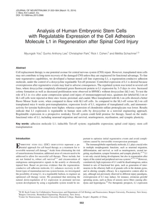
FIG. 1. Schematic illustration of experimental procedures for cell lines under in vitro and in vivo conditions. H9 human embryonic
stem cells (H9ESCs) (A). Predifferentiated human neural stem cells (H9NSCs) that had been subjected for 7 days to a differentiation
protocol are described in the Methods section (B). Cells were transfected with the pTet-off-hL1 plasmid and maintained under two
different conditions: cell line for hL1-on (B, without doxycycline) and cell line for hL1-off (b, with doxycycline). For selection of
transfected cells, cultures were treated with G418 (200 lg/mL) in the culture medium, which was changed every other day for 4 weeks
and expanded for storage (C, c, and D, d). Before transplantation into acutely spinal cord injured and cyclosporine-immunosuppressed
mice, cells were labeled for quantum dot analysis (E, e). Mice were tested by Basso Mouse Scale every week for 6 weeks without
doxycycline (F) and with doxycycline (f ) in the drinking water. DOX, doxycycline; SCI, spinal cord injury.
554 YOO ET AL.
3. Construction of the vector system
The pTet-off-GFP plasmid is a nonviral single-entity system
containing two CAG promoters driving the expression of green
fluorescent protein (GFP) and the transactivator in opposing di-
rections (Fig. 2). The vector system was assembled as follows: The
pd2EGFP plasmid (Clontech, Mountain View, CA) was con-
structed as the backbone in three steps. First, the pd2EGFP was
linearized using SalI and BglII and ligated with the SalI and BamHI
fragment of pCX-EGFP containing the CAG promoter driving
enhanced GFP (eGFP) expression (pCAG-EGFP). Second, an
XbaI-digested fragment with seven repeat tetracycline response
FIG. 2. Schematic representation of the pTet-off-GFP and pTet-off-hL1 systems. This system uses a single plasmid doxycycline
Tet-off promoter containing seven tetracycline response elements (TREs) located between two oppositely oriented CAG promoters.
The CAG promoters drive expression of GFP and the hybrid tetracycline-KRAB repressor. In the absence of doxycycline, the
7 · TREs are silenced, allowing activity of the CAG promoters as well as transcription of GFP and the repressor. In this condition, the
cells are ‘‘ON’’ for the target genes (A). In the presence of doxycycline, no gene expression is observed, because now the 7 · TREs
are bound by the Tet portion of the hybrid repressor, and the CAG motifs are blocked by the KRAB portion. This allows for tight
regulation of gene expression and is referred to as ‘‘OFF’’ for the target genes (B). Construction of the nonviral single pTet-off-hL1
plasmid, where the GFP gene is replaced by flag tagged for measuring hL1 for hL1-on (C) and hL1-off (D) cells. GFP, green
fluorescent protein; DOX, doxycycline; CAG, chimeric cytomegalovirus and chicken b-actin; TREs, tetracycline response elements;
KRAB, Kruppel-associated box.
REGULATED L1 EXPRESSION AND REGENERATION 555
4. elements (7 · TREs) was cloned using polymerase chain reaction
from pLVCT-rtTR2SM221
and ligated into SpeI-cut pCAG-EGFP
(pCAG-EGFP-TRE). Last, the SpeI fragment from pLVCT-
rtTR2SM2 containing the transactivator was ligated together with
XbaI/SpeI-cut pCAG-EGFP-TRE (pCAG-Tet-off-GFP, named
pTet-off-GFP). Then, the reverse tetracycline transcriptional re-
pressor was fused with the Kruppel-associated box (KRAB) do-
main, a transcriptional repressor protein of the eukaryotic
ubiquitous zinc finger family. Thus, the plasmid system is expected
to enhance repressor functions by the KRAB domain. For L1 ex-
pression, the inducible human L1 sequence was exchanged for the
GFP sequence by inserting the Klenow-blunted human L1 com-
plementary DNA into the EcoRI/blunted site of pCAG-Tet-off-
GFP named pTet-off-hL1.
Predifferentiation of H9NSC-ESCs into H9NSCs
and immunocytology
We followed a slightly modified adherent monolayer protocol,
first published by Smith and coworkers.22,23
The following pro-
tocol has been shown to produce the best results: First, the un-
differentiated H9ESCs were preconditioned at 80–90%
confluence with neural induction medium (NIM), which consisted
of a 1:1 ratio of Dulbecco’s modified Eagle’s medium (DMEM)/
F12 and neurobasal medium (Life Technologies, Carlsbad, CA).
This medium was supplemented for 2 days with B27 supplement
(1%, without retinoic acid; Life Technologies) and N2 supple-
ment (0.5%; Life Technologies). Preconditioned cells were then
passaged using Accutase (Life Technologies) and transferred onto
10-cm dishes coated with Matrigel (BD Biosciences, San Jose,
CA) at a passaging ratio of 1:3. Cells were then maintained for 2
more days in NIM. Five days after induction using NIM, the
medium was changed to neural precursor media (NPM), which
consisted of a 1:1 ratio of DMEM/F12 and neurobasal medium,
supplemented with B27 supplement (0.5%) and N2 supplement
(0.5%), as well as 20 ng/mL of basic fibroblast growth factor
(FGF-2; Peprotech, Rocky Hill, NJ). Upon 90–100% confluence,
cells were passaged at a ratio of 1:2 (harvested vs. plated cells)
and plated onto Matrigel-coated dishes. The culture medium was
changed every other day. After 2–4 days in NPM, cells had as-
sumed a flattened, bipolar morphology, typical of neural stem
cells (NSCs). To characterize the predifferentiated NSCs and
hL1-on and -off cells before transplantation, cells (1.5 · 104
) were
plated for indirect immunofluorescence (IF) into four-well glass
chamber slides coated with Matrigel (2 h, 37°C). After 2 days,
cells were fixed with 4% paraformaldehyde (PFA) in phosphate-
buffered saline (PBS, pH 7.4) for 15 min at room temperature and,
after washing in PBS, for 10 min with PBS containing 0.5% Triton
X-100 for permeabilization. After washing in PBS, primary an-
tibodies (Abs) were added (diluted in 0.1% Triton X-100, 1%
bovine serum albumin [BSA], and 3% nonimmune goat serum)
and incubated with cells for 1 h at room temperature. Primary Abs
were mouse monoclonal anti-nestin (1:200, catalog no.:
MAB3526; Millipore, Temecula, CA), anti-A2B5 (1:300, catalog
no.: MAB312; Millipore), and rabbit polyclonal anti-glial fi-
brillary acidic protein (GFAP; 1:200, catalog no.: G4546; Sigma-
Aldrich, St. Louis, MO), anti-doublecortin (DCX; 1:250, catalog
no.: AB18723; Abcam, Cambridge, MA), anti-octamer-binding
transcription factor 4 (Oct4; 1:500, catalog no.: AB3209; Milli-
pore), anti-Ki67 (1:200, catalog no.: AB833; Abcam), anti-beta
III tubulin (1:300, catalog no.: PRB435P; Covance, Emeryville,
CA). Secondary Abs were Alexa 555–conjugated goat anti-mouse
immunoglobulin G (IgG) or Alexa 555–conjugated goat anti-
rabbit IgG (1:400, catalog no.: 115-001-003; Jackson Im-
munoresearch, West Grove, PA) diluted in the buffer used for
dilution of primary Abs and incubated for 30 min at room tem-
perature. After washing in PBS, 4’,6’-diamidino-2-phenylindole
(DAPI; 1 lg/mL) was added for 10 min at room temperature.
Slides were rinsed with PBS and mounted in Aqua Poly/Mount
medium (Polysciences, Warrington, PA), sealed with nail polish,
and stored at 4°C. Images were captured with an Axiovert200
Fluorescence Live Cell Imaging Workstation (Carl Zeiss AG,
Jena, Germany).
Transfection and generation of stable cell lines
Sequence-verified, endotoxin-free pTet-off-GFP and pTet-off-
hL1 plasmids were transfected into mouse neuroblastoma N2a
and rat C6 glioma cells for measuring regulatable GFP and hL1
expression, respectively. After transfection into N2a and C6 cells
using Fugene HD (Roche Applied Science, Indianapolis, IN) at a
ratio of 5:2 of Fugene HD versus plasmid DNA, cells were treated
24 h later with G418 (200 lg/mL) for selection of stable cell
lines. One week thereafter, 12 pTet-off-GFP-N2a clonal cell lines
were isolated and expanded in DMEM high-glucose, 1-mM
Na-pyruvate, 10% fetal bovine serum (FBS), 1% penicillin/
streptomycin with G418 to analyze regulation of GFP expression.
To analyze regulatable hL1 expression, stably transfected and
selected pTet-off-hL1-C6 cells were maintained with or without
DOX (1 lg/mL) to generate hL1-off and -on cells in DMEM/F12,
GlutaMAX, 10% FBS, 1% penicillin/streptomycin, and then
probed for inducible expression of hL1 by Western blot analysis.
To generate clonal pTet-off-GFP-H9NSC lines, the rat primary
Nucleofector kit (Lonza, Allendale, NJ) was used according to the
manufacturer’s instructions. In brief, after passaging, 4 · 106
cells
in 100 lL of Nucleofector solution were incubated for 10 min at
room temperature with 2 lg of plasmid DNA. The mixture of cells
and DNA was transferred to 96-well plates and electroporated
using Lonza software at a setting of ‘‘Neuron Rat High Effi-
ciency’’ (Lonza). After transfection, 80 lL of warm culture me-
dium was added to each well, and the cell suspension was
transferred to new six-well dishes with FGF-2 (20 ng/mL). G418
was added 2 days after transfection at a concentration of 50 lg/
mL, being increased to 200 lg/mL after 4 days, when the culture
medium was changed, and maintained for 20 days. The three cell
lines with the highest percentage (approximately 95% GFP-
positive cells with high, middle, and weak fluorescence intensi-
ties) of GFP-positive cells were collected as colonies under a
fluorescence microscope. To generate the stable inducible hL1-
expressing cell lines, the same procedure as for the generation of
the pTet-off-GFP-H9NSC line was used. H9NSCs were trans-
fected with plasmid pTet-off-hL1, except that cells were
maintained with or without DOX (1 lg/mL) to generate hL1-off
and -on cells, respectively. After five passages, clones were
expanded and conserved in liquid nitrogen.
Animals and spinal cord injury
Eight-week-old C57BL/6 female mice, purchased from the
Charles River Laboratories (Wilmington, MA), were deeply an-
esthetized with ketamine-xylazine (ketamine, 160 mg/kg; xyla-
zine, 24 mg/kg; Butler Schein Animal Health, Chicago, IL) and
subjected to spinal cord compression injury, as detailed before.24–26
Animals were maintained in the core animal facility at the
Division of Life Science and the W.M. Keck Center for Colla-
borative Neuroscience (Rutgers University). After surgery, mice
were kept on a warm mat (35°C) for several hours to prevent hy-
pothermia, being thereafter singly housed in a temperature- and
humidity-controlled room with water and standard food provided
ad libitum. Bladders were manually voided once- or twice-daily,
depending on the palpability of the bladder. Animals were trans-
cardially perfused under anesthesia with 4% PFA in PBS for his-
tological and immunohistological analyses, as previously
described.7,27
All experimental procedures were approved by the
animal care and facilities committee of Rutgers, The State Uni-
versity of New Jersey.
556 YOO ET AL.
5. Surgery and cell transplantation into cyclosporine-
immunosuppressed mice
Three days before transplantation, mice were injected intraperi-
toneally (i.p.) with cyclosporine (10-mg/kg dose) for immunosup-
pression, which was continued daily after transplantation. For
transplantation, mice were anesthetized by an i.p. injection of
ketamine/xylazine, and bupivacaine (0.1 mL of 0.125%; Hospira,
Lake Forest, IL) was injected around the incision site to provide
local anesthesia. A 3-cm skin incision along the median line on the
back of the animals was made and laminectomy was performed with
Mouse Laminectomy Forceps (Fine Science Tools, Heidelberg,
Germany) at the T7–T9 level, followed by a mechanically con-
trolled compression injury using a mouse spinal cord compression
device.24–26
The spinal cord was compressed for 1 sec for the severe
compression injury with a time- strength-controlled electromag-
netic device. Both hL1-expressing (hL1-on) cells and hL1-non-
expressing (hL1-off ) H9NSCs were labeled using the Qtracker Cell
Labeling Kit (Life Technologies), according to the manufacturer’s
instructions. Cell transplantation was performed immediately after
compression injury by inserting a 33-gauge needle connected to a
5-lL Hamilton syringe (Hamilton, Reno, NV) using a stereotactic
micromanipulator (Narishige, East Meadow, NY). One microliter of
the cell suspension (105
cells/lL) was injected 1 mm deep into the
cord mid-line of the lesion site and 0.5mm rostral and caudal to it
with each injection lasting for 7 min. The skin was closed with
wound clips. Mice injected with hL1-off H9NSCs were treated with
DOX by administration through the drinking water at a concentra-
tion of 250 lg/mL in 3% sucrose solution distributed in amber
bottles for protection from degradation by light. Mice injected with
hL1-on H9NSCs were supplied with 3% sucrose solution without
DOX. Solutions were changed and measured every other day during
the course of the experiment. To test for GFP inducibility in vivo, we
followed the same procedure as the one described above, but we
transplanted GFP-on cells into the injured spinal cord. Mice were
then maintained with or without DOX in the drinking water for 7
and 10 days to test for GFP induction in vivo.
Locomotor assessment
We assessed locomotor function by the BMS score25,28,29
1
week before and every week after injury. For assessment, mice
were allowed to move in an open field, 1 m in diameter, for 5 min.
Hindlimb movements were observed and scored according to the
BMS scale by two expert and independent observers, blinded to the
treatment.
Immunohistology
Animals were deeply anesthetized with an i.p. injection of keta-
mine/xylazine followed by vascular washout with PBS and transcar-
dial perfusion with 4% PFA in PBS. Spinal cords were removed and
cryoprotected by incubation in 20% sucrose in PBS overnight at 4°C,
frozen, and cut into 20-lm-thick serial sections in a sagittal plane
rostral and caudal to the lesion site. Sections were mounted on mi-
croscope slides and saved at -80°C. Sections of equivalent distance
from the lesion center of each group were thawed to room tempera-
ture, washed three times, and blocked with 10% goat serum in PBS for
2h at room temperature. Slides were incubated overnight at 4°C for
immunostaining with mouse monoclonal anti-human L1 Ab (1:400,
catalog no.: UJ127; Abcam), anti-chondroitin sulfate Ab (CS56;
1:200, catalog no.: C8035; Sigma-Aldrich), and rabbit polyclonal Abs
to tyrosine hydroxylase (TH; 1:500, catalog no.: AB152; Millipore),
and serotonin (5-HT; 1:400, catalog no.: 10385; Abcam). For negative
control, nonimmune mouse IgG (1:400, catalog no.: ab37355; Abcam)
was used instead of the specific primary Abs. After washing with PBS,
slides were incubated with Alexa 555–conjugated goat anti-mouse
IgG (1:800, catalog no.: 115-001-003; Jackson Immunoresearch) or
Alexa 555–conjugated goat anti-rabbit IgG (1:800; Jackson Im-
munoresearch) in PBS for 2h at room temperature. Some sections
were incubated with DAPI, rewashed with PBS, mounted with Aqua
Poly/Mount medium (Polysciences), and tile imaged with an Ax-
iovert200 Fluorescence Live Cell Imaging Workstation (Carl Zeiss).
Quantification of immunofluorescence, and Western
blot and cell migration analysis
Fluorescence intensities of spinal cord areas immunolabeled for
hL1 were quantified using four serially spaced (400 lm apart) mid-
sagittal sections in the rostrocaudal direction from 4 animals. Pho-
tographic documentation was performed with the Axiovert200
Fluorescence Live Cell Imaging Workstation (Carl Zeiss), AxioVi-
sion software (Carl Zeiss), and ImageJ software (National Institutes
of Health, Bethesda, MD). Both immunostaining and imaging were
performed under identical conditions. Staining intensity thresholds
for Ab were determined after all images were acquired to optimize the
signal-to-noise ratio for a particular Ab. The threshold selected was
55 (within the full range of intensities extending from 0 to 255) for
q-dot, 85 for hL1, 90 for sections 0.5 mm away from the injury center
to evaluate migration of hL1 immunopositive H9NSCs, 85 for 5HT,
75 for TH, and 70 for CS56. Mean fluorescence intensity (MFI) of the
area of immunoreactivity was at 0.8 mm equidistant rostral and
caudal from the center of the injury site for hL1 and CS56. Then,
serial sections, 400 lm spaced apart, were evaluated starting from the
injury center up to a rostal and caudal distance of 1.5 mm to analyze
the MFI of hL1. For 5HT and TH, mean immunofluorescence in-
tensities were measured 0.5mm caudal to the injury site, with in-
tensities higher than the thresholds stated above. These values were
normalized to the total tissue areas.
Western blot analysis for hL1 has been described previously.27
Briefly, stably transfected pTet-off-hL1 C6 and H9NSC lines were
generated with or without DOX (1 lg/mL) and saved as pellets at
- 80°C until use for Western blot analysis. Cells were thawed on ice,
lysed by triturating in radioimmunoprecipitation assay buffer
(Sigma-Aldrich), and centrifuged at 1000 · g and 4°C for 20 min to
remove insoluble matter. Concentration of extracted proteins was
tested by bicinchoninic acid (Pierce Biotechnology, Rockford, IL).
A total of 30 lg of protein solution was boiled for 5 min in sodium
dodecyl sulfate (SDS) sample buffer and separated by 4–12% gra-
dient SDS/polyacrylamide gel electrophoresis (Life Technologies).
Proteins were electroblotted onto polyvinylidene difluoride mem-
branes, blocked, and probed with hL1 monoclonal Ab (1:400,
FIG. 3. Nonviral single pTet-off-GFP and pTet-off-hL1 systems efficiently silence gene expression in a doxycycline dose- and time-
dependent manner. The pTet-off-GFP plasmid vector was transfected into N2a cells, which were then treated with doxycycline at 0, 0.5,
and 1 lg/mL (A, B, and C, respectively). GFP was efficiently silenced by 1 lg/mL of doxycycline in N2a cells within 3 days (G). The
pTet-off-GFP system was transfected into H9NSCs and a clonally selected line was treated with doxycycline at 1 lg/mL for 0, 4, and 8
days (D, E, and F, respectively). GFP was silenced 8 days after starting the doxycycline treatment of H9NSCs (H). Western blot
analysis from the selected cell line after pTet-off-hL1 system transfection shows that hL1 expression was silenced by doxycycline in C6
cells and H9NSCs (I and J, respectively; n = 3 experiments). Asterisks indicate significant differences between the groups. **p < 0.01, as
assessed by one-way ANOVA, followed by Tukey’s post-hoc analysis. Data represent means – standard error of the mean; n = 4 images
for each cell line. Scale bar, 100 lm for all panels. GFP, green fluorescent protein; GAPDH, glyceraldehyde 3-phosphate dehydro-
genase.
‰
REGULATED L1 EXPRESSION AND REGENERATION 557
7. catalog no.: UJ127; Abcam) or polyclonal flag Ab (1:3000, catalog
no.: ab1162; Abcam). Secondary mouse or rabbit Abs conjugated to
horseradish peroxidase with enhanced chemiluminescence intensi-
fication (Pierce Biotechnology) were used for detection of hL1.
Statistical analysis
All numerical data are presented as group mean values with
standard error of the mean. The statistical significance of the BMS
score and mean immunoreactivity intensity for each group were es-
timated by one-way analysis of variance, followed by Tukey’s post-
hoc test. p values <0.05 were considered statistically significant.
Results
Constructs of the pTet-off-GFP and pTet-off-hL1
systems
The novel nonviral single Tet-off plasmid systems, pTet-off-GFP
and pTet-off-hL1, were constructed as described in the Methods
section. To increase the efficiency of inducibility, we included seven
repeats of the TRE between two CAG promoters placed in opposite
transcriptional orientations. These two promoters drive expression
of GFP or hL1 and the tetracycline reverse transactivator (Fig. 2). In
the absence of DOX (on), the 7 · TRE are dormant, allowing the
activity of the CAG promoters to control the transcription of GFP or
hL1 and of the transrepressor (Fig. 2A,C, respectively). In the
presence of DOX (off ), the TREs are bound by the transrepressor,
being fused to a strong KRAB repressor, which leads to repression
of the CAG promoters and silencing of GFP or hL1 and the trans-
repressor (Fig. 2B,D, respectively). For construction of the nonviral
single pTet-off-hL1 plasmid, the GFP insert was replaced by the
full-length hL1 insert with flag tagged in the pTet-off-GFP plasmid.
The pTet-off-GFP-transfected clonal cell line, N2a-#12, was main-
tained in the presence of DOX at 0, 0.5, and 1 lg/mL (Fig. 3A, B, and
C, respectively). We observed that expression of GFP was reduced by
1 lg/mL of DOX in the N2a-#12 cell line within 3 days (Fig. 3C).
Time-dependent regulation by DOX of GFP in GFP-on and -off cells
in the pTet-off GFP system stably transfected cell line, N2a-#12, was
observed in GFP-off cells without DOX, with GFP-off cells expres-
sing GFP after 9 days (Supplementary Fig. 1) (see online supple-
mentary material at http://www.liebertpub.com). A bar graph in
Figure 3G shows that in the presence of 0.5 and 1lg/mL of DOX,
GFP was silenced, in comparison to cells maintained in its absence. A
clonal pTet-off-GFP-H9NSC line was clonally selected 4 weeks after
transfection with the GFP system. The clonal line was treated with
DOX at 1 lg/mL for 0, 4, and 8 days (Fig. 3D, E, and F, respectively).
The bar graph in Figure 3H illustrates the effect of the different
exposure times to DOX on GFP expression in H9NSCs, which was
significantly reduced by day 4 and not detectable by day 8. To test
FIG. 4. H9NSCs express stage-specific markers. Immunofluorescence staining of nestin, A2B5, and doublecortin (DCX) (A, B, and C,
respectively). A phase-contrast image of the cells demonstrating rosette formation (D). Bar graph showing the percentage of cells
positive for each of the neural stem cell markers (E; n = 3 experiments). More than 96% of all cells are positive for the neural stem cell
marker, nestin, approximately 18% of cells are positive for the neuroglial progenitor marker, A2B5, and 11% of cells are positive for the
early neuronal progenitor marker, DCX. The astrocyte marker, GFAP, and the embryonic stem cell marker, Oct4, were not detected (E).
Scale bar, 100 lm for all panels. GFAP, glial fibrillary acidic protein; DAPI, 4’,6-diamidino-2-phenylindole; Oct4, octamer-binding
transcription factor 4.
REGULATED L1 EXPRESSION AND REGENERATION 559
8. DOX specificity and toxicity, we constructed a nonregulatable plas-
mid pCAG-GFP to generate a clonal nonregulatable pCAG-GFP-
H9NSC line, showing that GFP expression and cell viability were not
affected (99% of all cells are GFP positive) by treatment with DOX
(1 lg/mL) during 2 weeks (Supplementary Fig. 2) (see online sup-
plementary material at http://www.liebertpub.com). Western blot
analysis of the stably hL1-expressing cell line with the pTet-off-hL1
system showed that expression was silenced by DOX in C6 cells and
in H9NSCs (Fig. 3I and J, respectively).
Characterization of H9NSCs and hL1-H9NSCs
The phenotype of H9NSCs was characterized by indirect IF for
expression of nestin, A2B5, and DCX (Fig. 4). Nestin, a marker for
neural stem cells, was strongly positive in H9NSCs. The neural
progenitor marker, A2B5, and the neuronal progenitor marker,
DCX, were only weakly expressed (Fig. 4A–C). Phase-contrast
microscopy showed rosette formation, characteristic of neural pro-
genitor cells derived from ESCs (Fig. 4D). A bar graph demon-
strates the percentage of cells immunoreactive for each marker (Fig.
4E), with more than 96% of all cells being positive for nestin, 18%
positive for the glial progenitor marker, A2B5, and 11% positive for
the early neuronal progenitor marker, DCX. The astrocyte marker,
GFAP, and ESC marker, Oct4, were not detected (Fig. 4E).
The pTet-off-hL1 plasmid was introduced into H9NSCs by elec-
troporation in the presence (off) or absence (on) of DOX (1lg/mL).
Within five passages, the index of each passage for hL1-on cells
showed more colonies 4 weeks after plating than hL1-off cells
(Fig. 5A). hL1-off cells showed a faster doubling time, starting within
the first passage, than the hL1-on cells (Fig. 5B). After five passages
under selective pressure of G418 to obtain stable cell lines for hL1-on
and -off, cells were characterized for differences in cell type and
developmental stage-specific markers using immunocytochemistry.
hL1-on cells were reduced by 28% for the proliferation marker, Ki67,
and by 8% for the NSC marker, nestin, compared with hL1-off cells
(Supplementary Fig. 3A and 3D and 3B and 3E, respectively) (see
online supplementary material at http://www.liebertpub.com). Ex-
pression of the neuronal progenitor marker, DCX, and the mature
neuronal marker, beta III tubulin, was not different between cells
(Supplementary Fig. 3C and F and G and H, respectively) (see online
supplementary material at http://www.liebertpub.com). The bar graph
showed differences for Ki67 and nestin expression by hL1-on versus
hL1-off cells (Supplementary Fig. 3I, representing the means of three
independent experiments, with nine images for each experiment)
(see online supplementary material at http://www.liebertpub.com).
Evaluation of regulatable expression of hL1 in vivo
using pTet-off-GFP-H9NSCs
Before we transplanted hL1-on and -off cells, we tested
regulatable expression of GFP using the stable clonal cell line,
FIG. 5. Expression of exogenous hL1 leads to increased colony
formation and lower cell proliferation. H9NSCs were transfected
with the pTet-off-hL1 plasmid system by electroporation in the
presence (off ) or absence (on) of doxycycline (1 lg/mL). After 4
weeks, numbers of colonies were counted (A) and the cells were
maintained for five passages. The doubling index of each passage
was measured (B). Asterisks indicate significant differences be-
tween the doxycycline-treated and nontreated groups at the same
time points (*p < 0.05), as assessed by the two-side t-test; n = 3
experiments. DOX, doxycycline.
FIG. 6. Increased functional recovery in mice transplanted with
hL1-on cells after severe spinal cord compression injury. One
microliter containing 105
hL1-on or -off cells were transplanted,
immediately after severe compression injury, into three locations:
the injury site and 0.5 mm rostral and caudal to the injury site. The
Basso Mouse Scale for analysis of locomotor activity was used to
score functional recovery for 6 weeks after severe spinal cord
injury (SCI). Asterisks indicate significant differences (*p < 0.05)
between the transplanted groups at the same time points, being
detectable at 5 and 6 weeks by one-way ANOVA for repeated
measurements, followed by Tukey’s post-hoc analysis. Data rep-
resent means – standard error of the mean. Numbers of mice are
indicated in brackets. DOX, doxycycline.
560 YOO ET AL.
9. pTet-off-GFP-H9NSC, for 7 and 10 days using DOX (Supplementary
Fig. 4) (see online supplementary material at http://www.liebertpub
.com). Mice maintained with without DOX in the drinking water
showed similar levels of GFP and red quantum dot expression after
7 days (Supplementary Fig. 4A) (see online supplementary material
at http://www.liebertpub.com). Mice maintained with DOX in
the drinking water showed a decrease of GFP expression, whereas
measurements for quantum dot showed no significant changes after
7 days between treatments with and without DOX (Supplementary
Fig. 4B) (see online supplementary material at http://www.liebertpub
.com). The bar graph (Supplementary Fig. 4C) (see online sup-
plementary material at http://www.liebertpub.com) indicates the
comparison of mice maintained with DOX and mice maintained
without DOX, as illustrated by the ratio of red to green. By 7 and 10
days after injection of cells, GFP expression was reduced by 61 and
56%, respectively.
Mice engrafted with hL1-on cells show better
locomotor recovery than hL1-off cells
Immunosuppressed mice were injected at three sites into the
acutely lesioned spinal cord with 1 lL containing 1 · 105
cells into
the lesion site and 0.5 mm rostral and caudal to the lesion site. DOX
(250 lg/mL) was included in the drinking water to maintain
silencing of hL1 expression. Body weight at 6 weeks after addition
of DOX into the drinking water and water consumption was not
different between the DOX-treated and nontreated groups
(Supplementary Fig. 5A and B, respectively) (see online supple-
mentary material at http://www.liebertpub.com). BMS was ana-
lyzed weekly to score for locomotor activity over the time period
6 weeks after injury. Mice engrafted with hL1-on cells showed
better recovery than mice engrafted with hL1-off cells, with a
marked difference at 5 and 6 weeks after injury (Fig. 6).
Expression of hL1, TH, chondroitin sulfate,
and migration of H9NSCs in engrafted mice
Six weeks after injury and injection, hL1-on and -off cells had
survived in the host spinal cord and had migrated away from the
injection sites. hL1 immunoreactivity was more intense in mice
engrafted with hL1-on cells not treated with DOX than in mice
treated with DOX, as quantified by ImageJ software (Fig. 7A vs. B).
MFIs of the area at 0.8 mm equidistant rostral and caudal to the
lesion center showed more hL1 immunoreactivity with hL1-on
cells versus hL1-off cells (Fig. 7C). Immunostaining for hL1 in
cells labeled in vitro before injection with red quantum dots showed
FIG. 7. hL1-on cells transplanted into severely compression-injured mouse spinal cords express hL1 for at least 6 weeks. One
microliter containing 105
hL1-on or -off cells were transplanted immediately after a severe compression injury into three locations: the
injury site and 0.5 mm rostral and caudal to the injury site. After 6 weeks, mice were perfused, and sagittal spinal cord sections were
analyzed by immunofluorescence using an antibody specific for human L1, not reacting with mouse L1. hL1 immunoreactivity is more
intense in spinal cords of mice that were not given doxycycline in their drinking water (hL1-on) (A), compared with mice given
doxycycline in their drinking water (hL1-off ) (B). Immunoreactivity of the entire image quantified above threshold using ImageJ
software (National Institutes of Health, Bethesda, MD), 0.8 mm equidistant rostral and caudal to the injury center (C). Arrows indicate
the injury site. Asterisks indicate significant differences between the groups: **p < 0.05, as assessed by two-side t-test. Data represent
means – standard error of the mean (n = 4 mice; in total, 12 slices were analyzed). Scale bar, 200 lm for all panels. DOX, doxycycline.
Color image is available online at www.liebertpub.com/neu
REGULATED L1 EXPRESSION AND REGENERATION 561
10. overlap for hL1 (Fig. 8A,B, purple) with quantum dots (Fig. 8C, D,
red), as observed in the merged images (Fig. 7E,F). The q dot
labeling intensity was less pronounced in hL1-on cells than in hL1-
off cells. It is likely that higher labeling intensity in aggregated
hL1-off cells resulted from charge transfer between closely
neighboring cells. In mice transplanted with hL1-on cells (Fig.
8A,C,E), migration away from the injury site was better than for
hL1-off cells (Fig. 8B,D,F). Also, hL1-on cells migrated better up
to 1.5 mm away from the injury site in the rostral and caudal di-
rection than hL1-off cells (Fig. 8G). We also tested the neuronal
markers, TH and 5-HT, as well as the glial scar marker, chondroitin
sulfate (CSPG). In both groups, immunoreactive 5-HT axons were
not detected in the caudal area 0.5 mm away from the injury center
(Fig. 9A,D). However, TH immunoreactive axons were more
abundant in this area, as indicated by arrowheads in Figure 9B, with
hL1-on cells versus hL1-off cells in Fig. 9E. In addition, the volume
of the glial scar was reduced in mice having received the hL1-on
cells, as compared with hL1-off cells (Fig. 9C,F). The bar graph
illustrates that mice having received hL1-on cells showed a higher
mean IF intensity of TH and lower CSPG IF intensity, when
compared with hL1-off cells (Fig. 9G).
Discussion
The aim of the present study was to demonstrate that regulatable
expression of the regeneration-conducive adhesion molecule, L1,
can be used as a mode to improve cell-based therapy for spinal cord
FIG. 8. H9NSCs expressing hL1 migrate better in injured mouse
spinal cords than H9NSCs not expressing hL1 after severe spinal
cord compression injury. Immediately after spinal cord compres-
sion, 1 lL containing 105
hL1-on and -off quantum dot (red) labeled
cells were transplanted at 0.5mm rostral and caudal from the injury
site. After 6 weeks, mice were perfused, and sagittal spinal cord
sections were analyzed by immunofluorescence using an antibody
against human L1. Mice transplanted with hL1-on cells in the ab-
sence of doxycycline (A, C, and E). Mice transplanted with hL1-off
cells in the presence of doxycycline (B, D, and F). hL1-on cells had
migrated better up to 1.5mm away from the injury site in the rostral
and caudal directions, compared with hL1-off cells (G). Merged
image showing colocalization of hL1 immunofluorescence and
quantum dot labeling (E and F). Data represent means – standard
error of the mean (n = 3 mice; in total, nine slices were analyzed).
Scale bar, 300 lm for all panels. Color image is available online at
www.liebertpub.com/neu
FIG. 9. hL1-expressing H9NSCs transplanted into the injured
spinal cords express higher levels of TH and show a smaller area
of chondroitin sulfate immunoreactivity. Immediately after spinal
cord injury, 1 lL containing 105
hL1-on or -off quantum dot (red)
labeled cells were injected 0.5 mm rostral and caudal from the
injury site. After 6 weeks, mice were perfused, and sagittal spi-
nal cord sections were analyzed by immunofluorescence with
antibodies against 5-HT and TH and the antibody, CS56. Mice
transplanted with hL1-on cells in the absence of doxycycline
(A, B, and C). Mice transplanted with hL1-off cells in the pres-
ence of doxycycline (D, E, and F). Immunoreactivity of an entire
image quantified above threshold using ImageJ software (National
Institutes of Health) (G). Mean fluorescence CS56 immunore-
activities in the area at 0.8mm equidistant rostral and caudal to the
lesion site for CS56 immunoreactiviy and 0.5 mm caudal to the
lesion site for 5-HT and TH were compared between hL1-on and
-off cells. Dotted lines in panels indicate the injury site. Rostral in
the panels is left. Asterisks indicate significant differences between
the groups: *p < 0.05, as assessed by the two-side t-test. Data rep-
resent means – standard error of the mean (n = 3 mice for 5-HT
and TH; in total, nine slices were analyzed; n = 4 mice for CS56;
in total, 11 slices were analyzed). Scale bar, 200 lm for all panels.
5-HT, 5-hydroxytryptamine (serotinin); TH, tyrosine hydroxylase.
Color image is available online at www.liebertpub.com/neu
562 YOO ET AL.
11. injury (SCI). Engineered derivatives of human ESCs comprising
the L1 sequence under the control of the tightly regulatable Tet-off
system were phenotypically advantageous in vitro and out-
performed their unmodified counterparts in vivo.
This novel Tet regulation system is also functional in other cell
types, such as N2a neuroblastoma and C6 glioma cells and wild-
type (WT) H9NSCs. As predicted, a critical increase in L1 ex-
pression over nominal basal levels normally found in H9NSCs
enhances their survival and migration as well as promotes loco-
motor recovery after injury in a mouse model of SCI. Here, we
avoided the use of viral expression systems to minimize con-
cerns for future use of this system in translational approaches to
ameliorate the consequences of devastating acute and chronic le-
sions to the human nervous system.
The benefits of the novel regulatable system include the fol-
lowing: 1) gene expression driven by two independent and strong
CAG promoters to regulate a range of gene expression; 2) inclusion
of 7 · TREs located between the two promoters to increase sensi-
tivity of regulation; 3) generation of a single plasmid system
combining TRE and transrepressor to reduce leakage of expression;
4) driving of promoter activity by the transrepressor is also reg-
ulatable by DOX, thus avoiding redundant transrepressor expres-
sion; and 5) selection of the transfected cells in vitro by G418
independently of regulation by DOX.
Compared with the Tet-on system, the Tet-off system has dis-
advantages regarding future therapeutic applications, because ap-
plication of DOX to patients is not feasible for prolonged times.
The aim of the present study was, however, designed only to
demonstrate the possibility to regulate overexpression of L1 in
human stem cells. We used the Tet-off in this study because it is less
leaky in expression of the regulated molecule than the Tet-on
system. Newer generations of inducible systems are currently be-
coming available and will be considered in the future. Presently,
this hybrid Tet-off system can be used with a strong promoter to
overexpress the target gene at crucial times after transplantation of
stem cells, but then be able to control the silencing of target mol-
ecule expression. Our aim in the present study was met because we
could demonstrate that L1 expression is regulatable.
The observation that L1-expressing cells tend to form more
colony-forming units (aggregates) in vitro and proliferate less than
the cells not induced to express L1 is interesting from two points of
view. Whereas L1 is expressed endogenously in H9NSCs, levels of
expression appear too low to enhance regeneration, in comparison
with hL1-on cells. Here, we postulate that the increased expression
and density of L1 at the cell surface allows enhanced homo- and
heterophilic cis- and transinteractions, which allows beneficial
consequences in vivo. It is noteworthy that, in vivo, cells that are
hL1-off proliferated better than hL1-on cells, as observed before for
neuronal cells in vitro and in vivo, with L1 expression being up-
regulated in postmitotic neurons. Similarly, Schwann cells lacking
expression of L1 proliferate more after a peripheral nerve lesion
than their WT counterparts.30,31
Interestingly, L1 expression by
tumor cells of different origin correlates positively with their mi-
gratory and metastatic potential. For tumor cells, it has not been
determined by which molecular mechanisms L1 may contribute to
enhanced or reduced proliferation, a question that appears to be
eminent in characterizing the functional roles of L1 in tumor bi-
ology.32
With the availability of an L1 construct that is capable of
regulating L1 levels and that can be expressed in different cell
types, it appears that this problem can now be tackled experimen-
tally in vitro and in vivo. Thus, with the plasmid system that
we have constructed, it may be feasible not only to engineer a
regeneration-conducive, but also precarious adhesion molecule
after injury in human stem cells as well as to use this regulatable
feature for gaining insights into the function of this molecule in
tumor cells and in the developing and adult nervous system, where
proliferation and differentiation of neuronal progenitors and neu-
rons are an important aspect of normal and abnormal functions.
Acknowledgments
The authors are very grateful to Dr. Jennifer Moore for providing
H9NSCs and generous advice and to the New Jersey Commission
for Spinal Cord Research for support. M.S. is New Jersey Professor
of Spinal Cord Research. R.I.C. is supported, in part, by the Satell
Foundation.
Author Disclosure Statement
No competing financial interests exist.
References
1. Abdel-Salam, O.M. (2011). Stem cell therapy for Alzheimer’s disease.
CNS Neurol. Disord. Drug Targets 10, 459–485.
2. Francis, K.R., and Wei, L. (2010). Human embryonic stem cell neural
differentiation and enhanced cell survival promoted by hypoxic pre-
conditioning. Cell Death Dis. 1, e22.
3. Sa´nchez-Pernaute, R., Studer, L., Ferrari, D., Perrier, A., Lee, H.,
Vin˜uela, A., and Isacson, O. (2005). Long-term survival of dopamine
neurons derived from parthenogenetic primate embryonic stem cells
(cyno-1) after transplantation. Stem Cells 23, 914–922.
4. Shindo, T., Matsumoto, Y., Wang, Q., Kawai, N., Tamiya, T., and
Nagao, S. (2006). Differences in the neuronal stem cells survival,
neuronal differentiation and neurological improvement after trans-
plantation of neural stem cells between mild and severe experimental
traumatic brain injury. J. Med. Invest. 53, 42–51.
5. Theus, M.H., Wei, L., Cui, L., Francis, K., Hu, X., Keogh, C., and
Yu, S.P. (2008). In vitro hypoxic preconditioning of embryonic stem
cells as a strategy of promoting cell survival and functional benefits
after transplantation into the ischemic rat brain. Exp. Neurol. 210,
656–670.
6. Roonprapunt, C., Huang, W., Grill, R., Friedlander, D., Grumet, M.,
Chen, S., Schachner, M., and Young, W. (2003). Soluble cell adhesion
molecule L1-Fc promotes locomotor recovery in rats after spinal cord
injury. J. Neurotrauma 20, 871–882.
7. Chen, J., Bernreuther, C., Dihne, M., and Schachner, M. (2005). Cell
adhesion molecule L1-transfected embryonic stem cells with en-
hanced survival support regrowth of corticospinal tract axons in mice
after spinal cord injury. J. Neurotrauma 22, 896–906.
8. Runyan, S.A., Roy, R., Zhong, H., and Phelps, P.E. (2005). L1 CAM
expression in the superficial dorsal horn is derived from the dorsal root
ganglion. J. Comp. Neurol. 485, 267–279.
9. Chen, S., Mantei, N., Dong, L., and Schachner, M. (1999). Prevention
of neuronal cell death by neural adhesion molecules L1 and CHL1. J.
Neurobiol. 38, 428–439.
10. Kleene, R., Yang, H., Kutsche, M., and Schachner, M. (2001). The
neural recognition molecule L1 is a sialic acid-binding lectin for
CD24, which induces promotion and inhibition of neurite outgrowth.
J. Biol. Chem. 276, 21656–21663.
11. Brummendorf, T. and Rathjen, F.G. (1994). Cell adhesion molecules.
1: immunoglobulin superfamily. Protein Profile 1, 951–1058.
12. Saghatelyan, A., Carleton, A., Lagier, S., de Chevigny, A., and Lledo,
P.M. (2003). Local neurons play key roles in the mammalian olfactory
bulb. J. Physiol. Paris 97, 517–528.
13. Cui, Y.F., Hargus, G., Xu, J.C., Schmid, J.S., Shen, Y.Q., Glatzel, M.,
Schachner, M., and Bernreuther, C. (2010). Embryonic stem cell-
derived L1 overexpressing neural aggregates enhance recovery in
Parkinsonian mice. Brain 133, 189–204.
14. Cui, Y.F., Xu, J.C., Hargus, G., Jakovcevski, I., Schachner, M., and
Bernreuther, C. (2011). Embryonic stem cell-derived L1 over-
expressing neural aggregates enhance recovery after spinal cord injury
in mice. PLoS One 6, e17126.
REGULATED L1 EXPRESSION AND REGENERATION 563
12. 15. Bernreuther, C., Dihne, M., Johann, V., Schiefer, J., Cui, Y., Hargus,
G., Schmid, J.S., Xu, J., Kosinski, C.M., and Schachner, M. (2006).
Neural cell adhesion molecule L1-transfected embryonic stem cells
promote functional recovery after excitotoxic lesion of the mouse
striatum. J. Neurosci. 26, 11532–11539.
16. Becker, C.G., Lieberoth, B.C., Morellini, F., Feldner, J., Becker, T.,
and Schachner, M. (2004). L1.1 is involved in spinal cord regeneration
in adult zebrafish. J. Neurosci. 24, 7837–7842.
17. Zhang, Y., Bo, X., Schoepfer, R., Holtmaat, A.J., Verhaagen, J.,
Emson, P.C., Lieberman, A.R., and Anderson, P.N. (2005). Growth-
associated protein GAP-43 and L1 act synergistically to promote re-
generative growth of Purkinje cell axons in vivo. Proc. Natl. Acad.
Sci. U. S. A. 102, 14883–14888.
18. Xu, G., Nie, D.Y., Wang, W.Z., Zhang, P.H., Shen, J., Ang, B.T., Liu,
G.H., Luo, X.G., Chen, N.L., and Xiao, Z.C. (2004). Optic nerve
regeneration in polyglycolic acid-chitosan conduits coated with re-
combinant L1-Fc. Neuroreport 15, 2167–2172.
19. Yamanaka, H., Obata, K., Kobayashi, K., Dai, Y., Fukuoka, T., and
Noguchi, K. (2007). Alteration of the cell adhesion molecule L1 ex-
pression in a specific subset of primary afferent neurons contributes to
neuropathic pain. Eur. J. Neurosci. 25, 1097–1111.
20. Suzuki, M., Kasai, K., and Saeki, Y. (2006). Plasmid DNA sequences
present in conventional herpes simplex virus amplicon vectors cause
rapid transgene silencing by forming inactive chromatin. J. Virol. 80,
3293–3300.
21. Szulc, J., Wiznerowicz, M., Sauvain, M.O., Trono, D., and Aebischer,
P. (2006). A versatile tool for conditional gene expression and
knockdown. Nat. Methods 3, 109–116.
22. Nat, R., Nilbratt, M., Narkilahti, S., Winblad, B., Hovatta, O., and
Nordberg, A. (2007). Neurogenic neuroepithelial and radial glial cells
generated from six human embryonic stem cell lines in serum-free
suspension and adherent cultures. Glia 55, 385–399.
23. Ying, Q.L., Stavridis, M., Griffiths, D., Li, M., and Smith, A. (2003).
Conversion of embryonic stem cells into neuroectodermal precursors
in adherent monoculture. Nat. Biotechnol. 21, 183–186.
24. Steward, O., Zheng, B., and Tessier-Lavigne, M. (2003). False res-
urrections: distinguishing regenerated from spared axons in the injured
central nervous system. J. Comp. Neurol. 459, 1–8.
25. Apostolova, I., Irintchev, A., and Schachner, M. (2006). Tenascin-R
restricts posttraumatic remodeling of motoneuron innervation and
functional recovery after spinal cord injury in adult mice. J. Neurosci.
26, 7849–7859.
26. Curtis, R., Green, D., Lindsay, R.M., and Wilkin, G.P. (1993). Up-
regulation of GAP-43 and growth of axons in rat spinal cord after
compression injury. J. Neurocytol. 22, 51–64.
27. Chen, J., Wu, J., Apostolova, I., Skup, M., Irintchev, A., Kugler, S.,
and Schachner, M. (2007). Adeno-associated virus-mediated L1 ex-
pression promotes functional recovery after spinal cord injury. Brain
130, 954–969.
28. Basso, D.M., Fisher, L.C., Anderson, A.J., Jakeman, L.B., McTigue,
D.M., and Popovich, P.G. (2006). Basso Mouse Scale for locomotion
detects differences in recovery after spinal cord injury in five common
mouse strains. J. Neurotrauma 23, 635–659.
29. Engesser-Cesar, C., Anderson, A.J., Basso, D.M., Edgerton, V.R., and
Cotman, C.W. (2005). Voluntary wheel running improves recovery
from a moderate spinal cord injury. J. Neurotrauma 22, 157–171.
30. Guseva, D., Zerwas, M., Xiao, M.F., Jakovcevski, I., Irintchev, A.,
and Schachner, M. (2011). Adhesion molecule L1 overexpressed
under the control of the neuronal Thy-1 promoter improves myeli-
nation after peripheral nerve injury in adult mice. Exp. Neurol. 229,
339–352.
31. Guseva, D., Angelov, D.N., Irintchev, A., and Schachner, M. (2009).
Ablation of adhesion molecule L1 in mice favours Schwann cell
proliferation and functional recovery after peripheral nerve injury.
Brain 132, 2180–2195.
32. Siesser, P.F., and Maness, P.F. (2009). L1 cell adhesion molecules as
regulators of tumor cell invasiveness. Cell Adh. Migr. 3, 275–277.
Address correspondence to:
Melitta Schachner, PhD
Center for Neuroscience
Shantou University Medical College
22 Xin Ling Road
Shantou 515041
Guandong Province
People’s Republic of China
E-mail: schachner@stu.edu.cn
564 YOO ET AL.