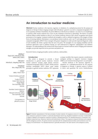
2. The Radiographer 2011 39
of the radiopharmaceuticals. It is important to note that the biological
processes take place with or without the radiopharmaceutical, the
radiopharmaceutical only makes it possible to image this physiologic
or pathophysiologic process by tagging or labelling the appropriate
compound (Figure 1). Since nuclear medicine employs compounds (or
their analogues) that are found naturally within the body, it is extremely
rare for a patient to have an adverse reaction to the radiopharmaceutical.
Exceptions to this do exist with some specific innovative tracers like
monoclonal antibodies that may elicit an immune response. Otherwise,
sensations experienced by patients at the time of administration
(particularly intravenously) are on the continuum describing a vasovagal
reaction.
How does nuclear medicine differ from x-ray? Fundamentally,
nuclear medicine principally evaluates the function (ie. physiology or
pathophysiology) of organs and systems while x-ray chiefly evaluates form
or morphology (ie. anatomy). It is true that x-ray, computed tomography
(CT), and MRI can image function and likewise nuclear medicine can
image morphology or structure. But it is fair to say that x-ray technology
is far better equipped to image anatomy than scintigraphy and that
scintigraphy provides a different level of physiological evaluation (Figure
2). Nuclear medicine is capable of tracing the most intricate biological
pathways by simply taking a compound that is known to (or should)
behave in a certain way within the body (ie. follow a particular biological
pathway) and attach it to a radioactive substance.
The history of nuclear medicine
The history of the individual disciplines that comprise the medical
radiationsciencesareintertwined.Thediscoveryandharnessingofthex-ray
and gamma rays in diagnosis and therapy have had a profound impact on
medicine and, indeed, the way people live their lives. From the irradiation
of food through to global security, the application of radiation has become
an integral part of our lives. The historical discoveries outlined briefly below
represent key events in transforming modern medicine and, indeed, in the
subsequent evolution and revolution that is enjoyed in medicine today.
While most of the scientific community were focusing their attention
on x-rays and the cathode tube, the French scientist Henri Becquerel
discovered radioactivity in 1896; spawning nuclear medicine. Unlike
x-rays (that were almost immediately and universally applied to medical
practice), some time would pass before any semblance of the industry
referred to as nuclear medicine developed. While working with uranium,
Becquerel noticed that photographic material would be exposed (fogged)
if in close proximity to uranium.1
The main difference between the
discovery of x-rays and radioactivity was that, unlike x-ray, other scientists
had not made similar observations. For Roentgen, his findings provided
science to the anecdotal evidence of many others and, was intuitive
and immediately adopted. Moreover, the very visual evidence conjured
immediate scientific, social and medical applications. Becquerel, however,
was not able to generate the same degree of enthusiasm for his discovery.
Several years later (1898), the Polish Marie Curie and her French
husband Pierre discovered radium and interest in radioactivity became
more widespread.1
Radium very quickly replaced x-rays for industrial
radiography. In 1899 Rutherford discovered alpha and beta particles and
in 1900 Villard discovered gamma rays.1
Alexander Graham Bell briefly dabbled in nuclear medicine,
suggesting the use of radioactive sources to treat tumours in 1903 and in
1913 it was reported as useful for various diseases.1
While these activities
represent the pioneering days in nuclear medicine, they also represent the
early years of radiotherapy. Nonetheless, radionuclide therapy remains a
central activity in nuclear medicine quite independent of the applications
of radiotherapy today.
Nuclear medicine itself tends to be characterised by the functional
assessment of a biological and metabolic process. In the 1920s,
radioactive phosphorus was used in animals to, for the first time, show
metabolic processes in an intact animal – the bone scan.1
Not long after,
phosphorus-32 (32
P) was used to treat a patient with leukemia. 32
P remains
a popular radionuclide for therapy and the bone scan (albeit without
phosphorus) is the most common nuclear medicine procedure today.
Radioactive iodine was used to study thyroid physiology in the late 1930s
and is used today for therapy and imaging.1
Strontium-89 (89
Sr) was first
evaluated in 1939 and is still used today for palliation of painful bone
metastases.1
While beta emissions are very useful for therapy, they are not
particularly good for imaging and radionuclides with gamma emissions
were required for nuclear medicine to make any advances.
The key breakthrough in nuclear medicine came in 1939 with
the discovery of Technetium-99m (99m
Tc) by Italian Emilio Segre and
American Glenn Seaborg.1
This allowed crude functional assessment
(eg. inject in left arm and measure intensity and timing for transit
to right arm with a detector). In 1951 the first scanner was developed
by Cassen (rectilinear scanner). A number of other key milestones
in nuclear medicine include; the first annual meeting of the Society of
Nuclear Medicine in 1954, the emergence of the scintillation camera
in 1958 (basis of current technology), commercial availability of 99m
Tc
generators in 1960, introduction of emission tomography in 1962, and
commercially available SPECT cameras in 1976.1
The emergence of
digital technology, computer technology, reconstruction algorithms,
multiple detector cameras and hybrid SPECT/CT devices, combined with
radiopharmaceutical developments, have all enhanced the capabilities of
nuclear medicine while the underlying principle remains fairly constant
since the late 1970s.
Since the discovery of x-rays and radium, knowledge / attitude
toward radiation safety has been pendulous; at the peril of patient and
health practitioner alike. While the mis-use of x-ray is well reported (hair
removal in beauty salons, shoe fittings, x-ray portraits, and even party
games), abuse of radium was more liberal in the early years.2
Radium
was popular as an ingested tonic, socialites served radium cocktails,
Figure 2: Anatomy (x-ray) and physiology (bone scan).
3. 40 The Radiographer 2011
radium toothpaste and radium contraceptive jelly were available, and
these represent but a fraction of the commercial exploitation of radium.2
The popularity of radium is perhaps typified by the use of radium paint
throughout the 1920s to produce luminescence; watch dials, dolls eyes,
religious artifacts to list but a few applications.2
Basic imaging principles
The basic principle of the gamma camera is to externally detect the
biodistribution of the administered radiopharmaceutical. Emission
of radioactivity, usually a gamma emission, facilitates this process. A
revision of the interactions of gamma, alpha and beta emission in tissue
(or matter) is beyond the scope of this article, however, it is an important
basis for the following discussion. Photons emitted from the patient
first encounter the collimator which acts as a physical discriminator to
eliminate photons from the resulting image whose angle of incidence
varies from approximately 90degrees to the image plane (Figure 3). Thus,
a single image reflects a ‘planar’ image. That is, one might expect the origin
of a detected photon to originate at some point along the line projected
back through the patient at 90 degrees to the detector face. The emission
of a photon from an unstable nuclei is random in direction and the lead
or tungsten septa of the collimator is designed to absorb photons not
incident at approximately 90 degrees. The size and/or length of the ‘hole’
or space between septa (generally a hexagonal array) govern the variation
from 90 degrees of the accepted angle of incidence (larger and/or shorter
holes have a larger window) and, thus, impact on image resolution and
sensitivity. The distance the object is from the detector has a significant
bearing on image resolution (Figure 3 and Figure 4).
Incident photons passing through the collimator interact with the
crystal. In the traditional gamma camera the crystal is inorganic sodium
iodide although there have been recent advances in solid state or semi-
conductor detectors (eg. CZT or cadmium zinc telluride). The energy of
the incident photon causes excitation in the crystal which, on de-excitation,
Figure 4: A series of images of a thyroid phantom with various size ‘nodules’,
positioned identically but positioned with the detector 0 cm (left), 5 cm (middle) and
10 cm (right) from the phantom. It is clear from these otherwise identical images that
a significant degradation in resolution and contrast results from increasing the object
to detector distance. This is evident in the ‘blurred’ boundaries, decreasing lesion
size and eventual lack of lesion detection.
Figure 5: Schematic representation of the basic operation of a gamma camera. Pho-
tons emitted from the body incident perpendicular to the detector are ‘trapped’ in the
crystal; converting the energy to a light emission. The light is detected by the pho-
tocathode of the photomultiplier tube (PMT) and converted to an electrical impulse
which is amplified through the PMT. Position circuitry provides an X and Y coordinate
for each event and that event is registered in the image if a Z signal is also received
from the pulse height analyser (PHA) where scatter events are eliminated.
Figure 3: Depiction of the operation of the collimator. The collimator is the interface
between the patient and the detector system; serving as a physical discriminator
of photons based on their angle of incidence. Photons incident perpendicular to the
imaging plane (A) generally pass through the collimator to interact with the detec-
tor. Photons incident at a small angle defined by the septal length and the ‘hole’
size are also detected (B). It is possible for events that are scattered to be incident
perpendicular to the detector (C) and this will mis-register the location of the event if
included in the image. Photons incident outside the small window about 90 degrees
(D) will be absorbed by the septa. Sensitivity of the system can be reduced through
smaller ‘holes’ (increased resolution) because it increases the cross sectional sur-
face area of the septa for elimination of events incident perpendicular to the detec-
tor but ‘in line’ with the septa (E). Other events, including those that might otherwise
be incident perpendicular to the detector, may undergo scatter events that take the
event outside the field of view of the detector (F). Clearly, lengthening the septa or
adding septa (smaller ‘holes’) will convert event B to a type D event. The impact of
the acceptable range of incident angles can be seen to the left of the figure with the
mis-registration error increasing with distance from the detector (G to H; front of
body to back of body) which highlights the importance of imaging an organ as close
to the detector as possible (see also Figure 4).
4. The Radiographer 2011 41
releases energy in the form of light; hence the term scintillation detector.
The amount of light emitted is proportional to the energy of the incident
photon which facilitates energy discrimination later in the process.
Theemittedlightisdetectedbyanarrayofphotomultipliertubes(PMTs).
PMTs are tubes that convert weak light outputs into amplified electrical
impulses (Figure 5). One end of the tube couples to the crystal and is coated
with a material which emits electrons in response to incident visible light
photons and is known as the photocathode. The signal is amplified through
the tube using dynodes until an impulse exits at the anode.
PositioningcircuitryprovidesanX,Ysetofcoordinatesfortheincident
photon and this coordinate is registered when a Z signal is received. The
X,Y coordinate represents a single ‘dot’ registered in the image. Each image
is comprised upwards of 500000 separate events yet there are billions of
other events that go undetected. A 200 MBq (megabecqueral) dose, for
example, represents 200 million events or disintegrations every second.
Doses in nuclear medicine are in the order, typically, of 150 MBq to 1000
MBq. The Z signal is produced by the pulse height analyser (PHA) when
the incident energy is within an acceptance window and, thus, the PHA
is an energy discriminator (Figure 6). The role of the PHA is to eliminate
events that are incident perpendicular to the detector after a scatter event
(event C in Figure 3). Following scatter, energy is lost and thus the amount
of light and subsequent signal amplitude is lower than the target events.
Advances in nuclear medicine imaging
Single photon emission computed tomography (SPECT)
Since its introduction into clinical nuclear medicine in the late 1970s
SPECT, has become a routine tool in the nuclear medicine. A significant
part of that transition was the advent of sufficient computing power for data
collection and subsequent reconstruction. In the late 1980’s in Australia,
the majority of gamma cameras in clinical departments did not have
SPECT capability. By the early 1990s in Australia, virtually all new gamma
cameras being purchased were SPECT systems. Nonetheless, the early
SPECT systems were based on modified planar cameras and these were not
optimised for SPECT creating reliability issues. Moreover, the limitations
associated with computing space and power meant sub-optimal acquisition
parameters were employed. The real value of SPECT emerged with the
advances in computer technology and gantry configurations
Computer technology allowed the use of optimal imaging parameters
(e.g. matrix) due to overcoming data storage limitations. Computing power
has significantly shortened reconstruction time from hours to seconds which
has allowed multiple re-slice. Certainly the advances in computing power
have allowed the introduction of gated SPECT which revolutionised cardiac
imaging in the late 1990s. The reconstruction process has been refined (pre-
and post-filter functions) which has allowed improved image quality. New
reconstruction approaches, like iterative reconstruction, are possible with the
improved computing technology. The use of 180 degree data collection and
reconstruction algorithms essentially halved the acquisition time.
Cameras were constructed with dedicated SPECT gantries which have
allowed greater physical integrity of the data collection (eg. less centre of
rotation issues typical of older generation cameras) and the introduction of
elliptical orbits to maximise spatial resolution. The introduction of digital
head technology has improved image quality. The duration of the imaging
time has been reduced with the introduction of multi-detector technologies
and cardiac configurations (90 degree). Image quality has been refined
with the development of high resolution/sensitivity collimator designs
Figure 6: Schematic representation of the energy photopeak. Application of a nar-
row acceptance window will eliminate scatter events that are incident perpendicular
to the detector but which have previously undergone a scatter event.
Figure 7: Schematic representation of the impact of attenuation on gamma emissions
that would otherwise be incident perpendicular to the detector. The organ may be of
greater or lesser density than surrounding body tissues and, indeed, some structures
will have very lower density or attenuation (eg. lung or gas pockets in the colon). Some
events pass from an organ structure, through surrounding body tissues and are de-
tected without attenuation (A). Events originating at the same depth within a structure
will have variable attenuation based on whether the pathway is through a lower den-
sity structure (B) or normal tissues (C). Independent of variations in the density along
the photon pathway, events originating from the same (or similar) locations within
a structure, based on probability, may be attenuated (D) or detected (E). In a dense
organ of interest, some events are attenuated within the organ itself (F), particularly if
the event originates deeper with the structure. Clearly in this schematic, imaging the
patient either posterior or laterally will change the fates of each of the hypothetical
events. The point of attenuation correction is to apply a uniform field along a series of
lines of response and estimate the probability of an event being attenuated along that
pathway, and then subsequently corrected for those loses.
5. 42 The Radiographer 2011
(eg. fan beam). The emergence of CT based attenuation correction has
improved image quality through attenuation correction of the data (Figure
7) while improving lesion localisation. More recently the introduction of
solid state detectors has improved resolution and sensitivity substantially
and subsequently reduced images times to a fraction of traditional SPECT
systems (eg. 20 minute SPECT reduced to just 3–5 minutes without loss
of quality). SPECT is a technique for generating images of single planes
within a volume of radioactivity from projections of that volume obtained
at a number of different angles.3
The principle is similar to that of PET and
CT (Figure 9). In a more practical sense, SPECT is a method of separating
overlying and underlying tissue from the source or target of interest by
reconstructing cut sections of the object, (traditionally transverse, sagittal
and coronal, but in cardiology, short axis, vertical long axis and horizontal
long axis). Unlike planar projections, SPECT does not have the problem
of superimposition of other structures in front of or behind the organ of
interest, thus improving image contrast and allowing smaller defects to be
accurately diagnosed. The advantages of SPECT over planar imaging are the
same as those for performing CT over x-ray. Clearly there will be situations
where SPECT (or CT) provides better quality images than the planar
equivalent without actually changing the outcome or patient management.
The difference is that the addition of CT to the patient work-up significantly
increases the radiation dose. For SPECT, there is no additional radiation
dose over that which was given for the planar study.
Single photon emission computed tomography/
computed tomography (SPECT/CT)
While hybrid imaging requires a dedicated article to adequately
address its emergence and role in clinical practice, in the context of
this discussion SPECT/CT represents an important development
in nuclear medicine. The boundaries demarcating the line between
disciplines have been blurred in the past; the original CT devices were
rotating radionuclide sources, the origins of radiation therapy and
radionuclide therapy are common and some modalities like MRI are
not a classical fit in either discipline. In recent times, there has been a
noticeable convergence to exploit synergies in diagnosis and therapy to
improve outcomes. Radiography has always been anatomy based and
nuclear medicine always been strongly physiology based. Advances in
instrumentation in both areas have seen significant clinical improvements
but also the emergence of hybrid systems where a single device operates
as both a SPECT system and CT, PET and CT or more recently PET and
MRI. These hybrid systems demand uniquely qualified individuals and
represent the cutting edge and future of imaging technology. As indicated
in Figure 9, the fusion of SPECT and CT has added a new dimension
allowing nuclear medicine to have anatomical definition and CT to have
sensitive physiological information.
Radiopharmacy
The radiopharmacy is an important aspect of all nuclear medicine
departments but not all nuclear medicine departments have a fully
functional radiopharmacy with on site 99m
Tc generator. Some departments
will accept delivery of radionuclides (99m
Tc) from a centralised
radiopharmacy and reconstitute their own radiopharmacueticals (kits)
on site for use on a daily basis. Others rely on unit dose deliveries from a
centralised pharmacy. Still others rely on a mix of the two.
The main radiopharmaceutical used in general nuclear medicine is 99m
Tc
and this is produced by a generator from decay from the Molybdenum-99
(99
Mo) parent. A single generator tends to provide sufficient radionuclide
activity for one week because the parent nuclide has a half-life of
approximately 67 hours (99m
Tc has a 6 hour half-life). The half-life is one
of the key issues in nuclear medicine. It governs how long a radioactive
source is useful for imaging. Every half-life, the activity is halved. Thus, for
99m
Tc our imaging window is typically around 6 hours and by 24 hours there
is very little to image. One needs to consider that the half-life is but one
issue, once administered to the patient, elimination (e.g. urinary system)
introduces the biological half-life which can further reduce the effective
window for imaging. A bone scan has relatively slow biological clearance
and consequently the patient is imaged several hours after administration
and a long window of opportunity for imaging (many hours) is available.
Conversely, renal imaging using perfusion tracers are rapidly excreted
(by the nature of the test) and imaging is performed immediately upon
administration and in a narrow window exists (minutes).
99
Mo and some other radionuclides used in nuclear medicine (eg.
Iodine-131 [131
I]) are produced in a fission nuclear reactor like the one at
Lucas Heights in Sydney. Others are produced by a cyclotron (Iodine-123
[123
I], Gallium-67 [67
Ga], Thallium-201 [201
Tl]); these are imported to
Australia commercially since the de-commission of Australia national
medical cyclotron. The cyclotrons operational in Australia are small (lower
Figure 8: Schematic representation of the principals of SPECT. Individual planar pro-
jections are acquired at regular intervals around the patient’s body (typically 60-120)
for either 360 or 180 degrees depending on the organ of interest. The subsequent im-
age profiles are fed into the reconstruction algorithm (typically filtered backprojection
or iterative reconstruction) to reconstruct the object of interest. The object generally
suffers some loss of detail over the object like CT or even a photograph.
Figure 9: High resolution CT, poor resolution Indium-111 (111
In) octreotide SPECT and
fusion SPECT/CT for neuroendocrine tumour detection and localisation.
6. The Radiographer 2011 43
maximum energy) and geared toward production of PET radionuclides
(eg. Flourine-18 [18
F], Nitrogen-13 [13
N]) only. Some radionuclide therapy
sources are produced in a nuclear reactor by neutron activation (eg. 32
P, 89
Sr).
One of the key strengths of nuclear medicine has been the innovation
of various radiopharmaceuticals. While the radionuclides have been fairly
static (Table 1), the biological products labelled to those radionuclides has
progressed rapidly. 99m
Tc labelling of somatostatin receptor tracers is one
example that has revolutionised the imaging of neuroendocrine tumours.
The development of tracers that cross the blood brain barrier reinvented
cerebral imaging and, indeed, broadened the clinical efficacy and utility.
Well established tracers, like 201
Tl chloride for myocardial perfusion
imaging, have had limitations associated with physical properties (eg.
half-life, photon energy) overcome by development of perfusion tracers
labelled to 99m
Tc. Even the humble bone scan has seen refinement of
radiophamraceuticals for improved localisation, improved target to
background ratios and more rapid localisation (earlier imaging times).
Radiopharmacy is a dynamic and very liquid environment that ensures
nuclear medicine remains cutting edge, providing an integrative and
adjunctive nexus to both radiography and radiation therapy.
There are several key strategies for safety in the radiopharmacy. Time,
distance and shielding (TDS) reduces radiation exposure to staff and other
visitors to the department. Reduce the time exposed, increase the distance
from the source and use shielding between yourself and the source. As
low as reasonably achievable (ALARA) is a concept that protects patients
and staff. In essence, and not too dissimilar to approaches in x-ray, the
lowest dose that can be reasonably used to provide diagnostic quality
results is used. While minimal, the risk of a radiation based adverse effect
is generally of greater importance in managing the patients than the risk of
adverse reaction to the radiopharmaceutical (with the notable exception
of monoclonal antibodies). The concepts of justification, optimisation
and limitation are also applied. Contrary to popular belief outside nuclear
medicine, the radiation doses to patients and staff are very low. For many
procedures, the radiation dose to the patient is lower than the x-ray
equivalent; especially if that equivalent is fluoroscopy, angiography or CT.
Doses to staff are typically higher than that of a general radiographer or
radiation therapist but only a fraction of the occupational exposure limits.
Clinical indications
There are a number of key reasons for choosing a general nuclear
medicine procedure either as an adjunct to other imaging modalities or as
an alternative to them (Table 2). These include, without being limited to:
• Diagnosis (detection and localisation) of conditions or diseases which
for some reason have gone undetected by other simpler imaging
techniques (e.g. fractures may not be visible on x-ray if there is no
separation of the bone and in small or complex structures).
• In some cases, nuclear medicine offers a simple, cost effective
alternative to more invasive, high-risk procedures (e.g. myocardial
perfusion study instead of a coronary angiogram).
• Nuclear medicine is extremely sensitive and in some cases can
detect and localise disease processes that other imaging modalities
cannot successfully image (e.g. cerebral perfusion studies where the
radiopharmaceutical actually crosses the blood brain barrier and is
trapped in the brain cells to allow perfusion mapping).
• Nuclear medicine studies can offer lower radiation doses than
traditional alternatives. This can be of significant advantage in
paediatrics,womenofchildbearingage,pregnantpatientsandpatients
having repeated follow up studies (e.g. radionuclide cystogram versus
the radiographic cystogram and the ventilation / perfusion (V/Q)
lung scan versus CT pulmonary angiography).
Table 1: Radionuclides typically employed in nuclear medicine.4,5
The table includes PET radionuclides but excludes radionuclide therapy.
Nuclide Emission
Gamma energy for
imaging
(MeV)
Half-life
Maximum particle
energy (MeV)
Production
Technetium 99m
Tc Gamma 0.140 6 hours – 99
Mo/99m
Tc generator
Thallium 201
Tl Gamma 0.80 & 0.167 73 hours – Cyclotron
Gallium 67
Ga Gamma 0.093, 0.184 & 0.296 78 hours – Cyclotron
Indium 111
In Gamma 0.173 & 0.247 2.8 days – Cyclotron
Iodine 123
I Gamma 0.159 13.3 hours – Cyclotron
Iodine 131
I Gamma 0.364 8 days 0.61 Fission
Fluorine 18
F Positron 0.511 109 min 0.64 Cyclotron
Carbon 11
C Positron 0.511 20 min 0.97 Cyclotron
Nitrogen 13
N Positron 0.511 10 min 1.20 Cyclotron
Oxygen 15
O Positron 0.511 2 min 1.73 Cyclotron
Gallium 68
Ga Positron 0.511 68 min 2.919 68
Ge/68
Ga generator
Rubidium 82
Rb Positron 0.511 75 seconds 3.15 82
Sr/82
Rb generator
Xenon 133
Xe Beta & Gamma 0.080 5.2 days 0.364 Fission
Krypton 81m
Kr Gamma 0.190 13 seconds 1.29 81
Rb/81m
Kr generator
7. 44 The Radiographer 2011
Organ / Pathology Radiopharmaceutical Indications
Bone 99m
Tc MDP or HDP
Metastases, metabolic disease (eg. Paget’s), trauma (particularly when xray is normal or difficult), infection, sports injuries (eg. pars
articularis, osteitis pubis, shin splints, stress fractures), avascular necrosis, assess prosthetic joints for loosening or infection, degenerative
change or arthritis, pain in bone or joints, reflex sympathetic dystrophy to name a few.
Bone marrow 99m
Tc sulphur colloid Define marrow distribution in tumour, biopsy, harvest and post joint replacement.
Brain
99m
Tc DTPA flow
99m
Tc HMPAO or ECD perfusion
The flow study is used to assess the integrity of the blood brain barrier (BBB). It is useful although superseded by CT and SPECT perfusion
in; cerebrovascular accident, tumour, other mass, infection and brain death.
Cerebral perfusion SPECT uses tracers that cross the BBB and are useful for functional assessment in epilepsy, stroke, transient ischaemic
attacks, carotid artery stenosis, infection and brain death.
Lacrimal glands
99m
Tc pertechnetate or DTPA – drops on eye
surface
Epiphora. The procedure is non invasive and assess the actual physiology without application of a catheter or pressure.
Salivary 99m
Tc pertechnetate Functional evaluation of salivary glands in suspected tumour, Sjogren’s syndrome or infection.
Thyroid
99m
Tc pertechnetate although 123I is used more
so in the USA
Grave’s disease, assess functional status of nodules, detect ectopic tissue, quantitate and differentiate causes of hyper- and hypo-
thyroidism, determine thyroid uptake for assessment and therapy planning, response to therapy.
Parathyroid
99m
Tc pertechnetate in combination with 99m
Tc MIBI
or 201
Tl chloride
Pre-operative localisation of hyperfunctioning parathyroid tissue (adenoma or hyperplasia). Locate residual hyperfunctioning parathyroid
tissue, including ectopic.
Oesophagus
99m
Tc sulphur colloid labelled in orange juice or
egg whites (liquid versus solid) - oral
99m
Tc pertechnetate - IV
Oesophageal transit (eg. spasm, achalasia), lung aspiration and oesophageal reflux studies for detection and quantitation.
Barrett’s oesophagus.
Stomach
99m
Tc sulphur colloid labelled in orange juice or
egg whites (liquid versus solid) - oral
Solid and liquid gastric empty studies to evaluate rapid or delayed transit. Physical or functional obstruction. Reflux studies can be
performed in conjunction.
Liver
99m
Tc sulphur colloid
99m
Tc red blood cells
99m
Tc IDA derivative (HIDA, DIDA, DISIDA etc)
Liver / spleen scan to determine size and shape of each, functional abnormalities of reticloendothelial system, focal nodular hyperplasia,
trauma and accessory spleen.
Haemangioma and focal nodular hyperplasia.
Assessment of biliary function including gallbladder ejection fraction following a fatty meal intervention. Detection of obstruction in the cystic
duct or common bile duct.
Colon
67
Ga citrate – oral
99m
Tc red blood cells - IV
99m
Tc pertechnetate - IV
Colon transit for constipation.
Gastrointestinal haemorrhage.
Meckel’s diverticulum
Kidneys
99m
Tc DTPA or MAG3
(renogram)
99m
Tc DMSA
Differential function and assessment of blood supply. Can be used instead of CT if contrast is contraindicated. Diuretic intervention for
assessment and quantitation of obstructive uropathy. Captopril intervention for detection of renal artery stenosis.
Cortical morphology imaging for assessment of pyelonephritis, scarring, trauma, space occupying lesion and in patients where contrast is
contraindicated.
Bladder
99m
Tc DTPA – instilled in bladder (direct method)
or IV (indirect)
Radionuclide cystogram is very sensitive and low doses radiation alternative to micturating cystogram for the detection and quantitation of
vesico-uretal reflux.
Testes 99m
Tc pertechnetate Evaluation of the acute scrotum for diagnosis of torsion. Largely superseded by ultrasound.
Heart
99m
Tc MIBI
99m
Tc red blood cells
Myocardial perfusion study to assess coronary flow reserve in known or suspect coronary artery disease. Includes quantitation of chamber
volumes and ejection fraction.
The blood pool study provides accurate quantitation of the left ventricular function, including volumes and ejection fraction. It can also be
employed to evaluation cardiac shunts (first pass study).
Lung
99m
Tc MAA (perfusion - IV) and 99m
Tc technegas
(ventilation - inhalation) although 99m
Tc DTPA
aerosols are also used in Australia.
Detection of pulmonary embolism (PE). Virtually 100% positive and negative predictive value. Intermediate probability generally need CT
angiogram. Differentiate old and new PE. Quantitative differential studies pre-surgical resection for emphysema. Can be sued to detect and
quantitate right to left cardiac shunts.
Sentinel node
99m
Tc antimony sulphur colloid - sub cutaneous,
inter-tumoural, peri-tumoural
Identification of lymphatic sentinel node prior to breast cancer surgery.
Breast 99m
Tc MIBI or 201Tl chloride
Scintimammography is a powerful tool for differentiating benign and malignant breast lesions. Particularly useful in monitoring response to
non surgical therapy. Is effective in assessing those not suitable for mammography.
CSF
99m
Tc DTPA (triple filtered) – intra-portal or
intrathecal
Hydrocephalis, shunt patency, cerebrospinal fluid (CSF) leak.
Lymphatics 99m
Tc antimony sulphur colloid - sub cutaneous Bilateral or unilateral lymphoedema, typically arms or legs but it has been performed in other structures (eg. penis or vulva).
Infection
67
Ga citrate
99m
Tc white blood cells
Fever of unknown origin, osteomyelitis, lung infection (eg. pneumocystis pneumonia in immune suppression).
Fever of unknown origin, infected joint prosthesis, inflammatory bowel disease, graft infection, abscess, diabetic infections.
Tumour
67
Ga citrate
111
In octreotide
123
I MIBG
Non-Hodgkin’s lymphoma and to a lesser extent Hodgkin’s disease. The latter superseded by PET.
Neuroendocrine tumours.
Pheochromocytomas, neuroblastoma, carcinoid tumours.
Table 2: A summary of the clinical indications of major nuclear medicine procedures.4,5
The list is not exhaustive but rather provides a snapshot of the main clinical proce-
dures performed in Australian nuclear medicine departments. The tabulated summary excludes PET, radionuclide therapy, research procedures and emerging techniques.
There are a number of procedures that can be modified for other purposes that are also not listed. Routes of administration are intravenous (IV) unless otherwise indicated.
8. The Radiographer 2011 45
• Computer quantitation of function is possible and is often the only
clue to detection of an abnormal process (eg. renograms in renal
artery stenosis and myocardial perfusion polar maps).
• Outside of trauma, many pathologies begin as a functional change
and that functional change eventually produces the structural change
seen in anatomical imaging. While widespread screening is neither
cost effective nor safe, nuclear medicine is capable of detecting disease
before the structural changes are apparent.
• Functional imaging allows earlier detection of disease, risk
stratification, tailoring of patient management, differentiation of
pathology (eg. benign or malignant) and assessment of response
to therapy. For example, response of a tumour to therapy can be
immediate from a functional perspective despite no ‘shrinkage’ on
anatomical imaging (indeed there can be some enlargement).
• Therapeutic doses can be performed for the treatment of a variety
of conditions. Scintigraphy can be used to map biodistribution of
therapeutic tracers before therapy is undertaken.
• AdvancedtoolslikeSPECTandSPECT/CTofferadditionaladvantages
which have been discussed above.
Conclusion
Nuclear medicine provides an important tool for an integrative
approach to medical radiation science. The synergies between
nuclear medicine and both radiography and radiation therapy are
evident historically and in the emergence of current best practice.
An understanding of the technical and clinical aspects of integrated
modalities affords advantage to clinical practitioners in all disciplines of
the medical radiation sciences. This article provides a basic insight into
nuclear medicine to extend the capabilities of radiographers, radiation
therapists and other health care professions engaged in the diagnostic
imaging industry.
References
1 Graham LS, Kereiakes JG, Harris C, Cohen MB. Nuclear medicine from Becquerel
to the present. RadioGraphics 1989; 9: 1189–202.
2 DiSantis D, DiSantis D. Radiologic history exhibit. Wrong turns on radiology’s road
of progress. RadioGraphics 1991; 11: 1121–38.
3 English R, Brown S. SPECT. Single photon emission computed tomography: A
primer (3rd edition). Virginia: Society of Nuclear Medicine; 1995. pp 23–148.
4 Christian PE, Waterstram-Rich KM (editors). Nuclear medicine and PET/CT:
technology and techniques (7th edition). Philadelphia: Elsevier Mosby; 2012.
5 Theobald T (editor.). Sampson’s textbook of radiopharmacy (4th edition). London:
Pharmaceutical Press; 2011.