colle's fracture.pptx
•Download as PPTX, PDF•
0 likes•161 views
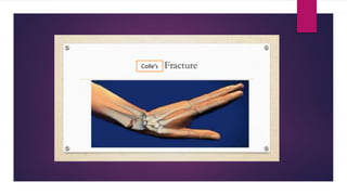
Colle's fracture is a fracture of the distal radius near the wrist joint. It most commonly occurs in postmenopausal women over 40 years old from falls on an outstretched hand. Clinical features include pain, swelling, and the classic "dinner fork deformity". Treatment involves closed or open reduction and casting for 4-6 weeks. Physiotherapy focuses on regaining range of motion, strength, and function. Complications can include nonunion, stiffness, or nerve damage if not properly treated.
Report
Share
Report
Share

Recommended
Recommended
More Related Content
What's hot
What's hot (20)
Student's Elbow (Olecranon Bursitis) - Dr Rohit Bhaskar
Student's Elbow (Olecranon Bursitis) - Dr Rohit Bhaskar
Physiotherapy Management for Wound Ulcers Rahul.AP BPT,MPT (CRD&ICU Managemen...
Physiotherapy Management for Wound Ulcers Rahul.AP BPT,MPT (CRD&ICU Managemen...
Similar to colle's fracture.pptx
Similar to colle's fracture.pptx (20)
More from BijayaSaha5
More from BijayaSaha5 (20)
Recently uploaded
❤️Call girls in Jalandhar ☎️9876848877☎️ Call Girl service in Jalandhar☎️ Jalandhar Call Girls Service ☎️
Call Girls In Jalandhar 8264406502 Jalandhar railway station Near Radisson Hotel Jalandhar, Majestic Grand Hotel, Ramada by Wyndham Jalandhar City Centre, Park Plaza Ludhiana, Windsor Fountain, G.T Road Jalandhar escort all Jalandhar service Russian available model female girls in Jalandhar VIP Lo price personal Jalandhar off class call girls payment high profile model and female escort 70% Off On Your First Booking Jalandhar Call Girls Service Cash Payment
Welcome to NehaChopra Jalandhar Call Girl Service, the Trusted call girl agency around. We Offer 70% Discount On Your First Booking For Jalandhar Call Girls Service Cash Payment is available.❤️Call girls in Jalandhar ☎️9876848877☎️ Call Girl service in Jalandhar☎️ Jal...
❤️Call girls in Jalandhar ☎️9876848877☎️ Call Girl service in Jalandhar☎️ Jal...chandigarhentertainm
(Deeksha) 💓 9920725232 💓High Profile Call Girls Navi Mumbai You Can Get The Service Of A Mumbai Call Girl At Any Time
WHATSAPP On Here:9920725232
Today call girl service available 24X7*▬█⓿▀█▀ 𝐈𝐍𝐃𝐄𝐏𝐄𝐍𝐃𝐄𝐍𝐓 CALL 𝐆𝐈𝐑𝐋 𝐕𝐈𝐏 𝐄𝐒𝐂𝐎𝐑𝐓 SERVICE ✅
⭐➡️HOT & SEXY MODELS // COLLEGE GIRLS
AVAILABLE FOR COMPLETE ENJOYMENT WITH HIGH PROFILE INDIAN MODEL AVAILABLE HOTEL & HOME
★ SAFE AND SECURE HIGH CLASS SERVICE AFFORDABLE RATE
★ 100% SATISFACTION,UNLIMITED ENJOYMENT.
★ >> 03-05-2024 (GRV)
★ All Meetings are confidential and no information is provided to any one at any cost.
★ EXCLUSIVE PROFILes Are Safe and Consensual with Most Limits Respected
★ Service Available In: - HOME & HOTEL 24x7 :: 3 * 5 *7 *Star Hotel Service .In Call & Out call SeRvIcEs :
★ A-Level (5 star escort)
★ Strip-tease
★ BBBJ (Bareback Blowjob)Receive advanced sexual techniques in different mode make their life more pleasurable.
★ Spending time in hotel rooms
★ BJ (Blowjob Without a Condom)
★ Completion (Oral to completion)
★ Covered (Covered blowjob Without condom
100% SAFE AND SECURE 24 HOURS SERVICE AVAILABLE HOME AND HOTEL SERVICES(Deeksha) 💓 9920725232 💓High Profile Call Girls Navi Mumbai You Can Get The S...
(Deeksha) 💓 9920725232 💓High Profile Call Girls Navi Mumbai You Can Get The S...Ahmedabad Call Girls
Hello, Guys welcome to Manalifun Goa Escort service. Are you want Top call girls in Goa at just ₹10000 then no further anywhere because we have a large number of local beautiful girls. We are a genuine platform to provide unlimited classification escort ads service without any commission. 9316020077
Here many Goa Independent call girls and ladies, publish their ads. Our call girl in Goa is well-known for real sexual fun in Goa. We are not allow any prostitute to work here without checking the details, Firstly all ads check by our team then we publish them here. So don’t hesitate to book Low rate call girls in Goa. 9316020077
Goa call girls: A real wonder in Goa
Who are the best Goa Escort Service provider for Goa call girls
High-Class call girls in Goa escort service for 100% Satisfaction
Choose a trusted call girl service in Goa with Us +91-9316020077
Goa Escorts Provide 100% Client Satisfaction
How Our Goa Call Girls Are Perfect For Instant Satisfaction
100% Guaranteed Goa call girls will make you excited
How to Find Cheap Call Girls in Goa
Our Reliable Escort Service in Goa Local Areas
Goa Escorts (cheap escort service in Goa)
Rate Chart of Goa call girls, (call girl Rate in Goa)
5-star hotel For Goa call girls service
Call girls in Goa are the ideal sex partner for you
BOOK YOUR FAVORITE Goa CALL GIRLS SERVICE WITH US CALL! US NOW~ 9316020077
Best way to Hire call girls in Goa
What’s the cost of escort service in Goa
North Goa Call Girls
Location :-
Baga , Caclangute , Candolim , Anjuna , Panaji Arpora , Vagator , Morjim , Siolim , Mandrem , Arambol , etc.
Vasco , Bambolim , Madgaon, Colva , EtcCall Girls Service In Goa 💋 9316020077💋 Goa Call Girls By Russian Call Girl...
Call Girls Service In Goa 💋 9316020077💋 Goa Call Girls By Russian Call Girl...russian goa call girl and escorts service
Recently uploaded (20)
Rajkot Call Girls 👙 6297143586 👙 Genuine WhatsApp Number for Real Meet
Rajkot Call Girls 👙 6297143586 👙 Genuine WhatsApp Number for Real Meet
Vip Call Girls Makarba 👙 6367187148 👙 Genuine WhatsApp Number for Real Meet
Vip Call Girls Makarba 👙 6367187148 👙 Genuine WhatsApp Number for Real Meet
Bareilly Call Girls 👙 6297143586 👙 Genuine WhatsApp Number for Real Meet
Bareilly Call Girls 👙 6297143586 👙 Genuine WhatsApp Number for Real Meet
❤️Call girls in Jalandhar ☎️9876848877☎️ Call Girl service in Jalandhar☎️ Jal...
❤️Call girls in Jalandhar ☎️9876848877☎️ Call Girl service in Jalandhar☎️ Jal...
Ernakulam Call Girls 👙 6297143586 👙 Genuine WhatsApp Number for Real Meet
Ernakulam Call Girls 👙 6297143586 👙 Genuine WhatsApp Number for Real Meet
Krishnagiri call girls Tamil Actress sex service 7877702510
Krishnagiri call girls Tamil Actress sex service 7877702510
Mangalore Call Girls 👙 6297143586 👙 Genuine WhatsApp Number for Real Meet
Mangalore Call Girls 👙 6297143586 👙 Genuine WhatsApp Number for Real Meet
Tirupati Call Girls 👙 6297143586 👙 Genuine WhatsApp Number for Real Meet
Tirupati Call Girls 👙 6297143586 👙 Genuine WhatsApp Number for Real Meet
Jaipur Call Girls 9257276172 Call Girl in Jaipur Rajasthan
Jaipur Call Girls 9257276172 Call Girl in Jaipur Rajasthan
(Deeksha) 💓 9920725232 💓High Profile Call Girls Navi Mumbai You Can Get The S...
(Deeksha) 💓 9920725232 💓High Profile Call Girls Navi Mumbai You Can Get The S...
bhubaneswar Call Girls 👙 6297143586 👙 Genuine WhatsApp Number for Real Meet
bhubaneswar Call Girls 👙 6297143586 👙 Genuine WhatsApp Number for Real Meet
Best Lahore Escorts 😮💨03250114445 || VIP escorts in Lahore
Best Lahore Escorts 😮💨03250114445 || VIP escorts in Lahore
bhopal Call Girls 👙 6297143586 👙 Genuine WhatsApp Number for Real Meet
bhopal Call Girls 👙 6297143586 👙 Genuine WhatsApp Number for Real Meet
Bhagalpur Call Girls 👙 6297143586 👙 Genuine WhatsApp Number for Real Meet
Bhagalpur Call Girls 👙 6297143586 👙 Genuine WhatsApp Number for Real Meet
Call Girls Service In Goa 💋 9316020077💋 Goa Call Girls By Russian Call Girl...
Call Girls Service In Goa 💋 9316020077💋 Goa Call Girls By Russian Call Girl...
Kolkata Call Girls Miss Inaaya ❤️ at @30% discount Everyday Call girl
Kolkata Call Girls Miss Inaaya ❤️ at @30% discount Everyday Call girl
palanpur Call Girls 👙 6297143586 👙 Genuine WhatsApp Number for Real Meet
palanpur Call Girls 👙 6297143586 👙 Genuine WhatsApp Number for Real Meet
Premium Call Girls Bangalore {7304373326} ❤️VVIP POOJA Call Girls in Bangalor...
Premium Call Girls Bangalore {7304373326} ❤️VVIP POOJA Call Girls in Bangalor...
Mathura Call Girls 👙 6297143586 👙 Genuine WhatsApp Number for Real Meet
Mathura Call Girls 👙 6297143586 👙 Genuine WhatsApp Number for Real Meet
Thoothukudi Call Girls 👙 6297143586 👙 Genuine WhatsApp Number for Real Meet
Thoothukudi Call Girls 👙 6297143586 👙 Genuine WhatsApp Number for Real Meet
colle's fracture.pptx
- 1. Colle’s
- 2. Definition • It was first described by Abraham colles in 1814. • Colles fracture is the fracture at the distal end of radius, at its cortico cancellous junction(about 2cm from the distal articular surface). It is not just the fracture of distal radius but the fracture dislocation of the inferior radio-ulnar joint. • Most common age group is above forty years, occuring most commonly in women.
- 3. Mechanism of Injury Fall on an outstretched hand.
- 4. Causes Have osteoporosis, a disease that weakens your bones Postmenopausal osteoporosis. Commonest skeletal injury In Elderly. Have low muscle mass, poor muscle strength, or lack agility (these conditions make you more likely to fall) Walk or do other activities in snow, on ice, or that require a lot offorward momentum, such as in-line skating and skiing Have an inadequate intake of calcium or vitamin D Road Traffic Accident, fall from height.
- 5. Clinical features: • Pain : arm was held close to body • Swelling • Deformity- There is classic ‘dinner-fork deformity’ seen in colles’ fracture. • Radial styloid process lies in the same level or little higher than the ulnar styloid process.
- 6. Types of Colle's Fracture • Open fracture: If the bone broke through yourskin •Comminuted fracture: If the bone broke into more than two pieces • Intra-articular fracture: If the bone broke inside your wrist joint • Extra-articular fracture: If your joint isn’taffected
- 7. Complications Early complication includes Swelling and Pain in the Finger Median Nerve compression Sudek's Atrophy Late Complication Malunion Non union of the ulnar styloid process. Rupture of the Extensor pollicislongus. Stiffness Shoulder hand syndrome
- 8. Treatment Undisplaced Fracture- A dorsal Splint for 1-2 days to remove swelling and cast will be Placed for 4 week to stabilize joining.
- 9. Treatment • Displaced Fracture – Reduction of the fracture under anesthesia and correct the Bone alignment. Then Dorsal Plaster Slab is applied. • Comminuted Fracture- Percutaneous K-wire fixation along with Plaster Immobilization. It then Remove after 5 weeks.
- 10. Physiotherapy Treatment Main role of Physiotherapy is in rehabilitation Stage. • Objectives of rehabilitation 1. Maintain joint range of motion 2. Increase muscle strength 3. Improve functional ability
- 11. Exercise Programme During reduction plaster cast – Uninvolved joint active rang of motion Wrist joint passive supination & pronation •Isometric hand muscle exercise Continue After removal the cast - Mobilized the affected wrist - - Start some strengthening exercise - - Start weight bearing exercise - Later stage Advance exercise ,full weight bearing exercise.
- 12. Prognosis • Expected time for healing 6th to 8th weeks until the fracture is stable. • Earlier treatment usually improves the result . • Chronic disease such as - Osteoporosis and Diabetes may slow the healing time