2. ECTOPIC PREGNANCY
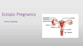
An ectopic or extrauterine pregnancy is one in which the blastocyst
implants anywhere other than the endometrial lining of the uterine
cavity. 98% of ectopic pregnancies implant in the fallopian tube, with
80% occurring in the ampullary segment.Other locations include, but
are not limited to, the ovary, cervix,
and abdomen.
3. Risk Factors
Specific examples of an inflammatory process include salpingitis and
salpingitis isthmica nodosa.
An acute chlamydial infection causes intraluminal inflammation and
subsequent fibrin deposition with tubal scarring. Despite negative cultures,
persistent chlamydial antigens can trigger a delayed hypersensitivity
reaction with continued scarring. Whereas endotoxin-producing Neisseria
gonorrhoeae causes virulent pelvic inflammation with a rapid clinical
onset, chlamydial inflammatory response is indolent and peaks at 7 to 14
days. The incidence of ectopic pregnancy has increased consistently with
the rise in chlamydial infections.
4. Risk Factors
Pregnancy after tubal sterilization is rare, but, when it does occur, there
is a substantial risk that the pregnancy will be ectopic due to the distorted
tubal anatomy created by the tubal ligation. Previous concerns that
intrauterine device use and pregnancy termination are predisposing risks
for ectopic pregnancy have been dispelled. A history of infertility,
independent of tubal disease, and ovulation induction also appear to be risk
factors in ectopic pregnancy. Additional risk factors include prior ectopic
pregnancy, smoking, prior tubal surgery, diethylstilbestrol exposure, and
advanced age.
5. Symptoms
The classic symptoms associated with ectopic pregnancy are
amenorrhea followed by vaginal bleeding and abdominal pain on the
affected side; however, there is no constellation of symptoms that are
diagnostic.
Normal pregnancy symptoms, such as breast tenderness, nausea, and
urinary frequency, may accompany more ominous findings. These include
shoulder pain worsened by inspiration and caused by
phrenic nerve irritation from subdiaphragmatic blood
as well as vasomotor disturbances, such as vertigo and
Syncope from hemorrhagic hypovolemia.
6. Clinical Findings
Prior to rupture, the diagnosis of an ectopic pregnancy is primarily
based on laboratory and ultrasound findings. With rupture, however,
nearly three-fourths of women will have marked tenderness on both
abdominal and pelvic examination, and pain is aggravated with
cervical manipulation. A pelvic mass, including fullness posterolateral
to the uterus, can be palpated in about 20% of women. Fever is not
expected, although a mild elevation in temperature in response to
intraperitoneal blood may occur. A temperature of 38°C may suggest an
infectious cause of a patient’s symptoms.
7. Clinical Findings
Abdominal distention and tenderness, with or without rebound, rigidity, or
decreased bowel sounds may be seen in cases of intra-abdominal bleeding.
Abdominal tenderness is present in 50% to 90% of patients with ectopic
pregnancies. Cervical motion tenderness caused by intraperitoneal
irritation and adnexal tenderness are commonly found.
An adnexal mass is present in roughly one third of cases, but its
absence does not rule out the possibility of an ectopic implantation. The
uterus may enlarge and soften throughout the first trimester, thus
simulating an intrauterine pregnancy. A slightly open cervix with blood or
decidual tissue may be found and mistaken for a threatened and/or
spontaneous abortion.
9. Diagnostic Procedures
TVS and serial serum β-hCG measurements are the most valuable
diagnostic aids to confirm a suspicion of ectopic pregnancy. The initial
assessment in the otherwise hemodynamically stable patient must
include a pregnancy test. A negative pregnancy test excludes the
possibility of ectopic pregnancy. Urinary pregnancy tests, which detect
hCG levels to 20 IU/L, are now commonly available. These tests detect
hCG as early as 14 days after fertilization and are positive in more than
90% of cases of ectopic pregnancy. Serum assays can detect the
presence of hCG as early as 5 days after fertilization, that is, before the
missed menstrual cycle.
10. Serum Human Chorionic Gonadotropin Levels
In normal pregnancies, serum β-hCG levels rise in a log-linear fashion
until 60 or 80 days after the last menses, at which time levels plateau at
about 100,000 IU/L. During this early phase of pregnancy, a 53% or
greater increase in serum β-hCG levels should be observed every 48
hours. A rise of hCG levels less than this should raise suspicion for an
abnormal gestation, either intrauterine or ectopic.
Complicating this scenario is the recognition that approximately 15% of
normal intrauterine pregnancies are associated with less than a 53%
increase in hCG, and 17% of ectopic pregnancies have normal hCG
doubling times. Although inappropriately rising serum β-hCG levels
suggest (but do not diagnose) an abnormal pregnancy, they do not
identify its location.
11. Transvaginal Ultrasonography
Using TVS, a gestational sac is usually visible between 4½ and 5 weeks
from the last menstrual period (LMP). The yolk sac appears between 5
and 6 weeks, and a fetal pole with cardiac activity is first detected at 5½
to 6 weeks. With transabdominal sonography, these structures are
visualized slightly later.
It is not uncommon for TVS to demonstrate an intrauterine pregnancy
by the time the hCG level is 1,000 to 2,000 IU/L. Transabdominal
ultrasonography should be able to identify an intrauterine gestation by
the time the hCG level reaches 5,000 to 6,000 IU/L. The absence of an
intrauterine pregnancy with β-hCG levels above the discriminatory
value signifies an abnormal pregnancy—ectopic, incomplete abortion,
or resolving completed abortion. Care must be taken to differentiate
between a uterine gestation and a pseudogestational sac.
12. Serum Progesterone Level
Serum progesterone concentration is higher in a viable pregnancy than an
ectopic pregnancy. There is minimal variation in serum progesterone
concentration between 5 and 10 weeks of gestation; thus a single value is
sufficient. A serum progesterone level of <5 ng/mL has been used to
identify a nonviable pregnancy with 98% specificity and with a sensitivity
of 75%. Conversely, a serum progesterone of >20 ng/mL has a sensitivity
of 95%, with a specificity of approximately 40% to identify a healthy
pregnancy. Serum progesterone values cannot differentiate between an
ectopic and an intrauterine pregnancy.
13. Endometrial Curettage
Curettage of the uterine cavity can also help rule out ectopic pregnancy
but should only be undertaken after the possibility of interrupting an intact
pregnancy has been considered. Although intrauterine and ectopic
pregnancies can exist simultaneously in rare cases (heterotopic
pregnancy), identification of chorionic villi in tissue samples identifies an
intrauterine location of the pregnancy and essentially rules out ectopic
pregnancy. The Arias-Stella reaction, a hypersecretory
endometrium of pregnancy seen on histologic examination, occurs with
both ectopic and intrauterine pregnancies and, therefore, is not useful in
identifying an ectopic pregnancy.
14. Culdocentesis
Culdocentesis can identify hemoperitoneum which may indicate a ruptured ectopic
pregnancy, although it is also consistent with other causes, such as a ruptured
corpus luteum cyst. An 18G needle is inserted posterior to the cervix, between the
uterosacral ligaments, and into the cul-de-sac of the peritoneal cavity. Aspiration of
clear peritoneal fluid (negative culdocentesis) indicates no hemorrhage into the
abdominal cavity but does not rule out an unruptured ectopic pregnancy.
Aspiration of blood that clots can indicate either penetration of a vessel or such
rapid blood loss into the peritoneal cavity that the blood clot has not had time to
undergo fibrinolysis. Aspiration of nonclotting blood is evidence of
hemoperitoneum (positive culdocentesis) in which the blood clot has undergone
fibrinolysis. If nothing is aspirated (equivocal or nondiagnostic
culdocentesis), no information is obtained.Purulent fluid
suggests a number of infection-related causes, such as
salpingitis and appendicitis.
15. Laparoscopy
The most accurate technique of identifying an ectopic pregnancy is by
direct visualization, which is done most commonly via laparoscopy. Even
laparoscopy, however, has a 2% to 5% misdiagnosis rate. For example, an
extremely early tubal gestation may not be identified because it may not
distend the fallopian tube sufficiently to be recognized as an abnormality
(false negative). Conversely, a false-positive diagnosis may result from a
hematosalpinx (blood in the fallopian tube) being misinterpreted as an
unruptured ectopic pregnancy or tubal abortion.
16. Medical Management
Methotrexate is the medical treatment usually used as an alternative to
surgical therapy. Methotrexate is a folic acid antagonist that competitively
inhibits the binding of dihydrofolic acid to dihydrofolate reductase, which,
in turn, reduces the amount of the active intracellular metabolite, folinic
acid. It stops the growth of rapidly dividing placental, embryonic, and fetal
cells. Factors that can be assessed in predicting the success of medical
therapy include the initial β-hCG level, the size of ectopic pregnancy as
determined by TVS, and presence or absence of fetal cardiac activity.
The initial serum β-hCG level is the best prognostic indicator of treatment
success in women managed with a single-dose methotrexate protocol. An
initial serum value <5,000 IU/L is associated with a success rate of 92%,
whereas an initial concentration >15,000 IU/L has a success rate of 68%.
Ectopic pregnancy size also appears to have an effect on methotrexate
success rates. Success rates are reported as high as 93% in cases with
ectopic masses <3.5 cm.
17. Contraindications to Medical Therapy for Ectopic Pregnancy
Absolute
• Breastfeeding
• Overt or laboratory evidence of immunodeficiency
• Known sensitivity to methotrexate
• Active pulmonary disease
• Peptic ulcer disease
• Hepatic, renal, pulmonary, or hematologic dysfunction
• Heterotopic pregnancy with viable intrauterine gestation
• Unable to comply with management protocol
Relative
• Gestational sac greater than 3.5 cm
• Embryonic cardiac motion
• Free peritoneal fluid (possible hemoperitoneum)
18. The most common side effects of methotrexate include nausea,
vomiting, diarrhea, gastric distress, dizziness, and stomatitis. Intramuscular
methotrexate given as part of a single-dose protocol has been the most
widely used medical treatment of ectopic pregnancy. Close monitoring is
imperative. A serum β-hCG level is determined before administering
methotrexate and is repeated on days 4 and 7 following injection. Levels
may continue to rise until day 4. Comparison is then made between the day
4 and the day 7 serum values. If there is a decline by 15% or more, serum
β-hCG levels are measured weekly until they are undetectable. If the β-
hCG level does not decline, the patient may require either surgery or a
second dose of methotrexate if no contraindications exist. Surgical
intervention may be required for patients who do not respond to medical
therapy.
19. During the first few days following methotrexate administration, up to
half of women experience abdominal pain that can be controlled with
nonsteroidal anti-inflammatory drugs. This pain presumably results from
tubal distention, tubal abortion, and/or hematoma formation.
Methotrexate given in a multidose protocol has also been used
successfully, but the single-dose protocol described appears to reduce the
amount of potential complications while achieving similar success rates.
Other medical treatments that have been used include hyperosmolar
glucose, potassium chloride, prostaglandins, and the progesterone receptor
424antagonist mifepristone (formerly referred to as RU-486). In some cases,
an agent may be administered systemically, but sometimes it may be
injected directly into the ectopic pregnancy.
20. Surgical Management
Conservative surgical techniques have been developed that maximize
preservation of the fallopian tube. If removal is done through the
laparoscope, definitive diagnosis as well as treatment can be accomplished
at the same operation with minimal morbidity, cost, and hospitalization. In a
linear salpingostomy, the surgeon makes an incision on the fallopian tube
over the site of implantation, removes the pregnancy, and allows the incision
to heal by secondary intention. A segmental resection is the removal of a
portion of the affected tube.Salpingectomy is removal of the entire tube, a
procedure reserved for those cases in which little or no normal tube
remains. Rh-negative mothers with ectopic
pregnancy should receive Rh immunoglobulin
to prevent Rh sensitization.
21. Ovarian Pregnancy
Diagnosis is based on the classic sonographic description of a cyst with
a wide echogenic vascular outer ring located on or within the ovary.
Medical management as well as surgery can be used to conserve the
Ovary.
Interstitial Pregnancy
Also termed cornual pregnancy, interstitial pregnancies implant in the
proximal tubal segment that lies within the muscular uterine wall.
Swelling lateral to the insertion of the round ligament is the
characteristic anatomic finding. because the muscular cornu of the
uterus is better able to expand and accommodate an enlarging
pregnancy. As a result, rupture of a cornual pregnancy typically occurs
between the 8th and 16th gestational weeks and is often associated
with massive hemorrhage, sometimes requiring hysterectomy.
22. Cervical Pregnancy
Two diagnostic criteria are necessary for confirmation of cervical
pregnancy: (1) the presence of cervical glands opposite to the placental
attachment site and (2) a portion of or the entire placenta must be
located below either the entrance of the uterine vessels or the
peritoneal reflection on the anterior and posterior uterine surface.
Abdominal Pregnancy
Abdominal pregnancies may result from primary implantation onto the
peritoneal surface or secondary implantation via tubal rupture or tubal
abortion. The patient is given the option of continuing the pregnancy to
fetal viability with operative delivery or operative termination of the
pregnancy at the time of diagnosis. In either case, removal of the
placenta is usually not attempted because of the risk of uncontrollable
hemorrhage. Allowing the placenta to spontaneously regress is often
the management chosen. Alternative treatments include
administration of methotrexate and embolization of placental vessels.