2. The requirement for the Cld-catalyzed reaction by
perchlorate-respiring bacteria is absolute; without it, ClO2
−
rapidly accumulates to toxic levels.2
However, Clds are found in
almost all bacterial phyla and in many archaea, the over-
whelming majority of which are not involved in perchlorate
respiration.12
These Clds subdivide according to sequence into
groups that appear to be functionally distinct from their
respiration-associated counterparts and from each other (Figure
1), in spite of their high level of sequence similarity.13
Investigation of the Cld from Staphylococcus aureus, a Gram-
positive bacterium, provided a striking example. In its heme-
bound form, it is completely inactive in the conversion of
ClO2
−
to Cl−
and O2.14
Genetic and biochemical evidence
instead suggests that the protein is essential for a terminal step
in heme biosynthesis,14,15
a biological function that appears to
be common to the Clds from Gram-positive bacteria and
potentially other species.13,15
The Cld from Klebsiella pneumoniae strain MGH 78578
(KpCld) represents another subfamily that may have yet a third
biochemical function. These enzymes are found in non-
perchlorate-respiring bacteria from Gram-negative phyla
(Figure 1).12,13
As a consequence, they are not expected to
be involved in either perchlorate respiration or heme
biosynthesis. Unlike the respiration-associated Clds, they are
homodimers rather than homopentamers, leading to a very
different monomer−monomer interface and resulting structural
context for their bound hemes (Figure 2A−C). Locally,
however, they possess very similar active site residues and
share structural attributes essential for efficient ClO2
−
/O2
conversion. In particular, they share the functionally important
distal arginine above the ferric heme’s open coordination
position (Figure 2D).8,11
Consistent with chemical expect-
Figure 1. Phylogenetic tree illustrating the major subdivisions of the
Cld protein family discussed in the text. The respiratory Clds, coming
mostly from Proteobacteria with some exceptions caused by lateral
gene transfer, form the first group (red). Members of a second group
of dimeric Clds (green), including KpCld, come from non-perchlorate-
respiring species but retain active site features critical for ClO2
−
/O2
conversion. Members of a third broad group of Clds (blue) catalyze a
terminal step in heme biosynthesis in Gram-positive bacteria and
potentially some Archaea. These have consequently been renamed
HemQs.11
Sequence accession numbers are given in parentheses, with
PDB entries used to indicate where structures are available. The
phylogenetic tree was generated using MEGA6.69
The bootstrap
consensus tree inferred from 1000 replicates is taken to represent the
evolutionary history of the taxa analyzed. Branches corresponding to
partitions reproduced in <70% of bootstrap replicates are collapsed.
Initial trees for the heuristic search were obtained by applying the
Neighbor-Joining method to a matrix of pairwise distances estimated
using a JTT model. A discrete Gamma distribution was used to model
evolutionary rate differences among sites [five categories (+G,
parameter = 2.4895)]; 5% alignment gaps, missing data, and
ambiguous bases were allowed at any position.70
The resulting tree
is consistent with those previously reported by others.11,13,71
Figure 2. Clds from perchlorate-respiring and nonrespiring
Proteobacteria. (A) KpCld, like the NwCld structure shown (PDB
entry 3QPI),7
has a truncated monomer relative to the respiratory
Clds. Both form functional homodimers. Protein monomers are
rendered as cartoons in different colors. Hemes are rendered as sticks.
(B) Respiration-associated DaCld (PBD entry 3Q08) forms a
functional homopentamer, shown looking down the C5 axis.4
(C) A
monomer−monomer interface from the structure in panel B suggests
differences in heme accessibility in the pentameric and dimeric Clds.
(D) Active site environments for DaCld (cyan carbons) and NwCld
(magenta carbons) are superimposed with several conserved residues
around the heme labeled (DaCld numbering). The two active sites are
highly similar; two notable exceptions are the orientation of the distal
Arg side chain and the identity of the residue at the position of W227,
which is a conserved glutamate in NwCld and other dimeric Clds.
Biochemistry Article
dx.doi.org/10.1021/bi501184c | Biochemistry 2015, 54, 434−446435
3. ations, O2 production has been demonstrated for at least one
protein from this subgroup, the Cld from Nitrobacter
winogradskyi [NwCld (structure shown in Figure 2A,D)];11
however, the dimeric enzymes have presumably not had the
selection pressure to evolve to the same level of efficiency as
Clds that are essential for metabolizing perchlorate.
Why bacteria like K. pneumoniae MGH 78578, a respiratory
pathogen, possess a potential means for chlorite detoxification
is not clear. K. pneumoniae and other non-perchlorate-respiring
species generally do not encode perchlorate or chlorate
reductase enzymes,17
yet pathogenic strains of clinically
pervasive18
species, including K. pneumoniae, Acinetobacter
baumanii, and Pseudomonas aeruginosa, all possess genes from
this cld subtype, often on plasmids or as part of pathogenicity
islands. These genes appear unlikely to guard against
environmental chlorite, because of its chemical instability and
the inability of negatively charged chlorite to passively enter
cells.19
However, an indirect antioxidant function against ClO3
−
or ClO4
−
is possible, if these can enter the cell and be reduced
by endogenous enzymes. To test this hypothesis, and to
enhance our understanding of the influence of the unusual
monomer and oligomerization states on an otherwise similar
heme environment, KpCld was characterized in parallel with
the Δcld strain of K. pneumoniae MGH 78578. The results
presented here report on both the biological function of this
group of Clds and the biochemical properties that make them
unique.
■ EXPERIMENTAL PROCEDURES
Chemicals. Chemicals were purchased from Alfa Aesar,
Sigma-Aldrich, or VWR. Sodium chlorite, 2,2′-azino-bis(3-
ethylbenzothiazoline-6-sulfonic acid) diammonium salt
(ABTS), guaiacol, and 2-chloro-5,5-dimethyl-1,3-cyclohexane-
dione (MCD) stock solutions were made fresh daily in buffers
prepared from distilled and deionized water and their
concentrations determined spectrophotometrically: ε262 = 160
M−1
cm−1
(NaClO2), ε415 = 3.6 × 10⁴
M−1
cm−1
(ABTS), and
ε290 = 20100 M−1
cm−1
(MCD).20
Iodometric titration was
routinely used to quantify working solutions of sodium chlorite
or hypochlorite [diluted from a 10−14% (w/w) stock], as well
as unreacted chlorite remaining at the end of experiments.21
Each substrate was dissolved directly in the specified aqueous
buffers with the exception of guaiacol, for which a 0.9 M stock
was prepared in dimethyl sulfoxide (DMSO). Stocks of hemin
were prepared in DMSO.
Generation of the Δcld Strain. Mutagenesis of K.
pneumoniae MGH 78578 was conducted as described
previously.22
Briefly, a derivative of pKOBEG encoding
apramycin resistance, pKOBEGApra, was used to facilitate
lambda Red-based replacement of cld with a 1652 bp FRT site-
flanked hygromycin resistance cassette (FRT-hph-FRT)
amplified from pJTAG-hyg (Deenathayalaguptha and Rajaku-
mar, unpublished data) using primers GmF and GmR.23
Targeting flanking sequences TS1 (572 bp) and TS2 (527 bp)
were amplified using primer pair P1 and P2 and primer pair P3
and P4. TS1, TS2, and FRT-hph-FRT were joined by splicing
overlap extension PCR (SOE-PCR) to produce a 2702 bp
amplicon (Figure S1 of the Supporting Information).
Arabinose-induced electrocompetent K. pneumoniae MGH
78578/pKOBEGApra maintained at 30 °C was electroporated
with 100 ng of the SOE-PCR product, and transformants were
recovered on LB/200 μg/mL hygromycin agar at 37 °C after
overnight culture. Several hygromycin resistant colonies were
examined, and the Δcld mutant was validated by PCR analysis
(Figure S1 of the Supporting Information). Bacteria and
plasmids used for cld mutagenesis are listed in Table S1 of the
Supporting Information.
Phenotypic Characterization of the Δcld Strain. Frozen
(−80 °C) glycerol stocks of wild-type (WT) and Δcld strains of
K. pneumoniae were revived by streaking on Luria broth (LB)
agar and LB agar/hygromycin B (75 mg/mL) plates,
respectively. Hygromycin B was used in all growth experiments
to maintain the mutant free of contamination. Single colonies
were inoculated into 5 mL of LB or LB with hygromycin B and
grown to an OD600 of 0.4. Cells were harvested in
microcentrifuge tubes and washed three times with M9
medium [0.2% (v/v) glycerol, 58 mM K2HPO4, 22 mM
Na2HPO4, 85 mM NaCl, and 18.7 mM NH4Cl] supplemented
with the following trace elements in 134 μM EDTA: 6.15 μM
ZnO, 570 nM CuCl2·2H2O, 340 nM CoNO3·6H2O, and 1.6
μM H3BO3. Cells were resuspended to a final optical density at
600 nm (OD600) of 0.4 and used as a uniform inoculate (1:100)
for all experiments.
For determination of minimal inhibitory concentrations of
ClOX
−
(X = 2−4), cells were inoculated into 10 mL of
anaerobic, N2-purged medium in sealed glass crimp-topped
bottles containing increasing concentrations of NaClO2,
NaClO3, or KClO4 (0.5−200 mM) and 50 mM KNO3. The
sealed bottles were placed in a 37 °C incubator with gentle
shaking. The minimal inhibitory concentration (MIC) was
defined as the lowest concentration of a reagent that would
inhibit the visible growth of K. pneumoniae after incubation for
24 h.24
No-nitrate controls were conducted in tandem under
aerobic conditions. Finally, for generation of growth curves,
cells were inoculated into aerobic media and their growth was
monitored via periodic measurement of their OD600 over time.
Growth, Purification, and Characterization of Wild-
Type (WT) and Mutant KpCld. DNA containing the full-
length coding region of chlorite dismutase from K. pneumoniae
MGH 78578 (GenBank accession number CP000650.1) was
amplified via PCR with primers KpCldFor (5′-CGC CATATG
AAT ACA CGA TTA TTT ACG TTC GCT GG-3′) and
KpCldRev (5′-TTT GGATCC CTA GGC CGG CTC ATG
CA-3′) from a K. pneumoniae genomic DNA template (added
cut sites for NdeI and BamHI at the 5′ and 3′ ends underlined,
respectively). The product was subsequently cloned into the
pET-15b (Merck/Novagen) expression vector for production
of protein with an N-terminal His tag. The Y62F and W97F
mutants of KpCld and the Y118F mutant of DaCld were
generated from the WT via PCR-based single-codon
substitution using a QuikChange kit. The W227F, W155F,
and W156F mutants of DaCld were generated in a similar
manner and were available from prior work.25
All KpClds were expressed in Escherichia coli Tuner (DE3)
cells (Merck/Novagen) grown in Terrific Broth (TB) with
ampicillin (100 μg/mL). Expression cultures were grown at 37
°C in a shaker incubator (250 rpm) to midlogarithmic phase
(OD600 = 0.5). Isopropyl β-D-thiogalactopyranoside (IPTG, 1
mM) and δ-aminolevulinic acid (50 mg/L) were added to
induce heme protein expression, and the temperature was
lowered to 20 °C. After 16 h, cell pellets were collected by
centrifugation and stored at −80 °C. Cells were thawed and
resuspended [20 mM phosphate buffer, 500 mM NaCl, 20 mM
imidazole, and 1 mM phenylmethanesulfonyl fluoride (pH
7.4)] and lysed by pulsed sonication on ice (7 min). The lysates
were clarified by centrifugation and supernatants loaded onto a
Biochemistry Article
dx.doi.org/10.1021/bi501184c | Biochemistry 2015, 54, 434−446436
4. 20 mL HisTrap column. The protein was eluted using a 20 to
500 mM linear gradient of imidazole in resuspension buffer.
Eluted proteins were screened by sodium dodecyl sulfate−
polyacrylamide gel electrophoresis and pure fractions buffer-
exchanged into 0.1 M phosphate buffer (0.1 M, pH 6.8) using
Amicon centrifuge concentrators (molecular weight cutoff of
10000). Pure protein was concentrated to 10 mg/mL (20%
glycerol), frozen in liquid N2, and stored at −80 °C. All KpCld
concentrations are given as heme-bound monomer, where
[heme] and [protein] were determined by the pyridine
hemochrome and Bradford assays,26
respectively.
Measurement of Initial Rates of Chlorite-Decompos-
ing Activity in the Steady State. A luminescence-based
probe was used to measure O2 evolution by KpCld during
chlorite decomposition under steady state conditions. Samples
included 12 nM WT KpCld and 0.05−2 mM chlorite in the
following buffer solutions: 50 mM phosphate-citrate (pH <6),
100 mM phosphate (pH 6−8), or 100 mM glycine (pH >8).
The probe was equilibrated in the buffer/chlorite solution for
5−10 min prior to initiation of the reaction by introduction of
enzyme. Kinetic traces were recorded at 1 s intervals for 3−10
min.
Monitoring Heme Chromophore Loss Caused by
NaClO2 or NaOCl. Samples (10 μM, 200 μL) of heme-
containing KpCld were titrated in UV−visible cuvettes with a
concentrated chlorite or hypochlorite stock (600 mM) added in
1 μL increments [all solutions in 0.1 M citrate-phosphate buffer
(pH 6.6)]. Samples were allowed to come to equilibrium
following each addition and corrected for dilution.
Chromophore loss was also monitored over time following
the addition of varying amounts of NaOCl as a function of pH.
Reactions were conducted in 0.1 M citrate-phosphate at pH 6−
8. Samples (10 μM, 200 μL) of heme-containing KpCld in
UV−visible cuvettes were manually mixed with a NaOCl stock,
yielding final NaOCl concentrations of 0−1 mM (0−2000
equiv). Spectra were measured every 6 s after mixing. The
absorbance at the Soret band maximum (409 nm) was plotted
versus time and fit to a single-exponential equation to obtain a
first-order rate constant (kobs). Values of kobs measured at a
given pH were plotted versus NaOCl concentration to obtain
second-order rate constants (k is the slope).
Measurement of Residual Activities and Turnover
Numbers. Chlorite’s potency as a suicide substrate was
assessed by the method of Silverman.27
Briefly, 5 μM samples
of KpCld were incubated for 1 h at room temperature with an
increasing number of equivalents of chlorite up to 3 × 10⁴
, with
or without 0.5 mM added guaiacol, MCD, or ABTS [0.1 M
citrate-phosphate buffer (pH 6.6)]. Reaction mixtures were
dialyzed against chlorite-free buffer for two 1 h cycles to
remove any unreacted substrate. The remaining KpCld
reactivity was measured using a Clark oxygen electrode by
adding 5 μL of each dialysate to 1.5 mL of 2 mM chlorite and
measuring the initial rate of O2 production. Rates were
measured three times, corrected for dilution, and averaged.
Residual activity was computed by referencing the activity
remaining after incubation in a given chlorite concentration to
the activity of the zero chlorite control. The turnover number,
defined as the total number of molecules of chlorite catalyzed
per KpCld heme before the catalyst is irreversibly inactivated,
was obtained by extrapolating a plot of residual activity versus
[ClO2
−
]/[KpCld] to the x-intercept.
Vibrational Characterization of KpCld. Resonance
Raman (rR) spectra were obtained with 413.1 or 441.6 nm
excitation from a Kr+
or HeCd laser, respectively, using the
135° backscattering geometry for collection of Raman-scattered
light. The spectrometer was calibrated against Raman
frequencies of toluene, dimethylformamide, acetone, and
methylene bromide. Spectra were recorded at ambient
temperature from samples in spinning 5 mm NMR tubes.
UV−visible absorbance spectra were recorded from the rR
samples before and after spectral acquisition to assess whether
sample integrity had been compromised by exposure to the
laser beam. The laser power at ferric and ferrous samples
ranged from 5 to 10 mW; no spectral artifacts due to
photoinduced chemistry were observed with these irradiation
powers. Ferric KpCld samples for the rR pH dependence study
were prepared in the following 100 mM buffers: potassium
phosphate (pH 5.7−7.5), Tris-HCl (pH 8.6−9.7), and borate
buffer (pH 10.1).
Electron Paramagnetic Resonance (EPR) Spectrosco-
py. The 9 GHz EPR spectra were recorded on a Bruker
EleXsys E500 spectrometer equipped with a standard Bruker
ER 4102 X-band resonator and a liquid helium cryostat
(Oxford Instruments, ESR 900). The spectra for WT KpCld at
pH 6.0 were recorded at 4 K, a microwave power of 1 mW, a
modulation amplitude of 4 G, and a modulation frequency of
100 kHz. The spectra at pH 8.0 were recorded at 12 K, a
modulation amplitude of 10 G, a microwave power of 0.5 mW,
and a modulation frequency of 100 kHz. Spectra for the mutant
KpClds were recorded at 4 K, a microwave power of 1 mW, a
modulation amplitude of 4 G, and a modulation frequency of
100 kHz. To avoid significant changes in pH upon freezing,
Tris-maleate buffer, which covers the pH range from 5.2 to 8.6
with minor changes (0.1 pH unit) upon freezing, was used. No
effect on the ferric EPR signal was observed for the Tris-
maleate buffer as compared to that with phosphate buffer when
using the same pHs. Typically, EPR samples of ferric KpCld
and DaCld [in 0.1 mM Tris-maleate (pH 6.0 or 8.0) and at 0.6
mM enzyme] were measured in 4 mm quartz tubes. The buffer
exchange in the pH range of 5.0−8.0, conducted with
Centricon microconcentrators (Amicon), could be reversibly
obtained without enzyme precipitation, degradation, or iron
release as judged by the electronic absorption and EPR spectra
recorded after each buffer exchange. Freezing and thawing
cycles at high enzyme concentrations used for the EPR
characterization did not induce any enzyme precipitation or
changes in the heme environment.
■ RESULTS
Heme Environment in KpCld As Measured by UV−Vis
and rR. KpCld has a reversible pH-dependent transition in
UV−visible spectrophotometric titrations. Experiments in
which the enzyme was titrated with base (Figure 3A) and
acid (data not shown) yielded the same pKa. Fits of UV−vis
data at 390 nm (Figure 3A, inset), 413 nm, and 574 nm (not
shown) indicated a pKa of 8.3, the same as that reported for the
Azospira oryzae Cld28
and slightly lower than that for Clds from
Ideonella dechloratans (8.5)29
and D. aromatica (8.7).5
Heme
proteins with neutral proximal histidine ligands have pKa values
[heme oxygenase-1 and -2 (7.6 and 8.5, respectively), SmFixL
(9.6), and HmuO (9.0)] lower than the pKa values of those
with a proximal histidinate [peroxidases (pKa values of 11−
12)].30−32
Consistent with a pKa in this range, the Fe(II)−His
vibrational frequency measured by rR for KpCld suggests a
charge-neutral proximal histidine (Figure S2 of the Supporting
Information; discussed further below).
Biochemistry Article
dx.doi.org/10.1021/bi501184c | Biochemistry 2015, 54, 434−446437
5. At pH 6.0, ferric KpCld exhibits a Soret band maximum at
405 nm, a broad α/β band envelope at 505 nm with a small
shoulder at 540 nm, and a charge transfer (CT) band at 645 nm
(Figure 3A). This spectrum is similar to that reported for the
related dimeric NwCld (405, 506, 543, and 640 nm) and the
pentameric Cld from Candidatus Nitrospira defluvii at pH
7.0,10,11
but different from Clds from D. aromatica (393, 506,
and 648 nm, pH 7.0), A. oryzae (392−394 nm), and I.
dechloratans (392, 509, and 648 nm, pH 7).5,9,29
The latter
three proteins, which come from perchlorate respirers, share
similar five-coordinate, high-spin (5cHS) heme sites, pentame-
ric oligomerization states, and hydrophobic, solvent-enclosed
heme environments, to which their blue-shifted Soret bands
have been attributed. In contrast, the visible spectrum of acidic
KpCld corresponds to a mixture of coordination states; this is
confirmed by its rR spectra (below).
The rR spectra (Figure 3B) of ferric KpCld in a mildly acidic
solution have a broad HS ν3 envelope spanning 1482−1490
cm−1
, consistent with a mixture of six-coordinate, high-spin
(6cHS) and 5cHS heme. The temperature dependence of the
HS ν3 envelope supports the presence of 5cHS and 6cHS
hemes. When the temperature of ferric KpCld is decreased
from 19 °C at pH 6.0, the 6cHS intensity of the ν3 envelope at
1484 cm−1
increases at the expense of the 5cHS component at
1491 cm−1
. Below −14 °C, the envelope narrows to a single
band centered at 1484 cm−1
, indicating complete conversion of
the HS heme to a 6c complex that is likely an aqua complex,
whose formation is exothermic (Figure S3 of the Supporting
Information). The acid−base behavior of this complex (see
below) supports this assignment. These data indicate that a
considerable fraction of the resting HS enzyme is coordinatively
saturated at near-physiological temperatures. In contrast, the
acidic form of DaCld contains only 5cHS heme.5
Another small ν3 band occurs at 1505 cm−1
at 19 °C and pH
∼6 (Figure 3B). The intensity of this band increases in concert
with that of a new 6cHS band at 1478 cm−1
as the pH is
increased. Bands in the 1505 cm−1
frequency range can be
consistent with 6cLS species or 5cQS (5c quantum mechanical
spin admixture of S = 3
/2 and S = 5
/2 ferric heme). The
appearance of UV−visible bands near 572 nm (typical for 6cLS
species) and 646 nm (CT) at pH 7.0 argue for assignment of
the ν3 band to a 6cLS species. At pH 10, ferric KpCld has a
Soret maximum at 413 nm, α/β bands at 572 and 540 nm, and
small absorbance bands near 490 and 603 nm. The 572 and 540
nm bands are consistent with a 6cLS complex, while the bands
at 490 and 603 nm are suggestive of a 6cHS species (Figure
3A). These features are very similar to those reported for
alkaline ferric DaCld.5
The mixture of spin states suggested by
the UV−visible spectrum is confirmed by the appearance of
two ν3 bands at 1478 and 1505 cm−1
in the rR spectrum of
alkaline KpCld. Growth of these bands is accompanied by the
appearance of a shoulder at 1640 cm−1
, which can be attributed
to ν10 for 6cLS heme, and a 2 cm−1
upshift in the frequency of
ν4 (not shown), both indicating growth in the population of
6cLS heme. The formation of heme hydroxides is often
characterized by parallel growth in the intensity of bands arising
from 6cHS and 6cLS hemes, which indicate its presence as a
thermal spin state equilibrium. Similar mixtures attributed to
such equilibria have been reported for a number of heme
proteins, including the alkaline forms of DaCld, myoglobin,
hemoglobin, Mycobacterium tuberculosis HbN, and
SmFixL.5,33−36
To determine whether the sixth ligand in KpCld is hydroxide
at pH 9.8, the νFe−OH modes were identified by isotopic
substitution (Figure 3C). Bands at 509 and 442 cm−1
were
assigned to the Fe−OH stretching modes for the LS and HS
Figure 3. pH-dependent behavior of KpCld indicates a water-
accessible heme. (A) UV−visible pH titration of KpCld. Spectra
shown at pH 7.13, 7.38, 7.85, 8.1.4, 8.46, 8.78, 9.14, 9.61, 10.07, and
10.43. Arrows indicate the direction of the absorbance change with
pH. The inset shows the absorbance at 390 nm as a function of pH; a
least-squares fit to the data (●) gives a pKa of 8.3. (B) Speciation of
ferric KpCld as observed in rR pH titration. Resonance Raman spectra
were obtained with 413.1 nm excitation at the indicated pH values.
(C) Soret-excited rR spectra of the oxygen isotopologs of alkaline
KpCld [50 mM Ches (pH 9.8)]. The top three spectra were measured
in the indicated solvent at identical acquisition times. The bottom
trace is the difference spectrum generated by a 1:1 digital subtraction
(H2O − H2
18
O).
Biochemistry Article
dx.doi.org/10.1021/bi501184c | Biochemistry 2015, 54, 434−446438
6. heme hydroxides, respectively. The LS νFe−OH band shifts to
494 cm−1
in D2O and to 488 cm−1
in H2
18
O. In the case of the
HS heme, no deuterium shift is detected. In H2
18
O, the νFe−
18
OH
band shifts by 22 cm−1
to 420 cm−1
. Thus, the pKa of 8.3
corresponds to the formation of the KpCld−OH complex.
Consistent with our previous report about the DaCld−OH
complex,5
the νFe−OH bands of both HS and LS KpCld−OH
complexes fall at the low end of the Fe−OH stretching
frequency range for heme hydroxides.
Finally, the UV−visible spectrum of ferrous KpCld (pH 7.0)
generated by reduction of the ferric protein with a 12-fold
redox excess of sodium dithionite exhibited a Soret band
maximum at 433 nm, typical of 5cHS ferrous heme, and α/β
bands at 586 and 555 nm, respectively (Figure S2 of the
Supporting Information). The 413.1 nm excited Fe(II) KpCld
spectrum at pH 7.0 has a ν4 at 1356 cm−1
, typical of ferrous
heme, and a ν3 at 1472 cm−1
, typical of 5cHS ferrous heme.
The 441.6 nm excited Fe(II) KpCld spectrum revealed an Fe−
His stretching band at 229 cm−1
, 7 cm−1
higher than that
observed for DaCld (222 cm−1
at pH 6.8),5
consistent with the
Fe−His bond being slightly stronger in KpCld assuming similar
normal mode compositions in the two enzymes.
Structural Features of the KpCld Heme Environment,
Including Second-Sphere Interactions As Revealed by 9
GHz EPR Spectroscopy. EPR spectroscopy is a sensitive
probe of the extended hydrogen bonding network of heme
active sites, including structural water molecules as well as
second-sphere (and beyond) amino acid residues.37,38
In the
absence of a crystal structure for KpCld and to expand the
structural view of the heme site provided by rR, the ferric EPR
spectra of wild-type KpCld and key variants in its heme
extended environment were examined as a function of pH.
These were compared to their DaCld counterparts, for which
the WT crystal structure has been determined.8
Figure 4 shows the 9 GHz EPR spectra of wild-type KpCld
recorded at cryogenic temperatures (4 and 12 K). The ferric
EPR spectrum of KpCld remained invariant in the pH range of
5.5−7.9 (top, black trace) but showed a dramatic change at pH
8.0 (top, gray trace). At pH 6.0 (Figure 4, top, black trace), the
EPR spectrum of KpCld showed an axial signal, with observed
effective g values of g⊥ = 5.92 and g∥ = 1.99, consistent with a
ferric high-spin species. At pH 8.0 (Figure 4, top, gray trace),
the KpCld EPR spectrum showed a distinct LS ferric species,
with effective g values of gCx = 2.54, gCy = 2.19, and gCz = 1.87.
The almost complete conversion of the ferric EPR signal from
high-spin to low-spin at pH 8.0, observed in frozen solutions,
differs from the more gradual effect observed in the rR
experiments in solution (Figure 3B) and most possibly reflects
the preferential configurations of the heme environment locked
in the frozen samples. The ferric LS EPR spectrum overall is
consistent with a nitrogenous amino acid side chain not directly
coordinated to the heme iron, in the vicinity of the Fe−OH
bond revealed by the rR characterization described in the
previous section. The relatively smaller contribution of another
LS signal with a large gmax of 3.10 (the other two components
being very weak and too broad to be detected) consistent, in
this case, with a nitrogenous ligand on the heme distal side8
was
also observed. The identity of this ligand is not known. It is
possible that, even after thorough dialysis, some imidazole used
in the purification of the protein remained in the active site.
Alternatively, repositioning of the distal arginine, with a
deprotonated guanidinium group replacing water molecules
close to the iron [for example, in the structural water molecules
shown in the NwCld structure (Figure 2)], could be envisioned
if considering the crystal structures of the N. defluvii Cld with
thiocyanate bound in the sixth coordination position of the
heme.10
The EPR spectra of DaCld differed substantially from those
of KpCld. Specifically, the spectrum at pH 6.0 showed the
contribution of two ferric HS EPR signals (Figure 4, labeled
with subscripts A and B), with effective g values of (gAx = 6.67,
gAy = 5.20, gAz = 1.96) and (gBx = 6.25, gBy = 5.24, gBz = 1.99).
Two distinct LS ferric species (Figure 4, subscripts C and D),
with effective g values of (gCx = 2.56, gCy ≈ 2.18, gCz = 1.87) and
(gDx = 2.64, gBy ≈ 2.18, gBz = 1.82), were consistently observed
at pH 8.0. These observations of two HS and two LS EPR
signals are consistent with our earlier reports that closed (more
active) and open (less active) DaCld conformers are present
between pH 5.6 and 9.1.16
To improve our understanding of the lack of pH-induced
changes in the HS EPR spectra as well as the difference in ferric
heme signals of the various Clds, structurally conservative
mutations at highly conserved Trp and Tyr residues within
hydrogen bonding distance of the heme propionates were
constructed. Specifically, the crystal structures of the Da and
Nw Clds show that a highly conserved tyrosine (Tyr118, Figure
2D; Tyr62 in KpCld) appears to make hydrogen bonding
contacts to both heme propionates via its phenol oxygen and
amide nitrogen. The indole N atom of a conserved tryptophan
(Trp155 of DaCld or Trp97 of KpCld) lies within hydrogen
bonding distance of one heme propionate. A second strictly
conserved Trp (Trp156 of DaCld or Trp98 of KpCld) is
sterically close to the same propionate but oriented such that
the indole nitrogen cannot form a hydrogen bond to it.
Accordingly, we anticipated that substitutions of these
conserved residues could considerably affect the orientation
Figure 4. 9 GHz EPR spectra of ferric Clds from K. pneumonia (top)
and D. aromatica (bottom) as a function of pH. A spin switch from
high-spin ferric (black trace) to low-spin ferric (gray trace) is observed
at pH ≥8.0. The conversion of the high-spin ferric signal was estimated
to be 95% in DaCld and 85% in KpCld. The spectra at pH 6.0 were
recorded at 4 K, a microwave power of 1 mW, a modulation amplitude
of 4 G, and a modulation frequency of 100 kHz. The spectra at pH 8.0
were recorded at 12 K, a modulation amplitude of 10 G, a microwave
power of 0.5 mW, and a modulation frequency of 100 kHz.
Biochemistry Article
dx.doi.org/10.1021/bi501184c | Biochemistry 2015, 54, 434−446439
7. of the propionate(s) and/or heme planarity, resulting in
measurable changes in the ferric EPR spectra.
Figure 5 shows dramatic changes observed in the ferric EPR
spectra of KpCld and DaCld upon mutation of these active site
residues. The axial HS EPR signal of WT KpCld (Figure 5, top,
dotted black trace) fully converted to a rhombically distorted
spectrum in the Y62F mutant (Figure 5, top, magenta trace),
with the contribution of two species [effective g values of (6.46,
5.39, 1.98) and (6.57, 5.54, 1.99)]. The W97F mutation in
KpCld induced a partial conversion of the wild-type axial signal
to one of the rhombically distorted HS forms of the Y62F
variant, with effective g values of (6.57, 5.54, 1.99), with a
contribution of the 6cLS form with a large gmax of 3.10 (Figure
5, top, purple trace). The same LS high-gmax form was observed
in WT KpCld, but only at basic pH (Figure 4, top, gray trace).
In the Y118F mutant of DaCld, positionally equivalent to Y62F
in KpCld, the EPR component with the largest rhombic
distortion disappears (Figure 5, bottom, magenta trace). The
ferric EPR spectrum of the Y118F DaCld variant then becomes
very similar to the previously reported rhombic EPR spectra of
the Clds from A. oryzae and I. dechloratans,9,29
and also similar
to that of Y62F KpCld. Accordingly, these results show that
breaking the hydrogen bonds to the propionates allows more
flexibility of the heme.
To explore these differences further, we investigated the
DaCld W227F mutant. This residue is conserved among the
Clds from known perchlorate respirers but not in the dimeric
Clds (Figures 1 and 2) and could represent a key differentiation
between the two groups. Trp227 is not connected to the heme
by hydrogen bonds yet is sterically close to those other residues
having hydrogen bonding interactions to the heme. Interest-
ingly, two significant changes were observed in the ferric EPR
spectrum of W227F DaCld relative to WT: the disappearance
of the component with the largest rhombic distortion, as in
DaCld Y118F, and an increase in the contribution of the axial
EPR signal (Figure 5, bottom, violet trace). Moreover, the ratio
of the axial and rhombically distorted signals of the W227F
DaCld EPR spectrum became pH-dependent, as in the case of
Burkholderia pseudomallei KatG,39
fully converting to the axial
signal at pH 5.0 (Figure 5, bottom, green trace). Hence, the
resulting EPR spectrum for DaCld W227F at pH 5.0 is the
same as that of wild-type KpCld (Figure 5, top, dotted black
trace). Notably, the mutant protein could be isolated in a stably
heme-bound state at low pH if concentrated and frozen
immediately following purification (see Experimental Proce-
dures).25
These results reinforce the idea that second-sphere
coordination influences the electronic structure of the heme
iron. They also suggest that the conserved residue Trp227 is
indeed important for distinguishing the heme environments of
DaCld and KpCld even though it is not directly connected to
the heme via hydrogen bonding.
KpCld Exhibits Chlorite-Decomposing Activity. KpCld
is a competent catalyst of chlorite decomposition; values of kcat
and kcat/KM are maximal near pH 5.0, at (1.9 ± 0.2) × 103
s−1
and (2.5 ± 0.4) × 106
M−1
s−1
, respectively (20 °C) (Figure S4
of the Supporting Information). These values are approximately
10-fold lower than the corresponding parameters for DaCld
measured at its pH 5.2 optimum (4 °C): kcat = (2.0 ± 0.6) ×
104
s−1
, and kcat/KM = (3.2 ± 0.4) × 107
M−1
s−1
.5
A pH
optimum between 5 and 6 was recently measured for the kcat
for the N. defluvii Cld.10,40
Steady state pH−rate profiles for KpCld (Figure S4 of the
Supporting Information) and DaCld5
are broadly similar, with
each protein possessing a more active acidic form and less
active alkaline form. Turning points in the plots of log kcat
versus pH were fit at pH 6.5 and 8.7 (DaCld).5
A transition
from a highly active low-pH form to a less active alkaline form
is apparent near pH 7 for KpCld. Turning points in log−log
plots of kinetic constants are associated with pKa values.
Though not explicitly assigned here for KpCld, the lower-pH
turning point in DaCld was previously assigned to the distal
arginine, first modeled as an explicit deprotonation and later
associated with movement of the distal pocket Arg between less
reactive “out” (alkaline) and more reactive “in” (acidic)
conformations (Figure 2). The latter model is supported by
both spectroscopic evidence and reactivity data with H2O2.5,6,16
Chlorite Acts as a Potent Suicide Substrate. Titration
of KpCld with chlorite (Figure 6A) demonstrated near-
complete elimination of the heme’s Soret band following
exposure to roughly (6.0 ± 0.3) × 103
equiv of the oxidant.
This value is intermediate between DaCld’s (2.0 × 10⁴
) and
SaCld’s (≤5).5,14
To relate heme destruction quantitatively to
the loss of catalytic activity, KpCld’s turnover number was
measured (Figure 6B). A line fit to a plot of residual activity
versus chlorite equivalents intercepts the x-axis at 5.3 × 10³
.
The same data could also be fit to an exponential decay curve,
consistent with some protection of the enzyme from
degradation by the accumulation of high product concen-
trations.41
The exponential curve predicts heme degradation
after exposure to even fewer equivalents of chlorite, that is, the
number of equivalents extrapolated from where the initial,
Figure 5. 9 GHz EPR spectra of selected mutations on the heme
environment of KpCld (top) and DaCld (bottom) at pH 6.0. Both
spectra of the wild-type Clds (black dotted traces) are shown for
comparison. Figure 2D shows the crystallographic structure of the
extended heme environment of NwCld (PDB entry 3QPI), in which
the amino acid residues at positions equivalent to those mutated in
KpCld (Y62F and W97F, at the same positions as Tyr118 and
Trp155) are shown. The other Trp mutated in DaCld (Trp227) is not
conserved in NwCld or KpCld. Spectra were recorded at 4 K, a
microwave power of 1 mW, a modulation amplitude of 4 G, and a
modulation frequency of 100 kHz.
Biochemistry Article
dx.doi.org/10.1021/bi501184c | Biochemistry 2015, 54, 434−446440
8. linear phase intercepts the x-axis. The number extrapolated
from a straight line fit to all of the data is therefore likely an
upper limit for turnovers.
The measured turnover number is similar to but slightly less
than the number of equivalents required to completely
eliminate the Soret band (Figure 6A, inset), suggesting that
loss of activity is correlated with destruction of the heme and
also some degree of protein damage. Consistent with these
results, a detailed study of chlorite-mediated damage to the
pentameric Cld from N. defluvii showed that chlorite effected
damage in a number of ways, including oxidation of methionine
residues, chlorination of aromatic side chains, and heme lysis.42
H Atom Donors and Chlorination Traps Increase
KpCld’s Turnover Number. The turnover number was
remeasured in the presence of excess guaiacol and ABTS,
which can act as sacrificial hydrogen atom donors toward highly
reactive oxidants. These could include ferryl heme species,
(H)OCl produced as intermediates in the O2-generating
reaction, or chlorine dioxide (ClO2) from the one-electron
oxidation of chlorite by a ferryl heme. In the presence of
guaiacol, the turnover number for DaCld increased by
approximately 10-fold.3
Here, in guaiacol or ABTS, the
turnover number increased more modestly, to 1.5 × 10⁴
(∼2-
fold larger). The same trend was observed in the presence of
MCD, a trap used to detect chlorinating agents (HOCl, ClO2,
or OCl−
) in enzymatic reactions.43,44
These results indicated
that a species capable of reacting with guaiacol, ABTS, and
MCD was responsible for chlorite-mediated inactivation of
KpCld.
(H)OCl Is Produced by KpCld during Steady State
Turnover with Chlorite. To directly detect the species
generated during turnover, the reaction between ClO2
−
(3
mM) and KpCld (0.1 μM) was monitored via UV−vis under
steady state conditions. Unreacted chlorite was quantified by
iodometric titration. ClO2
−
decomposition was incomplete: 36
± 5% of the initially present chlorite had degraded by the end
of the experiment, indicating (6.7 ± 0.3) × 103
turnovers per
heme; representative results are shown, and turnover numbers
are an average of three experiments (Figure 7A). Chlorite
decomposition moreover occurred with no observable accu-
mulation of ClO2 (ε262 = 160 M−1
cm−1
). When 50 μM MCD
was added to the same reaction mixture, conversion of the
MCD to DCD could be readily detected via the loss of the
MCD chromophore (Figure 7B). The reaction with ClO2
−
also
Figure 6. Chlorite acts as a suicide substrate and is rescued by H atom
donors and chlorination substrates. (A) ClO2
−
was added in 300 equiv
increments and the heme chromophore observed to diminish to
baseline after roughly 6 × 10³
equiv. The inset shows the Soret band
absorbance plotted vs added chlorite equivalents. (B) Residual
enzymatic activity following incubation with increasing equivalents of
chlorite. Linear extrapolation to the x-axis yields the turnover number:
no additives (■, 5.3 × 10³
), with added ABTS (●, , 1.5 × 10⁴
), and
with added MCD (○, −−−, 1.5 × 10⁴
).
Figure 7. Chlorite decomposition is enhanced by the presence of
(H)OCl trapping agents. (A) An inhibitory concentration of ClO2
−
(3
mM) was added to a catalytic amount (0.1 μM) of KpCld [0.1 M
citrate-phosphate (pH 6.6)]. Spectra were measured every 30 s (gray
lines) until the reaction had reached completion (black lines; t = 0 and
30 min); 36% of the initially present ClO2
−
decomposed without the
appearance of ClO2. (B) In the presence of 50 μM MCD and the same
amount of ClO2
−
, the turnover number more than doubles. MCD’s
chromophore disappears, consistent with conversion to the chlori-
nated product DCD. (C) Colorless DMSO, used as a HOCl-specific
trap, enhances ClO2
−
turnover to a similar extent.
Biochemistry Article
dx.doi.org/10.1021/bi501184c | Biochemistry 2015, 54, 434−446441
9. proceeded much closer to completion: 73 ± 5% consumed, or
(1.4 ± 0.1) × 104
turnovers per heme.
To distinguish definitively between HOCl or ClO2 as the
MCD-reactive species, DMSO was assayed for its ability to
enhance turnover. DMSO has previously been used as a highly
selective trapping agent for HOCl in aqueous ClO2/ClO2
−
mixtures.45
Neutral HOCl is the reactive species (HOCl ⇆
OCl−
+ H+
; pKa = 7.5), serving as an electrophilic oxidant via
transfer of OH+
to a nucleophilic acceptor substrate (here, the
lone pair of electrons on the sulfoxide).45
When the KpCld/
chlorite reaction shown in Figure 7A was monitored in the
presence of 50 μM DMSO (Figure 7C), the reaction proceeded
rapidly and nearly to completion [86 ± 7%; (1.6 ± 0.2) × 104
turnovers per heme]. This strongly suggested that HOCl and
not ClO2 is released from KpCld during turnover with ClO2
−
.
The results in panels A and B of Figure 6 are moreover
consistent with the measured turnover number for chlorite and
expected increases in turnover afforded by MCD (Figure 6),
and they support the conclusion that MCD’s protective
function is due to its reaction with HOCl.
Hypochlorous Acid Avidly Degrades KpCld’s Heme.
To quantify the influence of chlorite-derived HOCl on heme
degradation, the effect of exogenously added sodium
hypochlorite on the heme spectrum was measured titrimetri-
cally at pH 6.6, where it is expected to equilibrate rapidly to
form HOCl. The spectrum diminished to baseline following
addition of 800 ± 20 equiv of NaOCl (Figure S5 of the
Supporting Information), versus (6.0 ± 3) × 103
equiv of
ClO2
−
(Figure 3A). If HOCl alone were responsible for
catalytic inactivation, this would suggest that approximately one
in eight turnovers would lead to HOCl rather than O2 during
catalysis of ClO2
−
. This number is likely an upper estimate,
because the Fe(IV)O Por•+
generated concomitantly with
OCl−
(Scheme 1) would also likely lead to protein damage in
the absence of a reductant.
The measurements examining heme and catalytic stability
above (Figures 6 and 7) were conducted at a single pH (6.6).
However, both the enzyme and the HOCl suicide reactant are
known to undergo pH-dependent transitions. Second-order
rate constants for the heme/NaOCl reaction (k) were therefore
measured 1.5 pH units below and 0.5 pH unit above the
HOCl/OCl−
pKa of 7.5 to assess which is the more likely
reactive form. Heme decomposition was 1 order of magnitude
faster in the presence of HOCl (pH 6; k = 0.13 M−1
s−1
) than
in the presence of OCl−
(pH 8; k = 0.010 M−1
s−1
) (Figure S6
of the Supporting Information). However, the spin state of the
heme also clearly undergoes changes from HS to LS within this
pH range (Figures 3 and 4). Changes in the heme electronic
state could therefore also contribute to these differences in
reaction rate.
Measurement of MICs. K. pneumoniae exhibits substantial
ClO4
−
and ClO3
−
tolerance under aerobic conditions, with-
standing concentrations of ≥100 mM without significant
growth defects. It is much more strongly affected by ClO2
−
.
No growth was observed even at 20 mM NaClO2, the lowest
concentration tested. The mechanism of toxicity of ClO2
−
administered in the extracellular environment of bacteria has
not been described. Lipid epoxidation, protein unfolding, and
amino acid side chain modifications have all been observed in
the presence of (H)OCl and could occur in the presence of
ClO2
−
.46
However, the ClO2
−
/HClO2 pKa (1.8) is significantly
lower than that for OCl−
/HOCl.47,48
Hence, chlorite and not
hypochlorous acid is expected to predominate under biological
conditions, where its negative charge should bar its passive
entry into cells. Consistent with an expected extracytoplasmic
mechanism of toxicity, ClO2
−
is equally toxic to cells possessing
KpCld in the cytoplasm and those in which it is absent.
We hypothesized that ClO3
−
could be more toxic to K.
pneumoniae that actively reducing NO3
−
is as a respiratory
substrate. Consistent with that hypothesis, WT K. pneumoniae
appears to become sensitized specifically to ClO3
−
(not ClO4
−
or ClO2
−
) under nitrate-respiring conditions (anaerobic, 50
mM KNO3
−
), exhibiting a MIC near 70 mM (Table 1). This
shifts to 60 mM for the Δcld mutant. These results suggest that
K. pneumoniae metabolizes ClO3
−
under these conditions and
that KpCld affords some protection against ClO2
−
produced
endogenously from ClO3
−
. By contrast, ClO4
−
has no effect (up
to 80 mM) on either the WT or Δcld mutant strain of K.
pneumoniae, under either aerobic or nitrate-respiring con-
ditions. These results collectively suggest that ClO4
−
is not
incorporated and/or metabolized by K. pneumoniae.
■ DISCUSSION
Chlorite dismutases are heme-binding proteins catalyzing steps
in biological processes as divergent as perchlorate respiration
and heme biosynthesis. Genetic and biological evidence has
pointed toward a third, functionally distinct subgroup of the
Cld family (Figure 2), members of which are found in diverse
Gram-negative bacteria, including many obligate pathogens.11,17
These bacteria are not known to respire (per)chlorate, nor do
they lack any of the canonical genes required for making
heme.49
Their Cld proteins nonetheless share an almost
identical active site with the respiration-associated Clds, though
the overall monomer structure and oligomerization state differ.
The biological role and distinct molecular features of this
subgroup have been examined here, through biochemical
investigation of KpCld and phenotypic characterization of the
corresponding gene knockout in a pathogenic strain of K.
pneumoniae (MGH 78578).
The Δcld strain of K. pneumoniae had neither a growth defect
nor any obvious sign of impaired heme metabolism. This
contrasts sharply with the Δcld strain of S. aureus, a slow-
growing small colony variant with global deficiencies in its
cellular heme levels.14
We therefore investigated the hypothesis
that the cld acts as an antioxidant toward chlorite or a related
chlorine oxoanion. It was discovered that WT K. pneumoniae
gains a noticeable growth advantage over its Δcld counterpart
in the presence of ClO3
−
. This advantage is observed
Table 1. Determination of ClO3
−
MICs for WT and Δcld K.
pneumoniae MGH under Aerobic and Nitrate-Respiring
Conditionsa
WT Δcld
[ClO3
−
] (mM) aerobic anaerobic aerobic anaerobic
30 ++ ++ ++ ++
40 ++ ++ ++ +
50 ++ ++ ++ +
60 ++ + ++ −
70 ++ + ++ −
80 ++ − ++ −
100 ++ − ++ −
a
All cultures were grown in the presence of 50 mM added KNO3.
Legend: ++, robust growth; +, minor growth; −, no observable
growth.
Biochemistry Article
dx.doi.org/10.1021/bi501184c | Biochemistry 2015, 54, 434−446442
10. specifically under anaerobic, nitrate-respiring conditions (Table
1), even though K. pneumoniae does not have a dissimilatory
reductase for either ClO4
−
or ClO3
−
, nor have these anions
been shown to support anaerobic growth. It has been suggested
that ClO3
−
might be taken up and metabolized by widespread
bacterial nitrate-associated pathways.50,51
Catalytic reduction of
chlorate could be catalyzed by nitrate reductases because of the
favorable reduction potential and kinetic lability of ClO3
−
relative to those of ClO4
−
; indeed, some nitrate reductases
have been shown to accept chlorate as a substrate in vitro.52,53
The product ClO2
−
, trapped inside the cell because of its
charge, would be expected to quickly reach toxic levels if not
enzymatically removed.2
Hence, the Cld acts as an antioxidant
against the ClO2
−
generated endogenously from ClO3
−
.
Such an antioxidant function, even in the absence of
perchlorate or chlorate respiration, is consistent with both the
catalytic properties of KpCld and the potent toxicity of ClO2
−
.
Like superoxide dismutase or catalase, antioxidant enzymes
against reduced oxygen species, KpCld is relatively fast [kcat =
(1.9 ± 0.2) × 103
s−1
; kcat/KM = (2.5 ± 0.4) × 106
M−1
s−1
(25
°C, pH 5)]. This suggests that it may effectively outcompete
reactions between ClO2
−
and reactive intra- or extracellular
components. Potential targets of ClO2
−
-mediated damage are
not as well characterized as those of HOCl;46
however, ClO2
−
is known to act as an oxygen atom donor toward heme Fe(III)
and reactive double bonds, as well as a source of highly reactive
ClO2 and HOCl.42,43,54
These properties support its industrial
use as a microbicide and bleach.55
KpCld’s reactivity toward ClO2
−
, while rapid, is limited by
the instability of the heme cofactor in the presence of its
strongly oxidizing substrate. The heme spectrum and catalytic
activity are completely eliminated by exposure to roughly 6000
equiv of ClO2
−
(Figure 6). The turnover number is enhanced
by the inclusion of sacrifical reductants or chlorination traps in
the reaction mixture, including DMSO, a reagent that combines
rapidly and specifically with HOCl.45
This strongly suggests
that OCl−
, generated along with FeIV
O Por•+
from the
heterolytic cleavage of the O−ClO−
bond (Scheme 1), is
partially responsible for loss of the heme and its associated
catalytic activity. This is consistent with recently reported
results for a respiratory-type Cld from N. defluvii (discussed
further below).42
The results also indirectly support the mechanism for O2
generation proposed in Scheme 1, wherein the OCl−
leaving
group recombines with the Fe(IV)O Por•+
to produce O2
and Cl−
. Kinetic sluggishness in the recombination would
create the opportunity for release of OCl−
. Thus, the role of the
arginine likely involves both steering the OCl−
leaving group
and maintaining it in its more nucleophilic, anionic form,
thereby mechanistically favoring the O−O bond forming
reaction, as illustrated in Scheme 1. Evidence in support of
this model is found in the sensitivity of Clds lacking the distal
Arg to oxidative conditions. For example, the Cld from S.
aureus, which contains a glutamine at the homologous position,
undergoes complete beaching of the heme following exposure
to only 5 equiv of ClO2
−
.14
By the same token, substitution of
the arginine in the N. defluvii Cld with a neutral residue
strongly sensitizes its heme toward oxidant-mediated degrada-
tion.42
However, while HOCl is readily observed during chlorite
turnover with KpCld, no MCD-trappable HOCl could be
detected for DaCld under conditions similar to those used here
(Figure S7).3
DaCld is at the same time significantly less prone
to catalytic inactivation (20000 turnovers vs 6000) and (H)OCl
loss (Figure 6).3,4
Hence, in spite of their shared O2-evolving
activity and conserved active site residues, including the distal
Arg, the heme environment in dimeric KpCld and its
pentameric DaCld counterpart must be distinct in some ways.
The best insight into the nature of these distinctions comes
from spectroscopic analysis of DaCld and KpCld, which
provides a nuanced view of the heme environment. First, it is
clear from both EPR and rR that KpCld and DaCld are
different. The occurrence of a single axial ferric high-spin signal
in the EPR spectrum of KpCld at pH <8.0 strongly contrasts
with the case for DaCld, which exhibits two rhombically
distorted ferric EPR signals. In fact, among the few Clds for
which the ferric EPR spectra have been reported so far, only
NwCld shows the same axial signal as KpCld.39
While the rR
spectra of DaCld and KpCld at pH 6 indicate high-spin heme,
the enzymes differ in coordination state with DaCld being 5c
and KpCld having a significant population of 6c heme. Second,
both EPR and rR spectra are consistent with water as the sixth
ligand in KpCld at neutral pH. An increased level of access of
water to the active site pocket in KpCld relative to DaCld is
suggested by the crystal structures of these two enzymes
(Figure 8).
Figure 8. Surface rendering of NwCld and DaCld in their expected
native oligomerization states, showing differences in the entryway
leading to the heme: (A) DaCld pentamer (PDB entry 3Q08) and (B)
NwCld dimer (PDB entry 3QPI). The monomers are rendered in
different colors and the hemes in the green monomers shown as black
sticks. The tunnel leading from the surface to the heme is more open
in NwCld than in DaCld. Figures generated by PyMol (www.pymol.
com).
Biochemistry Article
dx.doi.org/10.1021/bi501184c | Biochemistry 2015, 54, 434−446443
11. Functionally, a more open catalytic pocket could explain the
postulated lower fidelity in the recombination of Fe(IV)O
Por•+
with OCl−
, leading to a lower turnover number and a
higher frequency of hypochlorous acid escape in KpCld (Figure
8). The Cld from N. defluvii shares spectral features with KpCld
that are consistent with a more solvated distal pocket.11
It
likewise produces readily trappable HOCl during turnover with
ClO2
−
,42
and the EPR spectrum of its bound heme becomes
undetectable after exposure to roughly 3000 equiv of ClO2
−
.
Third, EPR characterization of mutants positioned within
hydrogen bonding distance to the heme propionates (Y118F in
DaCld and Y62F KpCld) or within the second coordination
sphere of the heme (W227 and W156 in DaCld and Trp97 in
KpCld) clearly indicates that both direct and indirect
interactions influence the electronic structure of the heme.
The fact that some of these (W227) are conserved only in the
respiratory Clds suggests that they could contribute to the
unique functional characteristics of those enzymes. Finally, the
absence of second-sphere nitrogenous ligands that could
explain the spin switch observed in frozen solution at pH 8
[85% conversion of the high-spin to low-spin ferric EPR signal
(see Figure 4)] strongly argues for a repositioning of the
guanidinium group of the distal side Arg in concert with
formation of the Cld−OH complex and/or changes in
hydrogen bonding interactions to the heme propionates.
Effects of these kinds were reported for cytochrome c
peroxidase, in relation to the formation of the ferryl−oxo
intermediate.56
The relatively less robust hemes in KpCld and potentially
other proteins in this subfamily are consistent with a biological
role in providing intermittent protection against environmental
ClO3
−
. The same may perhaps be true of the N. defluvii Cld
that, though pentameric in structure like DaCld and the other
respiratory Clds (Figure 1, red), is extraordinary for being
found within a species that is both non-perchlorate- or non-
chlorate-respiring and phylogenetically distinct from any known
respirers.12
The gene’s presence in this organism appears to be
the result of lateral transfer; hence, as in KpCld, it may provide
protection against environmental chlorate that is metabolized
via nitrate-reducing pathways.
Of course, a chlorate-directed antioxidant role for KpCld and
related enzymes (Figure 1, green) depends on the availability of
chlorate in the natural environment. Perchlorate is used as an
oxidant and propellant for rockets and fireworks, while chlorate
is a common constituent of herbicides.57
Because of their
pervasive industrial use and the lack of a known geochemical
mechanism for their formation, perchlorate and chlorate have
long been thought to be largely unnatural in origin. However,
the discovery of widespread perchlorate in pristine ground
waters, ice cores, and undisturbed deserts prompted deeper
investigation of its natural sources.58−60
Potentially ancient
atmospheric mechanisms for the generation of both perchlorate
and chlorate are now favored on Earth and on Mars, where
abundant perchlorate has been discovered.61−66
Hence, Clds of
the KpCld subtype may presently play an unaccounted-for role
in Earth’s chlorine cycle and could have done so for some time.
It is likewise possible that they could serve a similar role against
other halogen oxide species (e.g., iodate) that are especially
abundant in nonterrestrial environments such as the open
ocean.67−69
■ ASSOCIATED CONTENT
*S Supporting Information
Further description of K. pneumoniae mutant generation
(Figure S1), rR spectra of ferrous KpCld (Figure S2),
temperature dependence of the rR spectrum of KpCld (Figure
S3), pH−rate profiles (Figure S4), NaOCl/KpCld titration data
(Figure S5), reaction between NaOCl and KpCld monitored
over time (Figure S6), and reaction between DaCld and
chlorite in the presence of MCD (Figure S7). This material is
available free of charge via the Internet at http://pubs.acs.org.
■ AUTHOR INFORMATION
Corresponding Authors
*E-mail: jdubois@chemistry.montana.edu. Telephone: (406)
994-2844. Fax: (406) 994-5407.
*E-mail: gudrun.s.lukat-rodgers@ndsu.edu. Telephone: (701)
231-8834.
Funding
Support by National Institutes of Health Grants
R01GM090260 (to J.L.D.), GM094039 (to G.S.L.-R.), and
AI072719 (to K.R.R.) and by the CNRS (UMR 8221), CEA-
Saclay, and the French Infrastructure for Integrated Structural
Biology (FRISBI, ANR-10-INSB-05-01) (to A. I.) is gratefully
acknowledged.
Notes
The authors declare no competing financial interest.
■ ACKNOWLEDGMENTS
Dr. Thomas Chen is thanked for the generation of the KpCld
Y62F and W97F mutants, as well as preliminary work related to
but eventually not published as part of this paper. Dr. Béatrice
Blanc is thanked for generating the DaCld W227F, W155F, and
W156F mutants used in this work. Garrett Moraski is thanked
for helpful discussions.
■ ABBREVIATIONS
Cld, chlorite dismutase; KpCld, K. pneumoniae Cld; DaCld, D.
aromatica Cld; NwCld, Ni. winogradskyi Cld; MIC, minimal
inhibitory concentration; rR, resonance Raman; MCD,
monochlorodimedone; EPR, electron paramagnetic resonance;
WT, wild type; Por, porphyrin; Fe(IV)O Por•+
, ferryl−oxo
porphyrin radical intermediate; HS, high-spin; LS, low-spin;
6cHS, six-coordinate, high-spin; 5cHS, five-coordinate, high-
spin; 6cLS, six-coordinate, low-spin; PCR, polymerase chain
reaction; PDB, Protein Data Bank.
■ REFERENCES
(1) Tagore, R., Crabtree, R. H., and Brudvig, G. W. (2008) Oxygen
evolution catalysis by a dimanganese complex and its relation to
photosynthetic water oxidation. Inorg. Chem. 47, 1815−1823.
(2) Coates, J. D., and Achenbach, L. A. (2004) Microbial perchlorate
reduction: Rocket-fuelled metabolism. Nat. Rev. Microbiol. 2, 569−580.
(3) Streit, B. R., and DuBois, J. L. (2008) Chemical and steady state
kinetic analyses of a heterologously expressed heme dependent
chlorite dismutase. Biochemistry 47, 5271−5280.
(4) Lee, A. Q., Streit, B. R., Zdilla, M. J., Abu-Omar, M. M., and
DuBois, J. L. (2008) Mechanism of and exquisite selectivity for O-O
bond formation by the heme-dependent chlorite dismutase. Proc. Natl.
Acad. Sci. U.S.A. 105, 15654−15659.
(5) Streit, B. R., Blanc, B., Lukat-Rodgers, G. S., Rodgers, K. R., and
DuBois, J. L. (2010) How active-site protonation state influences the
reactivity and ligation of the heme in chlorite dismutase. J. Am. Chem.
Soc. 132, 5711−5724.
Biochemistry Article
dx.doi.org/10.1021/bi501184c | Biochemistry 2015, 54, 434−446444
12. (6) Mayfield, J. A., Blanc, B., Rodgers, K. R., Lukat-Rodgers, G. S.,
and DuBois, J. L. (2013) Peroxidase-type reactions suggest a
heterolytic/nucleophilic O-O joining mechanism in the heme-
dependent chlorite dismutase. Biochemistry 52, 6982−6994.
(7) DuBois, J. L. (2014) The Metal-Driven Biogeochemistry of Gaseous
Compounds in the Environment (Kroneck, P. M. H., and Sosa-Torres,
M. E., Eds.) Chapter 8, Volume 15 of Metal Ions in Life Sciences (Sigel,
A., Sigel, H., and Sigel, R.K.O., Eds.) Springer Science + Business
Media B.V., Dordrecht, The Netherlands.
(8) Goblirsch, B. R., Streit, B. R., DuBois, J. L., and Wilmot, C. M.
(2010) Structural features promoting dioxygen production by
Dechloromonas aromatica chlorite dismutase. JBIC, J. Biol. Inorg.
Chem. 15, 879−888.
(9) de Geus, D. C., Thomassen, E. A. J., Hagedoorn, P.-L., Pannu, N.
S., van Duijn, E., and Abrahams, J. P. (2009) Crystal structure of
chlorite dismutase, a detoxifying enzyme producing molecular oxygen.
J. Mol. Biol. 387, 192−206.
(10) Kostan, J., Sjoeblom, B., Maixner, F., Mlynek, G., Furtmueller, P.
G., Obinger, C., Wagner, M., Daims, H., and Djinovic-Carugo, K.
(2010) Structural and functional characterisation of the chlorite
dismutase from the nitrite-oxidizing bacterium “Candidatus Nitrospira
defluvii”: Identification of a catalytically important amino acid residue.
J. Struct. Biol. 172, 331−342.
(11) Mlynek, G., Sjoeblom, B., Kostan, J., Fuereder, S., Maixner, F.,
Gysel, K., Furtmueller, P. G., Obinger, C., Wagner, M., Daims, H., and
Djinovic-Carugo, K. (2011) Unexpected diversity of chlorite
dismutases: A catalytically efficient dimeric enzyme from Nitrobacter
winogradsky. J. Bacteriol. 193, 2408−2417.
(12) Maixner, F., Wagner, M., Luecker, S., Pelletier, E., Schmitz-
Esser, S., Hace, K., Spieck, E., Konrat, R., Le Paslier, D., and Daims, H.
(2008) Environmental genomics reveals a functional chlorite
dismutase in the nitrite-oxidizing bacterium ‘Candidatus Nitrospira
defluvii’. Environ. Microbiol. 10, 3043−3056.
(13) Goblirsch, B., Kurker, R. C., Streit, B. R., Wilmot, C. M., and
DuBois, J. L. (2011) Chlorite dismutases, DyPs, and EfeB: 3 Microbial
heme enzyme families comprise the CDE structural superfamily. J.
Mol. Biol. 408, 379−398.
(14) Mayfield, J. A., Hammer, N. D., Kurker, R. C., Chen, T. K.,
Ojha, S., Skaar, E. P., and DuBois, J. L. (2013) The chlorite dismutase
(HemQ) from Staphylococcus aureus has a redox-sensitive heme and is
associated with the small colony variant phenotype. J. Biol. Chem. 288,
23488−23504.
(15) Dailey, T. A., Boynton, T. O., Albetel, A.-N., Gerdes, S.,
Johnson, M. K., and Dailey, H. A. (2010) Discovery and character-
ization of HemQ an essential heme biosynthetic component. J. Biol.
Chem. 285, 25978−25986.
(16) Blanc, B., Mayfield, J. A., McDonald, C. A., Lukat-Rodgers, G. S.,
Rodgers, K. R., and DuBois, J. L. (2012) Understanding how the distal
environment directs reactivity in chlorite dismutase: Spectroscopy and
reactivity of Arg183 mutants. Biochemistry 51, 1895−1910.
(17) Clark, I. C., Melnyk, R. A., Engelbrektson, A., and Coates, J. D.
(2013) Structure and evolution of chlorate reduction composite
transposons. MBio 4, e00379-13.
(18) Wisplinghoff, H., Bischoff, T., Tallent, S. M., Seifert, H., Wenzel,
R. P., and Edmond, M. B. (2004) Nosocomial bloodstream infections
in US hospitals: Analysis of 24,179 cases from a prospective
nationwide surveillance study. Clin. Infect. Dis. 39, 309−317.
(19) Fabian, I., and Gordon, G. (1991) Complex-formation reactions
of the chlorite ion. Inorg. Chem. 30, 3785−3787.
(20) Manoj, K. M., and Hager, L. P. (2006) A colorimetric method
for detection and quantification of chlorinating activity of hemeper-
oxidases. Anal. Biochem. 348, 84−86.
(21) Jeffery, G. H., Bassett, J., Mendham, J., and Denney, R. C.
(1989) Vogel’s Textbook of Quantitative Chemical Analysis, Vol. 5th,
Longman Scientific and Technical, New York.
(22) Zhang, Y., Jiang, X., Wang, Y., Li, G., Tian, Y., Liu, H., Ai, F., Ma,
Y., Wang, B., Ruan, F., and Rajakumar, K. (2014) Contribution of β-
lactamases and porin proteins OmpK35 and OmpK36 to carbapenem
resistance in clinical isolates of KPC-2-producing Klebsiella pneumo-
niae. Antimicrob. Agents Chemother. 58, 1214−1217.
(23) Chaveroche, M., Ghigo, J., and d’Enfert, C. (2000) A rapid
method for efficient gene replacement in the filamentous fungus
Aspergillus nidulans. Nucleic Acids Res. 28, e97.
(24) Andrews, J. (2001) Determination of minimum inhibitory
concentrations. J. Antimicrob. Chemother. 48, 5−16.
(25) Blanc, B., Rodgers, K. R., Lukat-Rodgers, G. S., and DuBois, J. L.
(2013) Understanding the roles of strictly conserved tryptophan
residues in O2 producing chlorite dismutases. Dalton Trans. 42, 3156−
3169.
(26) Berry, E. A., and Trumpower, B. L. (1987) Simultaneous
determination of hemes a, b, and c from pyridine hemocrome spectra.
Anal. Biochem. 161, 1−15.
(27) Silverman, R. B. (1995) Mechanism based enzyme inactivators.
Methods Enzymol. 249, 240−283.
(28) Hagedoorn, P. L., de Geus, D. C., and Hagen, W. R. (2002)
Spectroscopic characterization and ligand-binding properties of
chlorite dismutase from the chlorate respiring bacterial strain GR-1.
Eur. J. Biochem. 269, 4905−4911.
(29) Stenklo, K., Thorell, H. D., Bergius, H., Aasa, R., and Nilsson, T.
(2001) Chlorite dismutase from Ideonella dechloratans. JBIC, J. Biol.
Inorg. Chem. 6, 601−607.
(30) Bellelli, A., Antonini, G., Brunori, M., Springer, B. A., and Sligar,
S. G. (1990) Transient spectroscopy of the reaction of cyanide with
ferrous myoglobin: Effect of distal side residues. J. Biol. Chem. 265,
18898−18901.
(31) Chu, G. C., Tomita, T., Sonnichsen, F. D., Yoshida, T., and
Ikeda-Saito, M. (1999) The heme complex of HmuO, a bacterial heme
degradation enzyme from Corynebacterium diphtheriae: Structure of the
catalytic site. J. Biol. Chem. 274, 24490−24496.
(32) Smulevich, G., Neri, F., Marzocchi, M., and Welinder, K. (1996)
Versatility of heme coordination demonstrated in a fungal peroxidase.
Absorption and resonance Raman studies of Coprinus cinereus
peroxidase and the Asp245 → Asn mutant at various pH values.
Biochemistry 35, 10576−10585.
(33) Asher, S. A., and Schuster, T. M. (1979) Resonance Raman
examination of axial ligand bonding and spin-state equilibria in
metmyoglobin hydroxide and other heme derivatives. Biochemistry 18,
5377−5387.
(34) Feis, A., Marzocchi, M. P., Paoli, M., and Smulevich, G. (1994)
Spin state and axial ligand bonding in the hydroxide complexes of
metmyoglobin, methemoglobin, and horseradish-peroxidase at room
and low-temperatures. Biochemistry 33, 4577−4583.
(35) Yeh, S. R., Couture, M., Ouellet, Y., Guertin, M., and Rousseau,
D. L. (2000) A cooperative oxygen finding hemoglobin from
Mycobacterium tuberculosis: Stabilization of heme ligands by a distal
tyrosine residue. J. Biol. Chem. 275, 1679−1684.
(36) Lukat-Rodgers, G. S., and Rodgers, K. R. (1998) Spin-state
equilibria and axial ligand bonding in FixL hydroxide: A resonance
Raman study. JBIC, J. Biol. Inorg. Chem. 3, 274−281.
(37) Colin, J., Wiseman, B., Switala, J., Loewen, P., and Ivancich, A.
(2009) Distinct role of specific tryptophans in facilitating electron
transfer or as [Fe(IV)O Trp•
] intermediates in the peroxidase
reaction of Bulkholderia pseudomallei catalase-peroxidase: A multi-
frequency EPR spectroscopy investigation. J. Am. Chem. Soc. 131,
8557−8563.
(38) Deemagarn, T., Wiseman, B., Carpena, X., Ivancich, A., Fita, I.,
and Loewen, P. (2007) Two alternative substrate paths for compound
I formation and reduction in catalase-peroxidase KatG from
Burkholderia pseudomallei. Proteins: Struct., Funct., Bioinf. 66, 219−228.
(39) Singh, R., Switala, J., Loewen, P. C., and Ivancich, A. (2007)
Two [Fe(IV)O Trp*] intermediates in M. tuberculosis catalase-
peroxidase discriminated by multifrequency (9−285 GHz) EPR
spectroscopy: Reactivity toward isoniazid. J. Am. Chem. Soc. 129,
15954−15963.
(40) Hofbauer, S., Bellei, M., Sundermann, A., Pirker, K., Hagmuller,
A., Mlynek, G., Kostan, J., Daims, H., Furtmuller, P., Djinovic-Carugo,
K., Oostenbrink, C., Battistuzzi, G., and Obinger, C. (2012) Redox
Biochemistry Article
dx.doi.org/10.1021/bi501184c | Biochemistry 2015, 54, 434−446445
13. thermodynamics of high-spin and low-spin forms of chlorite
dismutases with diverse subunit and oligomeric structures. Biochemistry
51, 9501−9512.
(41) Silverman, R. B. (1988) Mechanism-Based Enzyme Inactivation:
Chemistry and Enzymology, Vol. I, CRC Press, Inc., Boca Raton, FL.
(42) Hofbauer, S., Gruber, C., Pirker, K., Sundermann, A., Schaffner,
I., Jakopitsch, C., Oostenbrink, C., Furtmuller, P., and Obinger, C.
(2014) Transiently produced hypochlorite is responsible for the
irreversible inhibition of chlorite dismutase. Biochemistry 53, 3145−
3157.
(43) Jakopitsch, C., Pirker, K., Flemmig, J., Hofbauer, S., Schlorke, D.,
Furtmuller, P., Arnhold, J., and Obinger, C. (2014) Mechanism of
reaction of chlorite with mammalian heme peroxidases. J. Inorg.
Biochem. 135, 10−19.
(44) Shahangian, S., and Hager, L. P. (1981) The reaction of
chloroperoxidase with chlorite and chlorine dioxide. J. Biol. Chem. 256,
6034−6040.
(45) Lehtimaa, T., Kuitunen, S., Tarvo, V., and Vuorinen, T. (2010)
Kinetics of aldehyde oxidation by chlorous acid. Ind. Eng. Chem. Res.
49, 2688−2693.
(46) Winter, J., Ilbert, M., Graf, P. C. F., Oezcelik, D., and Jakob, U.
(2008) Bleach activates a redox-regulated chaperone by oxidative
protein unfolding. Cell 135, 691−701.
(47) Shriver, D., and Atkins, P. (2009) Inorganic Chemistry, 5th ed.,
pp 830, W. H. Freeman, New York.
(48) Taylor, J. B., and Wohlers, D. W. (2004) Toxicological profile
for chlorine dioxide and chlorite. U.S. Environmental Protection
Agency, Washington, DC.
(49) Panek, H., and O’Brian, M. R. (2002) A whole genome view of
prokaryotic haem biosynthesis. Microbiology (Reading, U.K.) 148,
2273−2282.
(50) Parsonage, D., and Ferguson, S. (1983) Reassessment of
pathways of electron flow to nitrate reductase that are coupled to
energy-conservation in Paracoccus denitrificans. FEBS Lett. 153, 108−
112.
(51) Stewart, V. (1988) Nitrate respiration in relation to facultative
metabolism in enterobacteria. Microbiol. Rev. 52, 190−232.
(52) Yoshimatsu, K., Sakurai, T., and Fujiwara, T. (2000) Purification
and characterization of dissimilatory nitrate reductase from a
denitrifying halophilic archaeon, Haloarcula marismortui. FEBS Lett.
470, 216−220.
(53) Afshar, S., Johnson, E., de Vries, S., and Schroder, I. (2001)
Properties of a thermostable nitrate reductase from the hyper-
thermophilic archaeon Pyrobaculum aerophilum. J. Bacteriol. 183,
5491−5495.
(54) Jakopitsch, C., Spalteholz, H., Fürtmuller, P. G., Arnhold, J., and
Obinger, C. (2008) Mechanism of reaction of horseradish peroxidase
with chlorite and chlorine dioxide. J. Inorg. Biochem. 102, 293−302.
(55) Richardson, S. D. (2003) Disinfection by-products and other
emerging contaminants in drinking water. Trends Anal. Chem. 22,
666−684.
(56) Bonagura, C. A., Bhaskar, B., Shimizu, H., Li, H. Y.,
Sundaramoorthy, M., McRee, D. E., Goodin, D. B., and Poulos, T.
L. (2003) High-resolution crystal structures and spectroscopy of native
and compound I cytochrome c peroxidase. Biochemistry 42, 5600−
5608.
(57) Urbansky, E. T. (1998) Perchlorate chemistry: Implications for
analysis and remediation. Biorem. J. 2, 81−95.
(58) Rao, B., Anderson, T., Orris, G., Rainwater, K., Rajagopalan, S.,
Sandvig, R., Scanlon, B., Stonestrom, D., Walvoord, M., and Jackson,
W. (2007) Widespread natural perchlorate in unsaturated zones of the
southwest United States. Environ. Sci. Technol. 41, 4522−4528.
(59) Scanlon, B. R., Reedy, R. C., Jackson, W. A., and Rao, B. (2008)
Mobilization of naturally occurring perchlorate related to land-use
change in the southern high plains, Texas. Environ. Sci. Technol. 42,
8648−8653.
(60) Furdui, V. I., and Tomassini, F. (2010) Trends and sources of
perchlorate in arctic snow. Environ. Sci. Technol. 44, 588−592.
(61) Rao, B. A., Wake, C. P., Anderson, T., and Jackson, W. A.
(2012) Perchlorate depositional history as recorded in North
American ice cores from the Eclipse Icefield, Canada, and the Upper
Fremont Glacier, USA. Water, Air, Soil Pollut. 223, 181−188.
(62) Rao, B., Mohan, S., Neuber, A., and Jackson, W. A. (2012)
Production of perchlorate by laboratory simulated lightning process.
Water, Air, Soil Pollut. 223, 275−287.
(63) Catling, D., Claire, M., Zahnle, K., Quinn, R., Clark, B., Hecht,
M., and Kounaves, S. (2010) Atmospheric origins of perchlorate on
Mars and in the Atacama. J. Geophys. Res.: Planets 115, E00E11.
(64) Kounaves, S., Carrier, B., O’Neil, G., Stroble, S., and Claire, M.
(2014) Evidence of martian perchlorate, chlorate, and nitrate in Mars
meteorite EETA79001: Implications for oxidants and organics. Icarus
229, 206−213.
(65) Hecht, M. H., Kounaves, S. P., Quinn, R. C., West, S. J., Young,
S. M. M., Ming, D. W., Catling, D. C., Clark, B. C., Boynton, W. V.,
Hoffman, J., DeFlores, L. P., Gospodinova, K., Kapit, J., and Smith, P.
H. (2009) Detection of perchlorate and the soluble chemistry of
Martian soil at the Phoenix Lander site. Science 325, 64−67.
(66) Balaji Rao, B. R., Hatzinger, P. B., Bohlke, J. K., Sturchio, N. C.,
Andraski, B. J., Eckardt, F. D., and Jackson, W. A. (2010) Natural
chlorate in the environment: Application of a new IC-ESI/MS/MS
method with a Cl18
O3-internal standard. Environ. Sci. Technol. 44,
8429−8434.
(67) Lu, Z., Jenkyns, H., and Rickaby, R. (2010) Iodine to calcium
ratios in marine carbonate as a paleo-redox proxy during oceanic
anoxic events. Geology 38, 1107−1110.
(68) Xu, S., Xie, Z., Li, B., Liu, W., Sun, L., Kang, H., Yang, H., and
Zhang, P. (2010) Iodine speciation in marine aerosols along a 15000-
km round-trip cruise path from Shanghai, China, to the Arctic Ocean.
Environ. Chem. 7, 406−412.
(69) Tamura, K., Stecher, G., Peterson, D., Filipski, A., and Kumar, S.
(2013) MEGA6: Molecular Evolutionary Genetics Analysis version
6.0. Mol. Biol. Evol. 30, 2725−2729.
(70) Le, S. Q., Dang, C. C., and Gascuel, O. (2012) Modeling protein
evolution with several amino acid replacement matrices depending on
site rates. Mol. Biol. Evol. 29, 2921−2936.
(71) Melnyk, R. A., Engelbrektson, A., Clark, I. C., Carlson, H. K.,
Byrne-Bailey, K., and Coates, J. D. (2011) Identification of a
perchlorate reduction genomic island with novel regulatory and
metabolic genes. Appl. Environ. Microbiol. 77, 7401−7404.
Biochemistry Article
dx.doi.org/10.1021/bi501184c | Biochemistry 2015, 54, 434−446446
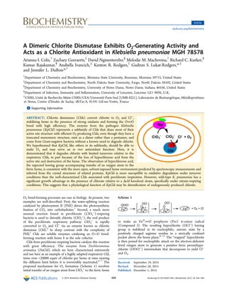
![The requirement for the Cld-catalyzed reaction by
perchlorate-respiring bacteria is absolute; without it, ClO2
−
rapidly accumulates to toxic levels.2
However, Clds are found in
almost all bacterial phyla and in many archaea, the over-
whelming majority of which are not involved in perchlorate
respiration.12
These Clds subdivide according to sequence into
groups that appear to be functionally distinct from their
respiration-associated counterparts and from each other (Figure
1), in spite of their high level of sequence similarity.13
Investigation of the Cld from Staphylococcus aureus, a Gram-
positive bacterium, provided a striking example. In its heme-
bound form, it is completely inactive in the conversion of
ClO2
−
to Cl−
and O2.14
Genetic and biochemical evidence
instead suggests that the protein is essential for a terminal step
in heme biosynthesis,14,15
a biological function that appears to
be common to the Clds from Gram-positive bacteria and
potentially other species.13,15
The Cld from Klebsiella pneumoniae strain MGH 78578
(KpCld) represents another subfamily that may have yet a third
biochemical function. These enzymes are found in non-
perchlorate-respiring bacteria from Gram-negative phyla
(Figure 1).12,13
As a consequence, they are not expected to
be involved in either perchlorate respiration or heme
biosynthesis. Unlike the respiration-associated Clds, they are
homodimers rather than homopentamers, leading to a very
different monomer−monomer interface and resulting structural
context for their bound hemes (Figure 2A−C). Locally,
however, they possess very similar active site residues and
share structural attributes essential for efficient ClO2
−
/O2
conversion. In particular, they share the functionally important
distal arginine above the ferric heme’s open coordination
position (Figure 2D).8,11
Consistent with chemical expect-
Figure 1. Phylogenetic tree illustrating the major subdivisions of the
Cld protein family discussed in the text. The respiratory Clds, coming
mostly from Proteobacteria with some exceptions caused by lateral
gene transfer, form the first group (red). Members of a second group
of dimeric Clds (green), including KpCld, come from non-perchlorate-
respiring species but retain active site features critical for ClO2
−
/O2
conversion. Members of a third broad group of Clds (blue) catalyze a
terminal step in heme biosynthesis in Gram-positive bacteria and
potentially some Archaea. These have consequently been renamed
HemQs.11
Sequence accession numbers are given in parentheses, with
PDB entries used to indicate where structures are available. The
phylogenetic tree was generated using MEGA6.69
The bootstrap
consensus tree inferred from 1000 replicates is taken to represent the
evolutionary history of the taxa analyzed. Branches corresponding to
partitions reproduced in <70% of bootstrap replicates are collapsed.
Initial trees for the heuristic search were obtained by applying the
Neighbor-Joining method to a matrix of pairwise distances estimated
using a JTT model. A discrete Gamma distribution was used to model
evolutionary rate differences among sites [five categories (+G,
parameter = 2.4895)]; 5% alignment gaps, missing data, and
ambiguous bases were allowed at any position.70
The resulting tree
is consistent with those previously reported by others.11,13,71
Figure 2. Clds from perchlorate-respiring and nonrespiring
Proteobacteria. (A) KpCld, like the NwCld structure shown (PDB
entry 3QPI),7
has a truncated monomer relative to the respiratory
Clds. Both form functional homodimers. Protein monomers are
rendered as cartoons in different colors. Hemes are rendered as sticks.
(B) Respiration-associated DaCld (PBD entry 3Q08) forms a
functional homopentamer, shown looking down the C5 axis.4
(C) A
monomer−monomer interface from the structure in panel B suggests
differences in heme accessibility in the pentameric and dimeric Clds.
(D) Active site environments for DaCld (cyan carbons) and NwCld
(magenta carbons) are superimposed with several conserved residues
around the heme labeled (DaCld numbering). The two active sites are
highly similar; two notable exceptions are the orientation of the distal
Arg side chain and the identity of the residue at the position of W227,
which is a conserved glutamate in NwCld and other dimeric Clds.
Biochemistry Article
dx.doi.org/10.1021/bi501184c | Biochemistry 2015, 54, 434−446435](data:image/gif;base64,R0lGODlhAQABAIAAAAAAAP///yH5BAEAAAAALAAAAAABAAEAAAIBRAA7)