
Recommended
More Related Content
Similar to ECG & Intro to Cardiac Arrhythmias.pptx
Similar to ECG & Intro to Cardiac Arrhythmias.pptx (20)
Recently uploaded
Recently uploaded (20)
ECG & Intro to Cardiac Arrhythmias.pptx
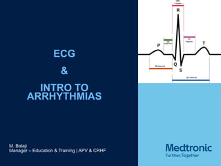
- 1. ECG & INTRO TO ARRHYTHMIAS M. Balaji Manager – Education & Training | APV & CRHF
- 2. SURFACE ECG The ECG is measured by placing skin electrodes on the body surface at different locations and connecting these electrodes in different configurations to a voltage amplifier and recorder
- 4. The Heart from All Directions
- 6. A QUESTION OF LOCATION……
- 7. Chest Leads V1 – V6
- 8. The Chest Leads: V1 - V6
- 9. NORMAL 12 LEAD ECG
- 10. 12 Lead ECG
- 11. VECTOR MAPPING
- 12. Vector Mapping
- 13. The ECG
- 14. NORMAL VALUES P waves ~ 100ms PR interval ~ 120 – 200ms QRS duration ≤ 120ms usually between 80 to 100ms QT interval is about 300 ms for rates of 100 bpm QT interval is about 400 ms for rates of 60 bpm Presentation Title (Edit on Slide Master) | June 1, 2015 | Confidential, for Internal Use Only 14
- 16. INTRODUCTION TO ARRHYTHMIAS Automaticity, Excitability & Conductivity are vital properties of cardiac cells and tissue These properties govern the rate at which the heart beats the sequence in which the different parts of the heart beat And thereby give the heart its natural rhythm Arrhythmia – Any variation from the natural, normal rate and rhythm of the heart Arrhythmias arise due to problems in automaticty or excitability or conductivity or any combination of these
- 17. CLASSIFICATION OF ARRHYTHMIAS Based on Heart Rate Bradyarrhythmia or Bradycardia – Any heart rhythm where the rate is less than 60 bpm Tachyarrhythmia or Tachycardia – Any heart rhythm where the rate is greater than 100 bpm
- 18. CLASSIFICATION OF ARRHYTHMIAS Based on the anatomical location of the heart that is the source of the arrhythmia Sinus Node Arrhythmias Atrial Arrhythmias AV Junctional Arrhythmias Ventricular Arrhythmias
- 19. SINUS ARRHYTHMIAS (SICK SINUS SYNDROME) Arrhythmias that arise from the Sino-Atrial node Sinus Bradycardia Sinus Tachycardia Sinus Arrest Sinus Block Brady Tachy Syndrome
- 20. NORMAL SINUS RHYTHM Atrial rate: 60-100 bpm, Ventricular Rate = Atrial Rate Normal P waves PR interval: 120-200 ms (.12-.20 seconds) QRS interval: 60-100 ms (.06-.10 seconds) QT interval: 360-440 ms (.36-.44 seconds) Rate = ?
- 21. SINUS BRADYCARDIA Identifying features Rhythm : Regular Rate : 40 –60 bpm P waves : Normal in shape and size, one P-wave precedes each QRS complex PR : Normal (120-200ms) QRS : Normal (less than 120 ms)
- 23. SINUS TACHYCARDIA Identifying features Rhythm : Regular Rate : 100 –160 bpm P waves : Normal in shape and size, one P-wave precedes each QRS complex PR : Normal (120-200ms) QRS : Normal (less than 120 ms)
- 25. SINUS/ARREST BLOCK Identifying features Rhythm : Regular underlying rhythm, irregular during pause Rate : Underlying rhythm normal or slow P waves : Normal in shape and size, one P-wave precedes each QRS complex; No P wave during pause PR : Normal (120-200ms), PR absent during pause QRS : Normal (less than 120 ms), QRS absent during pause
- 26. SINUS BLOCK
- 27. SINUS ARREST
- 28. ATRIAL FLUTTER
- 29. ATRIAL FLUTTER
- 32. ATRIVENTRICULAR BLOCKS AV Blocks – Disturbances in conduction of the impulse from the Atria to the Ventricles due to problems within the AV Junction Classifciation – based on location within the AV Junction and the severity of the block First Degree AV Block Second Degree AV Block – Type I, Type II Third Degree AV Block Typically occur due to degeneration of the AV nodal and HIS bundle cells
- 33. Identifying features Rhythm : Regular Rate : Normal, atrial & ventricular rate are same P waves : Normal Sinus PR : Prolonged, greater than 200 ms QRS : Normal (100ms or less) FIRST DEGREE AV BLOCK
- 34. FIRST DEGREE AV BLOCK
- 35. Identifying features Rhythm : Atrial - Regular : Ventricular - Irregular Rate : Atrial – Normal Sinus Rate : Ventricular – Less than atrial rate during block P waves : Normal Sinus PR : PR varies and successively prolongs until a P wave occurs without a QRS. A pause follows the dropped QRS QRS : Normal (100ms or less) SECOND DEGREE TYPE I BLOCK
- 36. SECOND DEGREE TYPE I BLOCK
- 37. Identifying features Rhythm : Atrial - Regular : Ventricular – Regular unless ratio of AV conduction varies Rate : Atrial – Normal Sinus Rate : Ventricular – Less than atrial rate during blocks P waves : Normal Sinus PR : PR may be normal or prolonged but does not vary QRS : Normal (100ms or less) if block is in the HIS bundle Broad or Wide if block is in the bundle branches SECOND DEGREE TYPE II BLOCK
- 38. SECOND DEGREE TYPE II BLOCK
- 39. Identifying features Rhythm : Atrial - Regular, Ventricular – Regular Rate : Atrial – Normal Sinus Rate : Ventricular – 30 to 40 (ventricular pacemaker) - 40 to 60 (AV nodal pacemaker) P waves : Normal Sinus PR : PR varies greatly, no relationship between P waves and QRS QRS : Normal (100ms or less) if block is in the HIS bundle Broad or Wide if block is in the bundle branches THIRD DEGREE AV BLOCK OR COMPLETE HEART BLOCK
- 40. THIRD DEGREE AV BLOCK OR COMPLETE HEART BLOCK
- 41. VENTRICULAR ARRHYTHMIAS Ventricular arrhythmias originate anywhere in the ventricles below the level where the HIS bundle branches out Types Bundle Branch blocks Idioventricular Rhythm or Ventricular Escape Rhythm Accelerated Idioventricular Rhythm Premature Ventricular Contractions (PVC, VPC, VPB) Ventricular Tachycardia (VT) Ventricular Fibrillation (VF) Ventricular Asystole or Standstill All ventricular arrhythmias except VF and standstill are characterised by a wide or broad QRS complex
- 42. BUNDLE BRANCH BLOCK One Ventricle depolarizes before the other
- 44. IDIOVENTRICULAR RHYTHM OR VENTRICULAR ESCAPE RHYTHM
- 46. Multifocal PVC’s PREMATURE VENTRICULAR CONTRACTION
- 47. VENTRICULAR TACHYCARDIA Arise from ectopic pacemaker circuits or (less commonly) sites in ventricular tissue, discharging impulses at a rate of 140-25 bpm Reentry or enhanced automaticity Sustained VT – VT that lasts for 30 seconds or more Non-sustained VT - VT that lasts for less than 30 s
- 48. Monomorphic Sustained VT VENTRICULAR TACHYCARDIA
- 50. VENTRICULAR FIBRILLATION Arises due to simultaneous action of several differnet ectopic circuits in the ventricles randomly, chaotically and uncoordinatedly depolarising different portions of ventricular tissue at different times The ventricles do not beat in a coordinated fashion – they quiver asynchronously and ineffectively There is little or no Cardiac output, blood pressure drops to zero and the patient becomes unconscious almost immediately Death is almost certain unless the arrhythmia is treated immediately
- 52. VENTRICULAR STANDSTILL OR ASYSTOLE
- 53. 53 THANK YOU