
Recommended
More Related Content
Similar to TUMORES RAROS DE LA MAMA.pdf
Similar to TUMORES RAROS DE LA MAMA.pdf (20)
Recently uploaded
Recently uploaded (20)
TUMORES RAROS DE LA MAMA.pdf
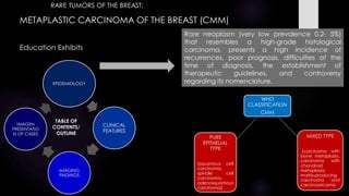
- 1. METAPLASTIC CARCINOMA OF THE BREAST (CMM) Education Exhibits TABLE OF CONTENTS/ OUTLINE EPIDEMIOLOGY CLINICAL FEATURES IMAGING FINDINGS IMAGEN PRESENTATIO N OF CASES Rare neoplasm (very low prevalence 0.2- 5%) that resembles a high-grade histological carcinoma, presents a high incidence of recurrences, poor prognosis, difficulties at the time of diagnosis, the establishment of therapeutic guidelines, and controversy regarding its nomenclature. WHO CLASSIFICATION CMM PURE EPITHELIAL TYPE (squamous cell carcinoma, spindle cell carcinoma, adenosquamous carcinoma) MIXED TYPE (carcinoma with bone metaplasia, carcinoma with chondroid metaplasia, matrix-producing carcinoma and carcinosarcoma RARE TUMORS OF THE BREAST:
- 2. TEACHING POINTS: Heterogeneous group of tumors characterized by the coexistence of adenocarcinoma with areas of differentiation towards other cell lines, non-glandular epithelial or mesenchymal, in a variable proportion. When the metaplastic component encompasses a significant proportion within the tumor, it is then termed metaplastic carcinoma. CLINICAL FEATURES Age of 55-60 years Radiotherapy Trauma Tamoxifen Lymphedema secondary to previous surgery RISK FACTORS Clinically presents as a palpable mass with no predilection for any quadrant and with rapid growth. Axillary lymph node involvement is rare (6 to 40%). Incidence of metastatic disease between 5-30%. Size (above 5 cm the prognosis worsens. Age Lymph node involvement Metastases at presentation Prognosis: survival is low (<50% at 5 years)
- 3. MAMMOGRAPHY Microlobulated or circumscribed tumor in a dense, heterogeneous or predominantly fatty breast pattern. There is usually no association with microcalcifications or distortion of the breast architecture. Large solid masses, complex internal echogenicity or with presence of hypoechoic and/or cystic areas, being related to necrosis and cystic degeneration, edges microlobulated and the margins well circumscribed; almost present posterior acoustic enhancement; lesions containing sarcomatous elements tend to create acoustic shadows. ULTRASOUND The definitive diagnosis is always anatomopathological Depending on its mesenchymal component, several subtypes of metaplastic carcinoma can be differentiated: sarcomatous, fibromatous, and angiosarcoma. The most common form of presentation is as squamous cell carcinoma.
- 4. Rare tumor of controversial histogenesis, aggressive behavior and unusual metastatic pattern to the gastrointestinal tract and serous surfaces Atypical morphological characteristics, high aggressiveness and poor prognosis. Represent around 0.7% of all breast tumors, some case series report prevalences of up to 1.28-1.5%. Rare variant of adenocarcinoma that can represent up to 8.7% of all lobular carcinomas EPIDEMIOLOGY WHO CLASSIFICATION 2012 Breast cancer with signet ring cell differentiation PRIMARYSIGNETRINGCELLCARCINOMAOFTHEBREAST RARE TUMORS OF THE BREAST:
- 5. CLINICAL FEATURES Painless, unclear mass located in the upper outer quadrant of the breast. Particularly, it may not be detected on manual examination or it may present as a small diffuse nodule, accompanied or not by changes in the nipple with edema, ulceration or telorrhoea, as well as skin changes such as dimpling and orange peel. Considered variants of lobular carcinoma, due to the histological, ultrastructural, and immunohistochemical similarities of both entities, but the presence of mucins is a rare finding in lobular carcinomas Unusual pattern of metastasis Gastroint estinal tract Serous Genital tract Immunohistochemical profile: positivity for CK 7, hormone receptors, and GCDFP protein help diagnose a breast origin, and the negativity of these markers, with positive immunostaining for CK 20 and CEA, support the diagnosis of gastrointestinal origin.