SlideShare a Scribd company logo
1 of 181
1
NoN CariouS TootH LesionS
(NCTL / NCCL)
Presented by: D r Ab h isek Gur ia
Dept. of Conservative Dentistry & Endodontics
2
Contents
 Introduction
 Definition
 Classification (Grippo)
Atrition
 Definition
 Classification
 Proximal surface attrition
 Occlusal surface attrition
 Etiology and Clinical features
 Treatment modalities
 The Dahl concept
 Schools of thought to increase the
V.D.
 Endodontic considerations
 Treatment strategies for Dentinal
hypersensitivity
 Nerve desensitization
 Anti-inflammatory agents
 Covering or plugging dentinal
tubules
 Restorative materials
 Periodontal surgery
 Lasers
5/11/2020 Management of Non-carious lesion
3
Management of Non-carious lesion
Abrasion, Erosion, Abfraction
 Definition
 Etiology and pathogenesis
 Clinical features
 Treatment modalities
 Monitoring of lesion progression
 Restorative decision making
 Perimolysis
Fracture line/tooth crack
 Etiopathogenesis
 Classification
 Characteristics
 Distribution
 Clinical diagnosis
 Radiographic examination
 Treatment modalities
 Management of peg laterals
 Management of palatogingival groove
5/11/2020
4
Management of Non-carious lesion
 Localized Non Hereditary Enamel
Hypoplasia
 Localized Non Hereditary Enamel
Hypocalcification
 Localized Non Hereditary Dentin
Hypoplasia
 Localized Non-hereditary Dentin
Hypocalcification
 Amelogenesis Imperfecta
 Dentinogenesis Imperfecta
 Definition
 Etiology
 Clinical features
 Treatment modalities
 Conclusion
 References
5/11/2020
5
Introduction
Non carious tooth tissue loss (NCTL):
 Surface loss due to a disease process other than dental
caries. (Pual A Brunton ,Decision making in Operative Dentistry )
 25% of tooth destruction does not originate from a
carious process.(Marzouk)
5/11/2020 Management of Non-carious lesion
6
Etiology of NCTL
1. Attrition
2. Abrasion
3. Erosion
4. Abfraction
5. Localized Non- Hereditary Enamel Hypocalcification
6. Localized Non- Hereditary Dentin Hypolpasia
7. Localized Non- Hereditary Dentin Hypocalcification
8. Fracture lines
9. Amelogenesis imperfect
10. Dentinogenesis imperfecta
5/11/2020 Management of Non-carious lesion
• John O Grippo,Marvin Simmering,JADA 2004 135;1109-1118
• Osborne-Smith KL, Burke FJ, Wilson NH. Int Dent J. 1999 Jun;49(3):139-43. Review
7
Attrition
• Mechanical wear of the incisal or occlusal surface as a
result of functional or parafunctional movements of
mandible (tooth to tooth contacts) (Sturdevant)
• Surface tooth structure loss resulting from
direct frictional forces between contacting
teeth. (Marzouk)
• It also includes the proximal surface wear at the contact
area because of the physiologic tooth movement
5/11/2020 Management of Non-carious lesion
8
Etiology
1. Bruxism
2. Developmental enamel defect
3. Physiologic
5/11/2020 Management of Non-carious lesion
9
Classification
10
A) Proximal surface attrition (proximal surface facets)
 Results from surface tooth
structure loss and flattening ,
widening of the proximal contact
areas.
 Mesiodistal dimension of the teeth is
decreased, leading to drifting , with
the possibility of overall reduction in
the dental arch.
5/11/2020 Management of Non-carious lesion
11
B) Occluding surface attrition ( OCCLUSAL WEAR)
 It is the loss ,flattening, faceting or reverse cusping of the
occluding elements.
 It leads to loss of vertical dimension of the tooth .
A) If the LOSS IS SEVERE & accomplished in a relatively short
time
5/11/2020 Management of Non-carious lesion
No chance for the alveolar bone to erupt occlusally to compensate for the
occlusal tooth loss
Loss of VD
overclosure during mandibular functional movements
strain areas on stomato-gnathic system.
12
Management of Non-carious lesion
B) If the loss occurs over a long period-
• The alveolar bone can grow occlusally, bringing the teeth to their
original occlusal termination.
• Vertical dimension loss will be confined to teeth but not imparted to
face.
• Deficient masticatory capabilities
• Cheek biting
• Decay
5/11/2020
13
Clinical presentation
• Flattened occlusal surfaces.
• The degree of wear in both arches is normally equal.
• Sometimes there may be presence of peripheral, ragged,
sharp enamel edges .
• The presence of hypertrophic masseter is a warning
sign of the impact of bruxism .
• TMJ problems can be elicited especially by the over
closure situation
5/11/2020 Management of Non-carious lesion
14
A. When surface attrition is SLOWER & compensated by,
intrapulpal deposition of secondary & tertiary dentin, then
there will be no pulpal exposure.
B. At other times, the attrition is faster than the intrapulpal
dentine deposition, leading to direct pulpal exposure.
5/11/2020 Management of Non-carious lesion
15
Treatment modalities
• Pulpally involved teeth →endodontic therapy /extraction
depending upon their restorability .
• Para functional activities ,( bruxism)-- be controlled with
protecting occlusal splints.
5/11/2020 Management of Non-carious lesion
16
Management of Non-carious lesion
 Myofunctional, TMJ/ any other symptoms in the stomato-
gnathic system -----diagnosed and resolved (modifying the
occlusal splint).
 Occlusal equilibration : should be performed by :
1. Selective grinding of tooth surfaces that includes
rounding and smoothening the peripheries of the
occlusal tables.
2. By creating adequate overlap between the working inclines
to prevent further cheek biting.
5/11/2020
17
Any exposed sensitive
dentin should be
protected and actual
carious lesion be
obliterated .
Periodontium be
examined and any
pathology be treated
.
Restorative
modalities can than
be initiated.
5/11/2020 Management of Non-carious lesion
18
Management of Non-carious lesion
 Restorations are only needed in the following situations:
 Noticeable loss of vertical dimension
 Or a progressive loss of tooth structure is observed
compromising the tooth strength .
 Caries ,if present
 Defect contributes to a periodontal problem.
 Worn tooth contour, (usually proximal ) which is not
conducive to the maintenance of periodontium .
 A tooth is cracked or endodontically treated.
5/11/2020
19
Procedure
• Amount of V.D. lost is estimated .
• It gives an estimate up to what should be the height of the
worn clinical crowns be increased .
Management of Non-carious lesion5/11/2020
20
 The additional V.D. that the stomognathic system can
accommodate without untoward effects is estimated.
 Composite temporary restorations are most frequently used
 Permanent restoration should be done in a cast alloy
 These restorations should be cemented only temporarily for
an extendedperiod of time ,until it is established that no
untoward symptoms would occur.
5/11/2020 Management of Non-carious lesion
21
 An acrylic splint( as a stabilization splint) may be necessary
5/11/2020 Management of Non-carious lesion
22
Restorative treatment
 Tooth wear can be followed and re-evaluated during
recall examinations.
 Less severe anterior wear can be treated with
adhesive composite resin.
(Strassler HE, Kihn PW, Yoon R. Conservative treatment of the worn dentition with adhesive composite resin.
Contemp Esthet Restor Pract. 1999)
5/11/2020 Management of Non-carious lesion
23
Management of Non-carious lesion
When the wear is more severe, a number of treatment
modalities are available.
1. Bonded porcelain veneers have been used to treat incisal wear.
(Ibsen RL, Ouellet DF. Restoring the worn dentition. J Esthet Dent. 1992;4:96-101.)
1. In some cases, the incisal edges can be restored to the original
vertical dimension with direct composite resin.
(Strassler HE, Kihn PW, Yoon R. Conservative treatment of the worn dentition with adhesive composite
resin. Contemp Esthet Restor Pract. 1999)
5/11/2020
24
Management of Non-carious lesion
 Hemmings and coworkers reported on the restoration of
severe anterior wear with composite restoration including re-
establishment of the occlusal vertical dimension.
 They reported a 89.4% success at 30 months.
(Hemmings KW, Darbar UR, Vaughan S. Tooth wear treated with direct composite restorations
at an increased vertical )dimension: results at 30 months. J Prosthet Dent. 2000;83:287-293.
5/11/2020
25
Management of Non-carious lesion
 Adhesive cast metal restorations have also been used to
replace missing tooth structure.
( Nohl FS, King PA, Harley KE, et al. Retrospective survey of resin- retained cast-
metal palatal veneers for the treatment of anterior palatal tooth wear. Quintessence
Int. 1997)
 In cases where the occlusion is severely altered by attrition,
the only treatment choice may be a reconstruction with crowns
and bridges.
(Stewart B. Restoration of the severely worn dentition using a systematized
approach for a predictable prognosis. Int J Periodontics Restorative Dent.
1998;18:46-57.)
5/11/2020
26
Management of Non-carious lesion
27
The Dahl concept
 Dahl and his coworkers (1975)
described the use of a partialbite
raising appliance‘ to create inter-
occlusal space in an 18-year-old
patient with severe localised
attrition.
 The removable appliance was cast in
cobalt-chromium, placed on the palatal
aspects of the upper anterior teeth,
and worn 24 hours a day.
5/11/2020 Management of Non-carious lesion
28
Management of Non-carious lesion
 The creation of inter- occlusal space significantly reduced the amount
of tooth preparation required, especially on the already compromised
palatal surface.
 Teeth were restored with full coverage porcelain bonded
crowns
5/11/2020
29
2 schools of thought to increase the V.D.
 Addition of increments : gradual increments by
progressively adding to the hard splint at 1mm /week, until
the patient reaches the increased V.D. for restorative
purposes----time consuming
 The second approach ----taking the patient immediately to
needed increase in V.D.----considerably lesser adjustments
are made ,lesser time consuming
Management of Non-carious lesion5/11/2020
30
Anterior Bite plane
 Used in the reduction of overbite.
 Occurs by altering the rate of eruption
of posterior teeth relative to the
eruption of lower incisors that are in
contact with the bite plane.
 Overbite reduction by this method ---
most successful in actively growing
patients
5/11/2020 Management of Non-carious lesion
31
Endodontic considerations
In certain cases intentional endodontic therapy has to be performed
 Hypererupted teeth
 Pulpal involvement
For location of calcified canal
 Use of magnification
 staining the pulp chamber floor with 1%methylene blue dye .
 searching for canal bleeding points .
 Performing the sodium hypochlorite ―champagne bubble test are helpful in locating
calcified.
 Long ,thin Ultrasonic tips can also be used
5/11/2020 Management of Non-carious lesion
32
Dentinal hypersensitivity
Dentin Hypersensitivity is a condition characterized by short , sharp
pain arising from exposed dentin in response to stimuli typically
thermal , evaporative , tactile, osmotic or chemical and which cannot
be ascribed to any other form of dental defect or pathology .
( Holland et al 1997)
5/11/2020 Management of Non-carious lesion
33
THEORIES
1. The transduction theory
2. Direct neural stimulation theory
3. The hydrodynamic theory
34
Treatment strategies for Dentinal
hypersensitivity ( DCNA 53,2009; 47-60)
1. Nerve desensitization
 Potassium nitrate
2 .Anti-inflammatory agents
 Corticosteroids
3. Covering or plugging dentinal tubules
• calcium hydroxide
• sodium fluoride
5/11/2020 Management of Non-carious lesion
35
Management of Non-carious lesion
• Sodium monoflourophosphate
• Stannous fluoride
• Oxalates
• Strontium chloride
 Protien precipitants
 formaldehyde
 glutaraldehyde
• Flouride iontophoresis
• Resins and Adhesives
4) Restorative materials
5) Periodontal surgery
6) Lasers
5/11/2020
36
Nerve desensitization
 Potassium nitrate
 5% concentration in a low abrasive tooth paste was able to
desensitize dentin for up to 4 weeks compared to a control
paste . (Tarbet et al )
 In bio adhesive gels at a concentration of 5% and 10% has
also been shown to be effective in reducing dentinal
hypersensitivity .(Freschoso SC,Menendez M,2003)
Management of Non-carious lesion5/11/2020
37
Anti inflammatory agents:
Corticosteroids
 glucocorticoids to the cavity preparation may reduce
dentinal hypersensitivity by their effect on pain
mediators
 Lawson and Huff found that paramethasone had a
significant desensitizing action.
 Furseth and Mjor reported complete obturation of dentinal
tubules after corticosteroid application to exposed dentin –
reduce dentin permeability.
 However there is a little experimental evidence to support
or refute the use of such agents
5/11/2020 Management of Non-carious lesion
38
Sodium fluoride
 Treatment of exposed root surface with
fluoride toothpaste (1.1%) and conc.
fluoride solutions(0.2%) is very efficient.
(Minkow B,1975;Kerns D G 1991)
 Mechanism- precipitated fluoride
compound mechanically blocking the
exposed dentinal tubules. (Tal et al)
5/11/2020 Management of Non-carious lesion
39
Sodium monoflourophosphate
• Tooth pastes containing sodium monoflourophospshates have
been shown to be effective in managing dentinal
hypersensitivity. (Hernandez F,Mohammed C,1972)
• Its mechanism of action is unclear (,Scherman A et al 1992)
5/11/2020 Management of Non-carious lesion
40
Stannous fluoride
 Stannous fluoride in aqueous solution or in glycerin gelled
with carboxymethylcellulose
(Miller JT et al 1969)
 Mode of action : induction of high mineral content which
creates a calcific barrier blocking the tubular openings on
the dentine surface .
(Furseth R ,1970)
5/11/2020 Management of Non-carious lesion
41
Fluoride iontophoresis
• It is the process of influencing ionic motion by an electric
current and has been used as a desensitizing procedure in
conjunction with sodium fluoride .
(Mc Fall WT,1986)
 Studies report that there is a immediate reduction in
sensitivity after treatment with iontophoresis, but the
symptoms gradually return over the next six months
(Kern DA et al 1989 )
5/11/2020 Management of Non-carious lesion
42
Procedure
5/11/2020 Management of Non-carious lesion
43
Oxalates
 potassium oxalates have both tubule obturation properties and
inhibitory effects caused by potassium ions on nerve activity
(Pashly DH ,1986)
 Oxalate ion reacts with calcium to form insoluble calcium
oxalate crystals that bind tightly to dentin and obturate
dentinal tubules (TrowbridgeHO,1990)
 Three types of oxalates are available :
1. 6% ferric oxalate (Sensodyne Sealant )
2. 30% di-potassium oxalate( Butler Protect )
3. 3% monohydrogen monopotassium oxalate
5/11/2020 Management of Non-carious lesion
44
Strontium chloride
 strontium ions have the capacity to reduce sensory nerve
activity, but less effectively than potassium ions.
(MarkowitzK , kim S 1990).
 Dentifrices containing 10% strontium chloride (Sensodyne) -
widely used as desensitizing agents and were one of the
first agents to be marketed for that purpose.
5/11/2020 Management of Non-carious lesion
45
Protein precipitants
(Formaldehyde and glutaraldehyde)
 Have ability to precipitate salivary proteins in the
dentinal tubules.
 However this effect has been questioned since various
formulations have been found to have little or no effect
on dentinal hypersensitivity(Addy M,Mostafa P,1988)
5/11/2020 Management of Non-carious lesion
46
Resins and Adhesives
• rationale – they seal the dentinal tubules and hence to
preclude the transmission of pain stimuli to the pulpal
nerve fibers.
• This mode of treatment is performed on localized
hypersensitive dentin.
• Resin-based materials have been reported to successfully
reduce dentinal hypersensitivity.(Kakaboura A ,2005)
5/11/2020 Management of Non-carious lesion
47
Management of Non-carious lesion
 Copeland reported successful treatment of dentinal
hypersensitivity for up to 18 months in 89% of
hypersensitive teeth treated by Scotchbond.
 5% glutaraldehyde and 35% hydroxyethyl methacrylate
(Gluma Desensitizer) has been reported to be an effective
desensitizing agent for up to 9 months. ( Kakaboura A ,2005)
5/11/2020
48
Bioactive glass:
 NovaMin - for remineralisation of teeth, treating
hypersensitivity.
 The active ingredient is called Calcium Sodium
Phosphosilicate.
 NovaMin is an ionic form of calcium, phosphorus, silica, and
sodium which are necessary for bone and tooth
mineralization.
5/11/2020 Management of Non-carious lesion
49
Lasers
 Nd:YAG laser irradiation in combination with 5% sodium
fluoride varnish has higher efficacy in the management of
DH than either treatment alone. (Kumar and Mehtas ,2005)
 Slutzky-Goldberg (2008) demonstrated that CO2 laser
treatment resulted in decreased permeability of dentinal
tubules as shown by a dye penetration test.
5/11/2020 Management of Non-carious lesion
50
Restorative materials
• The use of restorative materials is generally an invasive
solution to the problem of hypersensitivity.
• Commonly used materials include composite resins and
glass ionomer restorations.
5/11/2020 Management of Non-carious lesion
51
Abrasion
5/11/2020 Management of Non-carious lesion
52
Management of Non-carious lesion
 Abnormal tooth surface loss resulting from direct
frictional forces between the teeth and external objects
or from frictional forces between contacting teeth
components in the presence of abrasive medium.
(Sturdevant 5 th edition)
 It occurs most frequently on the cervical neck of the teeth.
 The labial or buccal surfaces. (tooth brush abrasion )
 Labial or buccal and lingual surfaces( in case of poorly fitted
clasps and artificial dentures ) .
5/11/2020
53
Causes of abrasion :
• Traumatic occlusion .
• Improper brushing technique .
• Occupational (Habits such as holding bobby pins in between the
teeth .)
• Tobacco chewing /tobacco pipe .
• Vigorous use of tooth picks between the adjacent teeth.
• Excessive mastication of coarse foods .
5/11/2020 Management of Non-carious lesion
54
 Tooth brushabrasion results in a
horizontal cervical notches on the
buccal surfaces of exposed radicular
cementum and dentin .
 Notching in right central incisor
caused by improper use of bobby
pins .
5/11/2020 Management of Non-carious lesion
55
clinical signs and symptoms
 The surface of the lesion is extremely smooth and polished and it
seldom has any plaque accumulation or caries activity in it .
 The surrounding walls tend to make a V shape ,by meeting at an
acute angle axially.
 Peripheries of the lesion are angularly demarcated from the adjacent
tooth surface.
 Probing or stimulating the lesion can elicit pain .
 Hypersensitivity may be intermittent in character appearing and
disappearing at occasional or frequently repeated periods .
5/11/2020 Management of Non-carious lesion
56
Treatment modalities
 Diagnose the cause of the presented abrasion.
 A detailed history is to be taken considering various factors such as:
• Oral hygiene techniques ( use of abrasive tooth cleaning techniques and
materials)
• Habits- pipe smoking, chewing tobacco, professional habits
• Iatrogenic causes,if any.
 Avoidance or counteraction of the causes which may lead to its
production.
 Instituting proper oral hygiene measures.
 incorporating correct method of tooth brushing .
5/11/2020 Management of Non-carious lesion
57
Management of Non-carious lesion
 Have the habit of chewing tobacco ,toothpick , etc
discontinued . If successful in breaking the habit proceed
with the restorative treatment as planned.
 Correcting or avoiding ill fitting metal clasps and dentures
5/11/2020
58
 Abrasive lesions at non-occluding tooth surfaces should be:
• Evaluated critically for the need for restoring them.
• If the lesions are multiple, shallow( not exceeding 0.5 mm in
dentin) and wide → no need to restore them .
• If there is involvement of cementum / enamel only → no need
to restore .
• If lesion is wedge (V) shaped and exceeds 0.5 mm into dentin →
restoration is performed .
Management of Non-carious lesion
59
Management of Non-carious lesion
 If restoration is not indicated for a lesion, then :
1. Edges of the defect should be eradicated to a smooth, non-
demarcating pattern relative to adjacent tooth surface.
2. Tooth surface then should be treated by fluoride solution
to improve caries resistance
5/11/2020
60
Restoring cervical abrasions
 Indicated when
1. Caries ,if present .
2. Sensitivity is present.
3. Lesion is esthetically objectionable .
4. If the defect contributes to a periodontal problem
5. The area to be involved in the design of a removable partial denture.
6. When the depth of defect is found to be close to pulp
7. Or a progressive loss of tooth structure is observed compromising the
tooth strength .
5/11/2020 Management of Non-carious lesion
61
Restorative materials :
• Glass ionomer restorative material.
• Resin modified glass ionomer.
• Polyacid-modified resin composites.
• Resin composites.
5/11/2020 Management of Non-carious lesion
62
Biomechanics
5/11/2020 Management of Non-carious lesion
63
Management of Non-carious lesion
 High modulus restorative materials are unable to flex in the
cervical regions when the tooth structure is deformed under
occlusal load and ,therefore the restorative materials can
be displaced from the cavity .
(Heymann HO ,Sturdevant Jr ,Baynes S ,JADA,122(2) 41- 57 )
 An intermediate material with reduced elastic modulus may
function as a stress absorbing layer and improve marginal
sealing .
(Kemp-Scholte CM ,Davidsson,CL complete marginal seal of class V resin composite restorations affected by
increased flexibility .JDR 1990 ;69:1240 -3 )
5/11/2020
64
Management of Non-carious lesion
 As a result materials with low elastic modulus such as :
1. Microfilled composites (Heymann and others ,1991 :Levitch and others ,1994 )
2. Flowable resins (Unterbink ,Liebenberg ,1999: Li and others 2006 )
3. Glass ionomer cements (Loguercio and others ,2003:Burgess and others ,2004)
Have been used in restoring cervical lesions ,with the aim of
absorbing the stresses generated during the polymerization
shrinkage of composites and mechanical loading in which the
teeth are subjected during function .
5/11/2020
65
Literature
 Two-year clinical evaluation of four polyacid- modified resin
composites and a resin-modified glass-ionomer cement
(Vitremer) in Class Vabrasion/erosion lesions. (Ermiş RB.. Quintessence Int.
2002 Jul- Aug;33(7):542-8)
Result
• Retention levels at 2 years were 90% for F2000, 90% for
Dyract AP, 89% for Compoglass F, 84% for Elan, and 95% for
the Vitremer restorations.
5/11/2020 Management of Non-carious lesion
66
Discussion
• GIC have an inhibitory effect on secondary caries,
and the release of fluoride is considered to be one
of the major benefits associated with GIC
• Polyacid- modified resin composites may not be
recharged again with fluoride as are GIC
67
Comparative analysis of techniques of restoring
cervical lesions- (Quintessence Int 1993:24:553- 559.)
• Aim - the sandwich technique and the GIC restoration, from the
standpoint of marginal leakage.
• While GIC may not have as good esthetic properties as resin materials
• the sandwich technique does not have universal application, because it
requires space : not the technique of choice in shallow cavities.
68
Result
1. The acid-etch technique was effective in reducing marginal
leakage along the tooth - composite resin interface in enamel.
2. None of the techniques studied consistently provided a complete
seal at the gingival aspect of cervical restorations.
3. Composite resin inserted over a GIC liner demonstrated
significantly less leakage than when the liner was not used.
4. The use of the sandwich technique or GIC alone provided the most
effective seals in cervical wedge-shaped cavities, compared to the
all- composite restoration.
5. There was no significant difference between the marginal leakage
of GIC and sandwich technique restoration
69
5-year clinical performance of resin composite versus
RMGIC in non-carious cervical lesions.
Franco Eb, Benetti A, SK Ishikiriama,SL Santiago ,JRP Lauris
METHOD AND MATERIALS: One operator placed 70
restorations (35 RMGIC and 35 resin composite restorations) in
30 patients under rubber dam isolation.
CONCLUSIONS : After 5 years of evaluation, the clinical
performance of resin modified glass ionomer restorations was
superior to resin composite restorations.
70
Erosion
71
Erosion
• Def- Loss of surface tooth structure by chemical
action in the continued presence of demineralizing
agents(acids). (Sturdevant- 5th edition)
• There is no convincing etiology ,and multiple factors
have been theorized for its pathogenesis:
Mechanical factors:
The action of the muscles of lips and cheeks , and of
tooth brush against affected surfaces .
72
Chemical factors :
• Ingested acids : citric acids esp. if use in large amounts , can precipitate or
initiate erosive lesion
• Secreted acids : the acidity of crevicular fluid has been correlated to
cervical erosion
(Bodecker CF. Local acidity: a cause of dental erosion- abrasion.Ann Dent 1945)
• Acid fumes : acid vapours from nitric acid and sulphuric acids, acting in the
mouths of workers in the factories ,where these acids are largely used or
manufactured ( Miller)
• Refused acids : as a result of chronic , frequent regurgitation ,the stomach‘s
hydrochloric acid can hit the teeth at specific locations ( atypical pattern of
erosion affecting buccal surfaces of lower posterior teeth)
73
Clinical presentation
• Extensive loss of buccal and
occlusal tooth structure
• Raised amalgam restoratins.
• Occlusal view of maxillary dentition
exhibiting concave dentin
depressions surrounded by
elevated rims of enamel
74
• Multiple cupped out
depressions corresponding to
the cusp tips
• Extensive loss of enamel and
dentin on the Buccal surface
of maxillary bicuspids. ( pt
had sucked chronically on
tamarinds )
75
Palatal surfaces of maxillary dentition
in which the exposed dentin exhibits a
concave surface and a peripheral
white line of enamel
• Perimylosis: decalcification of the teeth caused by exposure
to gastric acid in patients with chronic vomiting, as may occur
in anorexia or bulimia
• Loss of lingual enamel and dentin due to acid
regurgitation aggravated by circular movements of
tongue.
• Associated with stress reflux syndrome
76
• A similar appearance is found in patients with eating disorders-
• Anorexia - immoderate food restriction and irrational fear of
gaining weight, as well as a distorted body self-perception
• Bulimia nervosa - consuming a large amount of food in a short
amount of time followed by an attempt to rid oneself of the food
consumed, typically by vomiting
• Rumination - effortless regurgitation of most meals following
consumption
• Chronic alcoholism produces a similar pattern of erosion,
although usually more generalized.
• ( ND Robb and BGN Smith, Anorexia and bulimia nervosa (the eating disorders): conditions of
interest to the dental practitioner, J Dent (1996)
77
• Any food substance with a critical pH value of less than 5.5
can become a corrodent and demineralize the teeth.
( Stephan RM, JADA 1940) ,( Gray JA, J Dent Res 1962) , (Zero DT. Cariology. Dent Clin North
Am 1999)
• Holding ,swilling or retaining acidic drinks and foods in the
mouth prolongs the acid exposure on the teeth increasing the
risk of erosion .
(Mossazzez R ,Smith BGN,Barlett DW,Oral Ph and drinking habit during the ingestion of
carbonated drink in a group of adolescents with dental erosion ,J Dent 2000)
78
• The corrosive potential of an acidic drink does not depend
exclusively on its ph value, but also is strongly influenced by
its buffering capacity of the acid and by the frequency and
duration of ingestion.
(Lussi A. Dental erosion: clinical diagnosis and case histor taking. Eur J Oral Sci 1996 )
79
 The other substances that can corrode teeth.
 Chewable vitamin C tablets
 Aspirin tablets
 Aspirin powders
 Use of the amphetamine drug ecstasy
 Have been associated with corrosion on the occlusal
surfaces of posterior teeth.
80
The potential effects of pH and buffering capacity
on dental erosion.
Owens BM. Gen Dent. 2007 Nov-Dec;55(6):527-31
• This in vitro study evaluated five different soft drinks (Coca-
Cola Classic, Diet Coke, Gatorade sports drink, Red Bull high-
energy drink, Starbucks Frappucino coffee drink) and tap
water (control) in terms of initial pH and buffering capacity.
• Initial pH was measured in triplicate for the six beverages.
The buffering capacity of each beverage was assessed by
measuring the weight (in grams) of 0.10 M sodium hydroxide
necessary for titration to pH levels of 5.0, 6.0, 7.0, and 8.3.
81
Mean ph values for each beverage
• Coca cola
• Diet Coke
• Gatorade
• Red Bull
• Starbucks Frappucino
• Tap water
2.49
3.12
2.93
3.24
6.59
7.12
82
• Red Bull had the highest mean buffering capacity
(indicating the strongest potential for erosion of
enamel), followed by Gatorade, Coca-Cola Classic,
Diet Coke, and Starbucks Frappucino.
83
• Beverages with high buffering capacities compete with
natural buffering characteristics of saliva and resist ph
changes as a result .
• Greater the buffering capacity , more time it takes for saliva
to restore the pH value ,which causes beverage pH to decline
to a sustained level --------- prolonged periods of oral acidity
---- thus increasing the erosive potential .
84
Monitoring tooth wear
Most effective way to monitor wear is :
• comparing the dated study casts to the clinical
conditions of teeth over time .
• It can also be used as a part of preventive regime .
85
• Active wear → smooth and unstained ,clean surfaces.
→ erosion of tooth around the existing
restoration .
(Restoration is resistant to acid ,remains
unchanged ,but the tooth is gradually
dissolved leaving the restoration proud)
• Inactive wear — stained .
86
 Diminish the frequency and severity of acid challenge.
• ↓ the amount and frequency of acidic foods or drinks
• Acidic drinks should be drunk quickly rather than sipped.
• Use of straw reduces erosive potential
 Treating the underlying medical disorder or disease.
• GERD ,anorexia ,bulimia → refer to a physician
/psychologists
Protocol for the prevention of
progression of erosion
(Beatrice K,Edmond L ,J Contemp.Dental practise ,1999)
87
 Enhance the defense mechanisms of body:
• Saliva provides buffering capacity→ increases with
salivary flow rate.
• Saliva supersaturated with Ca, P → inhibits
demineralization of tooth structure.
• Stimulation of salivary flow → sugarless lozenge,
candy/gum is recommended
88
 Enhance acid resistance, remineralization and
rehardening of the tooth surfaces.
• Daily use topical flouride at home
• Fluoride application in office- 2-4 times a year
,flouride varnish recommended.
 Decrease abrasive forces.
• Use a soft bristled toothbrush and brush gently.
89
• No brushing should be done immediately after
consuming acidic food and drink as teeth will be
softened.
• Rinsing with water is better than brushing after
consuming acidic foods and drinks.
(Gandara, B.K; E.L Truelove ,Diagnosis and management of dental erosion. Journal
of Contemp.Dental Practice 1999)
90
91
Improve chemical protection
• Neutralize acids in mouth ---dissolving sugar free antacid
tablets 5 times a day ,particularly after an intrinsic or
extrinsic acid challenge.
• Dietary components- hard cheese ( provides Ca and PO4),
held in
 mouth after acidic challenge.
92
Mechanical protection
• By application of composites and direct
bonding
where appropriate – to protect exposed dentin
• Occlusal guard /Acrylic splint in the form of stabilization
splint
necessary to protect dentition from further damage
due to erosion .
Management of Non-carious lesion
93
 Monitor stability
• by use of casts /photos to document tooth wear
status.
• Regular recall examinations to review diet, oral
hygiene methods, compliance with medications, topical
flouride and splint usage.
94
Restoration
• Metallic restorations should be the choice of material
,if restoration indicated .
(more resistant to erosion )
• Tooth colored materials may also be used with minimal
or no tooth preparation, with the assumption that
restoration may require periodic replacement .
95
To be continued……..
96
NoN CariouS TootH LesionS
(NCTL / NCCL)
Presented by: D r Ab h isek Gur ia
Dept. of Conservative Dentistry & Endodontics
SHDCH , Hassan
Sem - 14
Date of Presentation : 26/02/T20
97
Contents
 Introduction
 Definition
 Classification (Grippo)
Atrition
 Definition
 Classification
 Proximal surface attrition
 Occlusal surface attrition
 Etiology and Clinical features
 Treatment modalities
 The Dahl concept
 Schools of thought to increase the
V.D.
 Endodontic considerations
 Treatment strategies for Dentinal
hypersensitivity
 Nerve desensitization
 Anti-inflammatory agents
 Covering or plugging dentinal
tubules
 Restorative materials
 Periodontal surgery
 Lasers
5/11/2020 Management of Non-carious lesion
98
Management of Non-carious lesion
Abrasion, Erosion, Abfraction
 Definition
 Etiology and pathogenesis
 Clinical features
 Treatment modalities
 Monitoring of lesion progression
 Restorative decision making
 Perimolysis
Fracture line/tooth crack
 Etiopathogenesis
 Classification
 Characteristics
 Distribution
 Clinical diagnosis
 Radiographic examination
 Treatment modalities
 Management of palatogingival groove
5/11/2020
99
Management of Non-carious lesion
 Localized Non Hereditary Enamel
Hypoplasia
 Localized Non Hereditary Enamel
Hypocalcification
 Localized Non Hereditary Dentin
Hypoplasia
 Localized Non-hereditary Dentin
Hypocalcification
 Amelogenesis Imperfecta
 Dentinogenesis Imperfecta
 Definition
 Etiology
 Clinical features
 Treatment modalities
 Conclusion
 References
5/11/2020
100
Abfraction
 Def- The pathological loss of tooth substance caused by the
biomechanical loading forces that result in flexure and failure of
enamel and dentin at a location away from the loading.(Grippo)
 Some authors explain the formation of cervical, wedge shaped
defect by the heavy force in eccentric occlusion resulting in
flexuring (elastic bending) of the tooth.
101
 Loss of tooth surface at the cervical areas caused
by tensile and compressive forces during tooth
flexure
Milosevic, 1998
5/11/2020 Management of Non-carious lesion
102
Etiopathogenesis
5/11/2020 Management of Non-carious lesion
103
 The size and the development of abfractive lesions are
proportional to the direction, magnitude, frequency, duration,
and location of the static and cyclic loading forces causing the
stress.
5/11/2020 Management of Non-carious lesion
104
Management of Non-carious lesion
 Enamel prisms and orientation of dentinal tubules.
 The cervical region depicts parallel prisms and tubules, which result in
less resistant dental tissue than in the occlusal/incisal regions.
5/11/2020
105
 Stress-strain pattern by finite element analysis.
 Stress distribution was performed using maximum principal stress,
identifying areas with tensile stress (positive) and/or compressive
stress (negative).
5/11/2020 Management of Non-carious lesion
106
 Microfractures can foster loss of tooth structure from
 tooth brush abrasion
 from acids in the diet or plaque or both .
 The resulting defect has a smooth surface .
 Also known as idiopathic erosion.
(Lee WC, Eakle WS, J Prosthet Dent 52(3): 374 -380, 1984.)
 Stresses that concentrate to produce abfractions in teeth usually
are transmitted by occlusal loading forces.
( Whitehead SA, Wilson NHF, Watts DC. J Esthet Dent 2000),(Pintado MR, DeLong R, Ko C, Sakaguchi RL,
Douglas WH. Correlation J Prosthet Dent 2000)
 Occlusal interferences, premature contacts, habits of
bruxism and clenching all may act as stressors.
107
Clinical features
 The lesion is typically wedge shaped with sharp line angles
 Affects buccal/labial cervical areas of teeth
 Deep, narrow V-shaped notch
 Commonly affects single teeth with excursive interferences or
eccentric occlusal loads
108
Types
1. Hairline Abfractions
2. Striation Abfractions
3. Saucerized Abfractions
4. Semilunar Abfractions
5. Cusp tip invagination Abfractions
Grippo. J of Esth Dent. 1991
Management of Non-carious lesion
109
Treatment modalities
Passive treatment
Active treatment
Passive treatment
 Monitoring
 Prevention
The form of the preventive treatment will be dependent on
the aetiology
5/11/2020 Management of Non-carious lesion
110
Active treatment
 Restorative – for Sensitivity, Aesthetics, Function,
 Orthodontic – for Aesthetics, Function
 Prosthodontic – for Aesthetics, Function, Space loss in
the vertical dimension.
5/11/2020 Management of Non-carious lesion
111
Monitoring abfraction lesions
• A novel method of determining the activity of abfraction
lesions over time ----Scratch test .
( Kaidonis JA.The tooth wear :view of anthropologists ,Clin Oral Investig 2008)
• A no.12 scalpel blade is used to superficially scratch the
tooth surface .
• Visual observation gives an indication of rate of tooth
structure loss
• Loss of scratch definition or loss of the scratch
altogether signifies active tooth structure loss.
112
Restoration
• When clinical consequences (e.g. dentin
hypersensitivity ) have developed or likely to be
developed .
• Aesthetics demands are a concern .
113
Tyas recommended the RMGIC should be the first preference
(Tyas MJ,the class V lesion –aetiology ,restoration,Aust. Dental Journal.1995)
• In esthetically demanding cases,
• RMGIC/GIC liner laminated with resin composite.
Vandelwalle and Vigil ( Gen Dent 1997)
• Recommended the use of microfilled resin composite(low modulus
of elasticity ) as it will flex with tooth and not compromise
retention .
114
Occlusal adjustments
 Occlusal adjustment may involve (Piotrowski BT
JADA 2001 and Ichim IP Dent Mater 2007):
• Altering cuspal inclines,
• Reducing heavy contacts
• Removing premature contacts.
 Occlusal splints
Aimed at reducing the amount of nocturnal bruxism
and non axial tooth loading when constructed properly
115
Fracture lines
The cause of fractures may include :
i. Physical trauma
ii. Occlusal prematurities
iii. Repetitive heavy and stressful chewing
iv. Resorption weakened teeth
v. Iatrogenic dental treatment
 It has been suggested that the determination of a fractured
tooth is often more of a prediction rather than a definitive
diagnosis based on a collective analysis of subjective and
objective findings.
116
• Five types of longitudinal fractures have been described
(American Association of Endodontists,2008 ):
(1) Craze line:
 Affect only the enamel,
 originate on the occlusal/labial surface
 typically from occlusal forces and are asymptomatic .
117
2) Fractured cusp:
 occur on the cusps and cervical margins
of the root
 can have acute pain to mastication and
cold.
3) Cracked tooth :
 occurs on the crown and may extend into
the root ,
 develop from damaging occlusal forces or
weakened tooth structure
118
 (4) Vertical root fracture:
 occur and originate only in the roots,
 have variable but a lesser degree of
signs and symptoms
 caused by wedging forces within the
roots (i.e. root canal obturation or posts)
119
5) Split tooth:
 a fracture through the crown and roots,
 developing from damaging occlusal forces or weakened
tooth structure,
 separating the tooth into two segments, with the tooth
typically being painful to mastication
120
• Cracked teeth are thought to occur as a result of
parafunctional habits or from weakened tooth structure
• The symptoms that develop subsequent to these cracks have
been termed as ―cracked tooth syndrome
• acute pain that results during the mastication (or release) of
small hard food substances and also exacerbates with cold.
(Cameron CE,JADA,1964 : American Associationof Endodontists,2008)
• However, the signs and symptoms of a cracked tooth may
also be consistent with an irreversible pulpitis or necrosis.
121
two main groups of cracked teeth :
A) Tooth infarctions:
(incomplete tooth fractures extending partially through a tooth )
that includes :
 Craze lines
 Cuspal fractures
 Cracked teeth
B) Vertical root fractures :
(that occur in endodontically treated teeth )
122
Characteristics of tooth infarctions
Problems in diagnosis :
• Infarctions typically originate internally and extend
peripherally → not likely to be identified by percussion until
the fracture extends to involve the periodontal ligament .
• The fractures are incomplete, tend to present in a mesial
distal orientation and are generally centered on the occlusal
table ,radiographs are not very diagnostic .
123
• Also difficult to differentiate masticatory pain /pain from
infarction /pain from microleakage associated with restorations .
• Infarctions ….not readily visualized without magnification (unless
they are at least 20um )
5/11/2020 Management of Non-carious lesion
124
Distribution
 Molars and premolars are the teeth are almost exclusively
involved
 Teeth with restorations are most likely to develop
infarctions
125
Pain characteristics
• Occurs when there is release of
pressure from biting (rebound
pain or relief pain )
• Can be duplicate diagnostically
by having the patient bite on a
moist cotton roll
126
Etiology
• Excessively large and incorrectly designed
restorations .
• Use of pins for supporting large restorations ,esp. self
threading and friction locked )
• Abrasion ,erosion ,caries ,along with age changes in dentin.
127
 Biting onto hard objects ,bruxism and clenching , wedging
effect of the cusp in the opposing fossa
 Use of both high speed handpieces and course diamond
burs can lead to infarctions
 Acute trauma to the teeth.
128
Clinical examination
• Begins with the chief complaint i.e. pain on chewing ,elevated
sensitivity to cold food and sweets
• Absence of carious etiology …trigger a suspicion of infarction
• Visual examination by …….Transillumination and …..Dyes
(methylene blue)
• Any existing restoration in the tooth should be removed
to reveal the infarction lines
129
 Removal of the restoration and
highlighting with the dyeto
detect infarction
 Use of optic light source to
identify an infarction .
 Note that the beam of light
does not cross the infarction
130
Biting test :
• Biting on rubber wheels, cotton tip applicators ,moist cotton,
commercial biting appliances like tooth slooth.
• Tooth slooth …….. Differentiates biting pain from restorations
with microleakage /pain from infarction……pressure is placed
first onto the restoration followed by tooth cusps .
131
 teeth with infarctions respond to a lower threshold to cold
and EPT as compared to the non cracked teeth.
 Cameron suggested the use of thin sharp explorer tip to
probe around the cervical circumference of the suspected
teeth….the click of the explorer‘s tip and the patient‘s
response can provide a clue
132
Radiographic examination
 radiographs are not very diagnostic .
 Even cone beam volumetric tomography (CBVT) scans
cannot consistently visualize these fractures, the coronal-
apical progression of fractures cannot always be objectively
assessed until the tooth has been extracted.
133
Treatment
 Aims :
 Preventing the separation of the hard tissue entities ,
 Keeping the bacteria's from colonizing the space
caused by infarction
It is not clear whether all the teeth with infarction
require root canal therapy, it depends on the extent of
the fracture .
134
 Orthodontic band was placed to bind the crown
together .
 After 3 weeks the tooth was completely
asymptomatic and the patient chose to restore it
with a crown
135
Localized Non Hereditary Enamel
Hypoplasia
5/11/2020 Management of Non-carious lesion
DEFINITION CAUSE Clinical presentation Treatment
During enamel formation ,
AMELOBLASTS are
injured/irritated ,their
metabolic product i.e
enamel matrix, would not
be properly formed
resulting in formation of
either hypoplastic or
hypomineralized enamel
When the teeth
erupt,these defects will
be apparent in the
crown portion of teeth
(tooth) which is called
as localized non
hereditary enamel
hypoplasia
a) Systemic disorders
b) Localized disorders
c) Fluorides
• Isolated pits to
widespread linear
defects,
• depressions, or loss
of a segment in the
enamel .
In contrast with the
caries and erosion and
abrasion lesions,
Enamel hypoplasia
does not Progress
a) Systemic disorders
Exanthematus diseases
NutritionaL
Deficiencies
(especially vitamins
A,C and D)
Hypocalcemia
Microbial process
e.g . (syphilis)
These defective
areas will have
different color
from the
surrounding enamel
If defects are
minimum ( narrow
lines /isolated pits
/shallow
depressions) -
then selective
odontomy/esthetic
reshaping can be
performed .
136
Management of Non-carious lesion
5/11/2020
CAUSE Clinical presentation Treatment
b) Localized disorders-
These include periapical infections of
the preceding deciduous tooth
(Turner‘s hypoplasia ),
traumatic intrusion of the
preceding deciduous tooth etc
If odontomy and esthetic
reshaping of the tooth
enamel can‘t produce a
pleasing functional
effect, then
Direct tooth colored
resinous material
(composite material)
is inserted with /without
tooth preparationc) Fluorides :
Metabolizing fluorides in
excessive amounts could poison
the ameloblasts and disturb
their activities to variable
degrees, leading too slightly
mottled enamel or a completely
disfigured crown in its enamel
137
Localized Non Hereditary Enamel
Hypocalcification
5/11/2020 Management of Non-carious lesion
DEFINITION CAUSE Clinical presentation Treatment
Hypomineralized
enamel results when
normal amount of
enamel matrix fails to
achieve full
mineralization and is a
usual consequence of
damage to ameloblasts
1.Childhood fever,
2.Trauma / Flourosis
During developmental
stages of tooth
formation
1.Appear chalky
2.soft to indentation.
3.Stainable.
4. If extensive- these
Lesions predispose to
attrition and abrasion.
5. Enamel chipped if
lesion involves the
entire surface
If diagnosis is made
early in tooth‘s life
,while the uncalcified
enamel is still intact
an attempt at
remineralization
should be made .
Can be done using-
A)Fluoride applications
B)Fluoride iontophoresis
C)strict prevention of
plaque accumulation
in these areas .
138
Treatment
 Vital bleaching
 Laminated veneering
 Composite
 Crowns
5/11/2020 Management of Non-carious lesion
139
Localized Non Hereditary Dentin
Hypoplasia
DEFINITION CAUSE Clinical presentation Treatment
Odontoblasts are
the specialized cells
,any disturbance in
their function deficient
Or complete absence
of dentin matrix
Deposition Leads to the
development of
localised non-hereditary
Dentin hypoplasia
It appears to be a
hereditary
disease,
transmitted as an
Autosomal dominant
characteristics
There would be NO
apparent
destruction to be
diagnosed or
treated ,till the
time the lesion is
covered with
enamel
Various
intermediary bases
that can be used
are :
Zinc oxide eugenol
Calcium hydroxide
Zinc phosphate
Cement
Polycarboxylate
cement
Varnishes
Glass ionomer
cement
During tooth
preparation for a
restoration , these
defects may get
exposed
5/11/2020 Management of Non-carious lesion
140
LOCALIZED NON-HEREDITARY
DENTIN HYPOCALCIFICATION
5/11/2020 Management of Non-carious lesion
DEFINITION CAUSE Clinical presentation Treatment
In hypocalcification,
there is failure of union of
many globules, the dentin
will be present in
substance ,but would be
softer ,more penetrable ,
and less resilient
a) Systemic disorders
b) Localized disorders
c) Fluorides
There would be NO
apparent
destruction to be
diagnosed or
treated ,till the
time the lesion is
covered with
enamel
Various
intermediary bases
that can be used
are :
Zinc oxide eugenol
Calcium hydroxide
Zinc phosphate
Cement
Polycarboxylate
cement
Varnishes
Glass ionomer
cementDuring tooth
preparation for a
restoration , these
defects may get
exposed
141
Amelogenesis Imperfecta
 Amelogenesis – enamel formation; imperfecta – improper
142
Amelogenesis Imperfecta
 Relatively rare group of inherited disorders
characterized by abnormal enamel formation
 AI can be inherited as an x-linked, autosomal recessive
(AR), or Autosomal dominant (AD) condition.
 AI classified into four main types based on the enamel
defects
143
Types
Type Clinical Appearance Enamel
Thickness
Radiographic
Appearance
Inheritance
Hypoplastic
(Type I)
Crowns size varies
from small to normal,
small teeth may lack
proxmial contacts,
color varies from
normal to opaque white
– yellow brown
Varies from thin
and smooth to
normal
thickness with
grooves, furrows
and/or pits
Enamel has
normal to slightly
reduced contrast/
thin
Autosomal
dominant,
recessive, or X-
linked
Hypomaturation
(Type II)
Varies from creamy
opaque to marked
yellow/brown,
surface of teeth soft
and rough, dental
sensitivity
Normal
thickness with
enamel that
often chips and
abrades easily
Enamel has
contrast similar to
or more than
dentin, unerupted
crowns have
normal
morphology
Autosomal
dominant,
recessive, or X-
linked
144
Types
Type Clinical Appearance Enamel Thickness Radiographic
Appearance
Inheritance
Hypocalcified
(Type III)
Opaque white to
yellow-brown, soft
rough enamel surface,
dental sensitivity and
open bite common,
heavy calculus
formation common
Normal thickness
with enamel that
often chips and
abrades easily
Enamel has
contrast similar to
or less than
dentin, unerupted
crowns have
normal
morphology
Autosomal
dominant,
recessive
Hypomaturation/
Hypoplasia/
(Type IV)
White/Yellow- Brown
mottled,
teeth can appear
small and
lack proximal contact
Reduced,
hypomineralized
areas and pits
Enamel contrast
normal to slightly
> dentin, large
pulp chambers
Autosomal
dominant
145
Management of Hypoplastic AI Types
 involves the use of bonding procedures to protect the malformed
teeth from caries and improve esthetics.
 Hypoplastic teeth usually have reasonably well mineralized enamel,
pitted, making them suitable for restorative therapies involving
bonding to the enamel
146
 Orthodontic therapy may be used to partially close the
interdental spaces prior to restoration
 Composite resin or porcelain veneers can be bonded to the anterior
teeth when the incisor shape, size and/or color requires
modification.
 However, if the enamel is extremely thin and malformed the teeth
can require full dental coverage with crowns.
147
Treatment of Hypomaturation and
Hypocalcified AI Types
 full coverage restorations should be placed.
 Esthetic anterior restorations can be made using a variety of
techniques.
 Open face stainless steel crowns with composite inserts
 composite restorations can greatly reduce tooth sensitivity and provide
reasonable esthetics.
148
 Resin crowns can be placed on permanent incisors soon after they
begin to erupt during the mixed dentition (about age 7 – 10 years).
 As the gingival margin becomes exposed during continued tooth erupt
the resins are easily modified by adding resin to the gingival margin of
the tooth.
 Ultimately, custom fabricated crowns can be placed on the dentition.
149
Dentinogenesis imperfecta
 It is a hereditary mesodermal defect
 It may be associated with
“Osteogenesis imperfecta”, also a
mesodermal defect.
 There is a wide range of difference in
manifestation of the lesions or
defects.
 It is classified in 3 types
150
Types
 Type I – Dentinogenesis imperfecta with Osteogenesis Imperfecta
 Autosomal dominant trait
 Clinical appearance varies greatly
 Type II - Dentinogenesis imperfecta without Osteogenesis imperfecta
 Also called as hereditary opalescent dentin
 Autosomal dominant trait
 Most common dominantly inherited disorder
 Type III - Brandywine type
 This is a racial isolate in Maryland
 Inherited as autosomal dominant trait
151
Types
Type Clinical Appearance Radiographic Appearance
Type I Clinical appearance varies
greatly
Deciduous teeth are mostly
affected
Partial or precocious obliteration of the pulp
chamber
Type II Both dentitions are equally
effected
Partial or precocious obliteration of the pulp
chamber
Type III Both dentitions are involved Variable
Changes similar to type I and II
In Brandywine population characteristic “Shell
teeth” are present
152
Clinical features
 Colour - gray to brownish violet or yellowish brown
 translucent or opalescent hue
 Enamel may be lost early through fracturing away because of
abnormal DEJ
 With the early loss of enamel the abnormal dentin undergoes
rapid attrition
 Teeth are prone for fracture
153
Histological feature
 The appearance of enamel is normal
 The pulp chamber is usually obliterated by the
continuous deposition of dentin.
 The dentin is composed of irregular tubules,
 Dentinal tubules are larger in diameter and
less in number
 Some times tubules may be absent
 Dentin often shows large areas of uncalcified
matrix
154
 The water content is greatly increased, as much as 60% above
normal
 Inorganic content is less than that of normal dentin
 Micro-hardness of dentin closely corresponds to that of cementum
155
Radiographic Features
 The Type I and Type II diseases are radiographically similar and
they often exhibit “bulb shaped” or “bell shaped” crowns of the
teeth with abnormally constricted cervical areas.
 The roots of the teeth are thin and spiked.
 varying degrees of obliteration of the coronal as well as the
radicular pulp chamber.
 The cementum, Pdl and the alveolar bone radiographically appear
normal.
156
 The type III - similar to those of the type I and type II,
 although in many cases the affected teeth exhibit extremely large
pulp chambers surrounded by a thin shell of dentin and enamel.
 Because of their typical appearance the affected teeth are often
called “shell” teeth.
 These teeth frequently exhibit multiple pulp exposure and associated
periapical pathology.
157
Management
 Any possible success for treatment depends upon early di-
agnosis and care.
 Only two treatment modalities : selective odontotomy and
permanent full veneering.
 Metal and ceramic crowns are given.
 These teeth are not suitable candidates for playing the role of
abutments for any bridge work since the roots are small and they
also tend to fracture under frictional stress.
 In case of severe generalized attrition, complete denture
prosthesis may be necessary.
158
Palatogingival groove (PGG)
159
Palatogingival groove (PGG)
 Radicular groove (1908): A developmental anomaly that begins
near the tooth cingulum and extends from the CEJ in apical
direction along the root with a wide range of depths and
lengths. (Black)
5/11/2020 Management of Non-carious lesion
160
History
 1958, Oehlers first reported radicular invagination of a
maxillary lateral incisor in a Chinese female
 1968, Lee et al. proposed the term PGG
5/11/2020 Management of Non-carious lesion
161
Other names
 Distolingual groove,
 Corono-radicular groove,
 Radicular lingual groove,
 Vertical developmental radicular groove,
 Cingulo-radicular groove,
 Developmental radicular anomaly,
 Interruption groove,
 Palatoradicular groove
5/11/2020 Management of Non-carious lesion
162
Etiology
a) consequence of an alteration in growth, such as an infolding of
the inner enamel epithelium and epithelial sheath of Hertwig,
b) variant of dens invaginatus
c) alteration of a genetic mechanism
d) attempt to form another root.
5/11/2020 Management of Non-carious lesion
163
Clinical charracteristics
 Prevalance -2-18%
 Commonly lateral incisor
 high prevalence in Sino-Americans and low prevalence in Sub-
Saharan Africans and Sahul-Pacific people
 Clinically, patients with a pathologic lesion related to PGG often
complain of dull intermittent or acute pain,
 mobility of the teeth, pain on percussion,
 Pus discharge, sinus tract formation and gingival swelling
5/11/2020 Management of Non-carious lesion
164
Management of Non-carious lesion
 They can also present with no symptoms.
 In most cases, the patient had no history of dental caries, trauma, or
discoloration of the teeth.
 Pulp vitality was retained or lost contingent upon pulpal nerve
involvement.
 Cases with advanced lesions along deep grooves frequently show no
response to thermal or EPT
 Funnel shaped hollow grooves with an accumulation of plaque and
calculus, along with loss of epithelial attachment, pocket formation, and
bleeding on probing.
 Observed bilaterally in the oral cavity.
5/11/2020
165
Classification
5/11/2020 Management of Non-carious lesion
166
Relationship between PGG and
periodontal/pulpal health
 Hypothesis 1-
 Al-Rasheed et al. studied the association between PGG and
periodontal health by investigating the plaque index (PI),
bleeding index (BI), and pocket depth (PD)
 They reported that teeth with an apical groove showed PI and
BI of 100%;
 In several case reports, PGG cases presented as a combined
type of primary periodontal lesion with secondary involvement
of the pulp
5/11/2020 Management of Non-carious lesion
167
Management of Non-carious lesion
 Hypothesis 2
 some authors have insisted that the pulpal and periodontal
lesions are unrelated with different etiologies that simply
coincide in time.
 the root apex, accessory foramina, lateral canals, and dentinal
tubules as possible communication channels between periodontal
defects
 pulpal tissues, and speculated that bacteria and necrotic debris
from a primarily infected root canal could extend into the apical
base of the fissure groove with subsequent coronal progression
along the groove.
5/11/2020
168
 Hypothesis 3
 irritants and microorganisms from the periodontal pocket could
progress along the hollow, funnel-shaped PGG surface, advancing
to the periodontal breakdown and root surface contamination
 resulting in retrogenic pulp necrosis even though PGGs do not
reach the apex and are not very deep.
5/11/2020 Management of Non-carious lesion
169
Diagnosis
 Radiographically, teardrop-like radiolucency can be observed in
cases with primary pulpal infection and pear shaped radiolucency
can be observed in the coronal aspect with apical periodontal
widening
5/11/2020 Management of Non-carious lesion
170
 CBCT can provide accurate, sensitive information to assess and
plan the treatment
5/11/2020 Management of Non-carious lesion
171
Treatment of PGG
1. Basic principle for treatment
a) complete eradication of microbials
b) permanent and thorough sealing of the root groove that
communicates between the root canal and the periodontium
c) periodontal regeneration and complete healing of the
periodontium.
5/11/2020 Management of Non-carious lesion
172
Treatment interventions needed in cases of relatively
shallow PGG (Kerezoudis et al 2003)
1. surgical removal of granulation tissue and irritants
2. a gingivectomy and apically positioned flap
3. surgical exposure and flattening of the groove by grinding,
with or without application of guided tissue regeneration
4. positioning a restoration in the groove
5. orthodontic extrusion of the tooth.
5/11/2020 Management of Non-carious lesion
173
2. Treatment of mild PGG
 ‘Saucerization’ to the level of the crestal bone with a rotary
cutting and polishing instrument
5/11/2020 Management of Non-carious lesion
174
3. Treatment of complex PGG
 granulation tissue removal through a flap,
 Saucerization with or without the guided tissue regeneration,
 Intentional extraction of a problematic tooth to achieve
complete removal of the groove and subsequent reimplantation
(intentional replantation),
5/11/2020 Management of Non-carious lesion
175
4. Various regenerative materials currently used to fill the
groove
 Dragoo et al. indicated that any subgingival restoration material
should include the following characteristics:
(i) biocompatibility, (ii) a dual-cure set, (iii) adhesiveness, (iv)
radiopacity, (v) compactness, (vi) surface hardness, (vii)
insolubility in oral fluids, (viii) absence of microleakage, (ix) low
coefficient of thermal expansion, and (x) low cure shrinkage.
 In addition, convenience of use and rapid setting that does not
interfere with hemostasis are important properties in clinical
circumstances
5/11/2020 Management of Non-carious lesion
176
 Several materials, including amalgam,
glass ionomer cement (GIC), composite
resin, and calcium silicate based-cement
such as mineral trioxide aggregate
(MTA), have been selected for PGG
treatment.
5/11/2020 Management of Non-carious lesion
177
5. Various regenerative materials currently
used to fill intrabony defects
 GTR membrane
 Bone graft
 PRF
 enamel matrix derivative (EMD, Emdogain,
Straumann, Basel, Switzerland)
5/11/2020 Management of Non-carious lesion
178
Prognosis
Depends on several factors:
 The location of the groove,
 severity of the accompanying periodontal disease,
 accessibility to the defect area, and
 type of groove (shallow/deep or long/ short)
5/11/2020 Management of Non-carious lesion
179
Management of Non-carious lesion
5/11/2020
180
Conclusion
 Depending on the degree of tooth wear, restorative treatment can
range from placement of bonded composites in a few isolated areas of
erosion, to full mouth reconstruction in the case of the devastated
dentition.
 Regardless of the type of restorative therapy provided, prevention of
the progression of tooth wear should be the basis of management.
 This will increase the likelihood of successful, long-term outcomes of
the restorative treatment.
 The patient should be monitored at regular intervals to determine
compliance and success of treatment.
181
References
1. Art and science of operative dentistry- Sturdevant 5 th edition
2. Operative Dentistry- Modern Theory and Practice: Marzouk
3. Shafer‘s Textbook of Oral Pathology- Shafer, Hine, Levy
4. Abfraction : separating fact from fiction --ADJ 2009
5. Fracture necrosis :Diagnosis ,Prognosis assesment and Treatment Recommendations –
Louis H Berman,Sergio Kettler
6. Non carious cervical lesions and abfractions :A re – evaluation –JADA 2003 ;134:845—850
7. Role of erosion in tooth wear :aetiology ,prevention and management ---IDJ(2005) 55,277-
284
8. Erosion –Chemical and biological factors of importance –IDJ (2005 ) 55 285-290
9. Removable Orthodontic Appliances—K.G.Isaacson
10. Quintessentials 3 – Decision–Making in Operative Dentistry Brunton, Paul A.

More Related Content

What's hot

Endodontic Periodontal Relationship, ENDO PERIO LESION
Endodontic Periodontal Relationship, ENDO PERIO LESIONEndodontic Periodontal Relationship, ENDO PERIO LESION
Endodontic Periodontal Relationship, ENDO PERIO LESIONDeepa jinan
 
CLINICAL DIAGNOSIS OF ENDODONTIC PATHOSIS
CLINICAL DIAGNOSIS OF ENDODONTIC PATHOSISCLINICAL DIAGNOSIS OF ENDODONTIC PATHOSIS
CLINICAL DIAGNOSIS OF ENDODONTIC PATHOSISShazeena Qaiser
 
ankylosis of teeth
ankylosis of teethankylosis of teeth
ankylosis of teethnesheenavk
 
pulpotomy procedures in primary dentition
 pulpotomy procedures in primary dentition pulpotomy procedures in primary dentition
pulpotomy procedures in primary dentitionParth Thakkar
 
TOOTH DISCOLORATION AND BLEACHING
TOOTH DISCOLORATION AND BLEACHINGTOOTH DISCOLORATION AND BLEACHING
TOOTH DISCOLORATION AND BLEACHINGDrDevanshiShrama
 
Endodontics periodontal lesions
Endodontics periodontal lesionsEndodontics periodontal lesions
Endodontics periodontal lesionsArshe Gs
 
Composite class 3 and class 5
Composite class 3 and class 5Composite class 3 and class 5
Composite class 3 and class 5Akshat Sachdeva
 
ABUTMENTS IN FIXED PARTIAL DENTURE
ABUTMENTS IN FIXED PARTIAL DENTUREABUTMENTS IN FIXED PARTIAL DENTURE
ABUTMENTS IN FIXED PARTIAL DENTUREAamir Godil
 
Restoration of endodontically treated teeth
Restoration of endodontically treated teethRestoration of endodontically treated teeth
Restoration of endodontically treated teethNivedha Tina
 
Root Resorption
Root ResorptionRoot Resorption
Root ResorptionIAU Dent
 
Amalgam &composite
Amalgam &compositeAmalgam &composite
Amalgam &compositeDrGhadooRa
 
Root fractures and its management
Root fractures and its managementRoot fractures and its management
Root fractures and its managementVasundhara naik
 
Procedural errors in endodontics
Procedural errors in endodonticsProcedural errors in endodontics
Procedural errors in endodonticsEdward Kaliisa
 
atraumatic restorative treatment
atraumatic restorative treatmentatraumatic restorative treatment
atraumatic restorative treatmentDrAmrita Rastogi
 
Clinical methods to compensate polymerization shrinkage
Clinical methods to compensate polymerization shrinkageClinical methods to compensate polymerization shrinkage
Clinical methods to compensate polymerization shrinkageDr-Faisal Al-Qahtani
 
Non carious cervical lesion
Non carious  cervical lesionNon carious  cervical lesion
Non carious cervical lesionRohan Vadsola
 

What's hot (20)

Endodontic Periodontal Relationship, ENDO PERIO LESION
Endodontic Periodontal Relationship, ENDO PERIO LESIONEndodontic Periodontal Relationship, ENDO PERIO LESION
Endodontic Periodontal Relationship, ENDO PERIO LESION
 
Non carious lesions
Non carious lesionsNon carious lesions
Non carious lesions
 
CLINICAL DIAGNOSIS OF ENDODONTIC PATHOSIS
CLINICAL DIAGNOSIS OF ENDODONTIC PATHOSISCLINICAL DIAGNOSIS OF ENDODONTIC PATHOSIS
CLINICAL DIAGNOSIS OF ENDODONTIC PATHOSIS
 
ankylosis of teeth
ankylosis of teethankylosis of teeth
ankylosis of teeth
 
Class II Inlay
Class II InlayClass II Inlay
Class II Inlay
 
pulpotomy procedures in primary dentition
 pulpotomy procedures in primary dentition pulpotomy procedures in primary dentition
pulpotomy procedures in primary dentition
 
TOOTH DISCOLORATION AND BLEACHING
TOOTH DISCOLORATION AND BLEACHINGTOOTH DISCOLORATION AND BLEACHING
TOOTH DISCOLORATION AND BLEACHING
 
Endodontics periodontal lesions
Endodontics periodontal lesionsEndodontics periodontal lesions
Endodontics periodontal lesions
 
Composite class 3 and class 5
Composite class 3 and class 5Composite class 3 and class 5
Composite class 3 and class 5
 
Apexification
ApexificationApexification
Apexification
 
ABUTMENTS IN FIXED PARTIAL DENTURE
ABUTMENTS IN FIXED PARTIAL DENTUREABUTMENTS IN FIXED PARTIAL DENTURE
ABUTMENTS IN FIXED PARTIAL DENTURE
 
Restoration of endodontically treated teeth
Restoration of endodontically treated teethRestoration of endodontically treated teeth
Restoration of endodontically treated teeth
 
Obturation materials ppt
Obturation materials pptObturation materials ppt
Obturation materials ppt
 
Root Resorption
Root ResorptionRoot Resorption
Root Resorption
 
Amalgam &composite
Amalgam &compositeAmalgam &composite
Amalgam &composite
 
Root fractures and its management
Root fractures and its managementRoot fractures and its management
Root fractures and its management
 
Procedural errors in endodontics
Procedural errors in endodonticsProcedural errors in endodontics
Procedural errors in endodontics
 
atraumatic restorative treatment
atraumatic restorative treatmentatraumatic restorative treatment
atraumatic restorative treatment
 
Clinical methods to compensate polymerization shrinkage
Clinical methods to compensate polymerization shrinkageClinical methods to compensate polymerization shrinkage
Clinical methods to compensate polymerization shrinkage
 
Non carious cervical lesion
Non carious  cervical lesionNon carious  cervical lesion
Non carious cervical lesion
 

Similar to Non carious tooth lesions

Non surgical management of gingival recession- Dr Harshavardhan Patwal
Non surgical management of   gingival  recession- Dr Harshavardhan PatwalNon surgical management of   gingival  recession- Dr Harshavardhan Patwal
Non surgical management of gingival recession- Dr Harshavardhan PatwalDr Harshavardhan Patwal
 
traumatic injuries management.ppt
traumatic injuries management.ppttraumatic injuries management.ppt
traumatic injuries management.pptAsheeshSawhny1
 
Principles of tooth preparation
Principles of tooth preparationPrinciples of tooth preparation
Principles of tooth preparationSayli Patil
 
Trauma from occlusion
Trauma from occlusionTrauma from occlusion
Trauma from occlusionAnkita Dadwal
 
Trauma from occlusion
Trauma from occlusionTrauma from occlusion
Trauma from occlusionAnkita Dadwal
 
Modern Treatment for Congenitally Missing Teeth : A Multidisciplinary Appro...
Modern Treatment for Congenitally Missing Teeth   : A Multidisciplinary Appro...Modern Treatment for Congenitally Missing Teeth   : A Multidisciplinary Appro...
Modern Treatment for Congenitally Missing Teeth : A Multidisciplinary Appro...Abu-Hussein Muhamad
 
Iatrogenic factors in periodontal disease
Iatrogenic factors  in periodontal diseaseIatrogenic factors  in periodontal disease
Iatrogenic factors in periodontal diseaselobna elsaadawy
 
Periodontal splinting
Periodontal splintingPeriodontal splinting
Periodontal splintingbibekjha
 
Restoration of endodontically treated teeth
Restoration of endodontically treated teethRestoration of endodontically treated teeth
Restoration of endodontically treated teethIAU Dent
 
Presentation1/ dental crown & bridge courses
Presentation1/ dental crown & bridge coursesPresentation1/ dental crown & bridge courses
Presentation1/ dental crown & bridge coursesIndian dental academy
 
Interceptive orthodontics: A short review
Interceptive orthodontics: A short reviewInterceptive orthodontics: A short review
Interceptive orthodontics: A short reviewKattyrv
 
Salvation of severely fractured anterior tooth: An orthodontic approach
Salvation of severely fractured anterior tooth: An orthodontic approachSalvation of severely fractured anterior tooth: An orthodontic approach
Salvation of severely fractured anterior tooth: An orthodontic approachAshok Ayer
 
Immediate denture (Dr.Noor Addeen Abo Arsheed)
Immediate denture (Dr.Noor Addeen Abo Arsheed)Immediate denture (Dr.Noor Addeen Abo Arsheed)
Immediate denture (Dr.Noor Addeen Abo Arsheed)Noor Addeen Abo Arsheed
 
OVERDENTURE department of prosthodontics.pdf
OVERDENTURE department of prosthodontics.pdfOVERDENTURE department of prosthodontics.pdf
OVERDENTURE department of prosthodontics.pdfSHAHEENSheikh19
 

Similar to Non carious tooth lesions (20)

Non surgical management of gingival recession- Dr Harshavardhan Patwal
Non surgical management of   gingival  recession- Dr Harshavardhan PatwalNon surgical management of   gingival  recession- Dr Harshavardhan Patwal
Non surgical management of gingival recession- Dr Harshavardhan Patwal
 
traumatic injuries management.ppt
traumatic injuries management.ppttraumatic injuries management.ppt
traumatic injuries management.ppt
 
Single complete denture final ppt - copy
Single complete denture final ppt - copySingle complete denture final ppt - copy
Single complete denture final ppt - copy
 
Canine impaction funal
Canine impaction funalCanine impaction funal
Canine impaction funal
 
Principles of tooth preparation
Principles of tooth preparationPrinciples of tooth preparation
Principles of tooth preparation
 
Trauma from occlusion
Trauma from occlusionTrauma from occlusion
Trauma from occlusion
 
Trauma from occlusion
Trauma from occlusionTrauma from occlusion
Trauma from occlusion
 
Modern Treatment for Congenitally Missing Teeth : A Multidisciplinary Appro...
Modern Treatment for Congenitally Missing Teeth   : A Multidisciplinary Appro...Modern Treatment for Congenitally Missing Teeth   : A Multidisciplinary Appro...
Modern Treatment for Congenitally Missing Teeth : A Multidisciplinary Appro...
 
Iatrogenic factors in periodontal disease
Iatrogenic factors  in periodontal diseaseIatrogenic factors  in periodontal disease
Iatrogenic factors in periodontal disease
 
Periodontal splinting
Periodontal splintingPeriodontal splinting
Periodontal splinting
 
Cracks in Endodontics
Cracks in EndodonticsCracks in Endodontics
Cracks in Endodontics
 
Restoration of endodontically treated teeth
Restoration of endodontically treated teethRestoration of endodontically treated teeth
Restoration of endodontically treated teeth
 
Diagnosis and treatment
Diagnosis and treatmentDiagnosis and treatment
Diagnosis and treatment
 
Presentation1/ dental crown & bridge courses
Presentation1/ dental crown & bridge coursesPresentation1/ dental crown & bridge courses
Presentation1/ dental crown & bridge courses
 
Interceptive orthodontics: A short review
Interceptive orthodontics: A short reviewInterceptive orthodontics: A short review
Interceptive orthodontics: A short review
 
Restorative dentistry and orthodontics by almuzian
Restorative dentistry and orthodontics by almuzianRestorative dentistry and orthodontics by almuzian
Restorative dentistry and orthodontics by almuzian
 
Salvation of severely fractured anterior tooth: An orthodontic approach
Salvation of severely fractured anterior tooth: An orthodontic approachSalvation of severely fractured anterior tooth: An orthodontic approach
Salvation of severely fractured anterior tooth: An orthodontic approach
 
Immediate denture (Dr.Noor Addeen Abo Arsheed)
Immediate denture (Dr.Noor Addeen Abo Arsheed)Immediate denture (Dr.Noor Addeen Abo Arsheed)
Immediate denture (Dr.Noor Addeen Abo Arsheed)
 
Oral surgery and orthodontic for orthodontists by Almuzian
Oral surgery and orthodontic for orthodontists by AlmuzianOral surgery and orthodontic for orthodontists by Almuzian
Oral surgery and orthodontic for orthodontists by Almuzian
 
OVERDENTURE department of prosthodontics.pdf
OVERDENTURE department of prosthodontics.pdfOVERDENTURE department of prosthodontics.pdf
OVERDENTURE department of prosthodontics.pdf
 

More from Dr. Abhisek Guria

More from Dr. Abhisek Guria (10)

Regenerative endodontics
Regenerative endodontics Regenerative endodontics
Regenerative endodontics
 
Obturation
ObturationObturation
Obturation
 
Forces acting on restoration
Forces acting on restorationForces acting on restoration
Forces acting on restoration
 
Pin retained restorations
Pin retained restorationsPin retained restorations
Pin retained restorations
 
Dental veneer
Dental veneerDental veneer
Dental veneer
 
Structure of dentine
Structure of dentineStructure of dentine
Structure of dentine
 
Infection controle in dentistry
Infection controle in dentistryInfection controle in dentistry
Infection controle in dentistry
 
Regressive changes of pulp
Regressive changes of pulpRegressive changes of pulp
Regressive changes of pulp
 
Anatomy of root apex
Anatomy of root apexAnatomy of root apex
Anatomy of root apex
 
Mandibular nerve
Mandibular nerveMandibular nerve
Mandibular nerve
 

Recently uploaded

Aspirin presentation slides by Dr. Rewas Ali
Aspirin presentation slides by Dr. Rewas AliAspirin presentation slides by Dr. Rewas Ali
Aspirin presentation slides by Dr. Rewas AliRewAs ALI
 
Russian Call Girl Brookfield - 7001305949 Escorts Service 50% Off with Cash O...
Russian Call Girl Brookfield - 7001305949 Escorts Service 50% Off with Cash O...Russian Call Girl Brookfield - 7001305949 Escorts Service 50% Off with Cash O...
Russian Call Girl Brookfield - 7001305949 Escorts Service 50% Off with Cash O...narwatsonia7
 
Russian Call Girls Chickpet - 7001305949 Booking and charges genuine rate for...
Russian Call Girls Chickpet - 7001305949 Booking and charges genuine rate for...Russian Call Girls Chickpet - 7001305949 Booking and charges genuine rate for...
Russian Call Girls Chickpet - 7001305949 Booking and charges genuine rate for...narwatsonia7
 
Housewife Call Girls Hoskote | 7001305949 At Low Cost Cash Payment Booking
Housewife Call Girls Hoskote | 7001305949 At Low Cost Cash Payment BookingHousewife Call Girls Hoskote | 7001305949 At Low Cost Cash Payment Booking
Housewife Call Girls Hoskote | 7001305949 At Low Cost Cash Payment Bookingnarwatsonia7
 
Kesar Bagh Call Girl Price 9548273370 , Lucknow Call Girls Service
Kesar Bagh Call Girl Price 9548273370 , Lucknow Call Girls ServiceKesar Bagh Call Girl Price 9548273370 , Lucknow Call Girls Service
Kesar Bagh Call Girl Price 9548273370 , Lucknow Call Girls Servicemakika9823
 
Call Girls Horamavu WhatsApp Number 7001035870 Meeting With Bangalore Escorts
Call Girls Horamavu WhatsApp Number 7001035870 Meeting With Bangalore EscortsCall Girls Horamavu WhatsApp Number 7001035870 Meeting With Bangalore Escorts
Call Girls Horamavu WhatsApp Number 7001035870 Meeting With Bangalore Escortsvidya singh
 
Call Girls Whitefield Just Call 7001305949 Top Class Call Girl Service Available
Call Girls Whitefield Just Call 7001305949 Top Class Call Girl Service AvailableCall Girls Whitefield Just Call 7001305949 Top Class Call Girl Service Available
Call Girls Whitefield Just Call 7001305949 Top Class Call Girl Service Availablenarwatsonia7
 
Call Girls Chennai Megha 9907093804 Independent Call Girls Service Chennai
Call Girls Chennai Megha 9907093804 Independent Call Girls Service ChennaiCall Girls Chennai Megha 9907093804 Independent Call Girls Service Chennai
Call Girls Chennai Megha 9907093804 Independent Call Girls Service ChennaiNehru place Escorts
 
Call Girls Colaba Mumbai ❤️ 9920874524 👈 Cash on Delivery
Call Girls Colaba Mumbai ❤️ 9920874524 👈 Cash on DeliveryCall Girls Colaba Mumbai ❤️ 9920874524 👈 Cash on Delivery
Call Girls Colaba Mumbai ❤️ 9920874524 👈 Cash on Deliverynehamumbai
 
Vip Call Girls Anna Salai Chennai 👉 8250192130 ❣️💯 Top Class Girls Available
Vip Call Girls Anna Salai Chennai 👉 8250192130 ❣️💯 Top Class Girls AvailableVip Call Girls Anna Salai Chennai 👉 8250192130 ❣️💯 Top Class Girls Available
Vip Call Girls Anna Salai Chennai 👉 8250192130 ❣️💯 Top Class Girls AvailableNehru place Escorts
 
VIP Call Girls Pune Vrinda 9907093804 Short 1500 Night 6000 Best call girls S...
VIP Call Girls Pune Vrinda 9907093804 Short 1500 Night 6000 Best call girls S...VIP Call Girls Pune Vrinda 9907093804 Short 1500 Night 6000 Best call girls S...
VIP Call Girls Pune Vrinda 9907093804 Short 1500 Night 6000 Best call girls S...Miss joya
 
Hi,Fi Call Girl In Mysore Road - 7001305949 | 24x7 Service Available Near Me
Hi,Fi Call Girl In Mysore Road - 7001305949 | 24x7 Service Available Near MeHi,Fi Call Girl In Mysore Road - 7001305949 | 24x7 Service Available Near Me
Hi,Fi Call Girl In Mysore Road - 7001305949 | 24x7 Service Available Near Menarwatsonia7
 
VIP Call Girls Pune Vani 9907093804 Short 1500 Night 6000 Best call girls Ser...
VIP Call Girls Pune Vani 9907093804 Short 1500 Night 6000 Best call girls Ser...VIP Call Girls Pune Vani 9907093804 Short 1500 Night 6000 Best call girls Ser...
VIP Call Girls Pune Vani 9907093804 Short 1500 Night 6000 Best call girls Ser...Miss joya
 
Russian Call Girls in Bangalore Manisha 7001305949 Independent Escort Service...
Russian Call Girls in Bangalore Manisha 7001305949 Independent Escort Service...Russian Call Girls in Bangalore Manisha 7001305949 Independent Escort Service...
Russian Call Girls in Bangalore Manisha 7001305949 Independent Escort Service...narwatsonia7
 
Sonagachi Call Girls Services 9907093804 @24x7 High Class Babes Here Call Now
Sonagachi Call Girls Services 9907093804 @24x7 High Class Babes Here Call NowSonagachi Call Girls Services 9907093804 @24x7 High Class Babes Here Call Now
Sonagachi Call Girls Services 9907093804 @24x7 High Class Babes Here Call NowRiya Pathan
 
Call Girls Service Chennai Jiya 7001305949 Independent Escort Service Chennai
Call Girls Service Chennai Jiya 7001305949 Independent Escort Service ChennaiCall Girls Service Chennai Jiya 7001305949 Independent Escort Service Chennai
Call Girls Service Chennai Jiya 7001305949 Independent Escort Service ChennaiNehru place Escorts
 
Call Girl Bangalore Nandini 7001305949 Independent Escort Service Bangalore
Call Girl Bangalore Nandini 7001305949 Independent Escort Service BangaloreCall Girl Bangalore Nandini 7001305949 Independent Escort Service Bangalore
Call Girl Bangalore Nandini 7001305949 Independent Escort Service Bangalorenarwatsonia7
 
Russian Call Girls in Chennai Pallavi 9907093804 Independent Call Girls Servi...
Russian Call Girls in Chennai Pallavi 9907093804 Independent Call Girls Servi...Russian Call Girls in Chennai Pallavi 9907093804 Independent Call Girls Servi...
Russian Call Girls in Chennai Pallavi 9907093804 Independent Call Girls Servi...Nehru place Escorts
 
Call Girl Service Bidadi - For 7001305949 Cheap & Best with original Photos
Call Girl Service Bidadi - For 7001305949 Cheap & Best with original PhotosCall Girl Service Bidadi - For 7001305949 Cheap & Best with original Photos
Call Girl Service Bidadi - For 7001305949 Cheap & Best with original Photosnarwatsonia7
 
Call Girls In Andheri East Call 9920874524 Book Hot And Sexy Girls
Call Girls In Andheri East Call 9920874524 Book Hot And Sexy GirlsCall Girls In Andheri East Call 9920874524 Book Hot And Sexy Girls
Call Girls In Andheri East Call 9920874524 Book Hot And Sexy Girlsnehamumbai
 

Recently uploaded (20)

Aspirin presentation slides by Dr. Rewas Ali
Aspirin presentation slides by Dr. Rewas AliAspirin presentation slides by Dr. Rewas Ali
Aspirin presentation slides by Dr. Rewas Ali
 
Russian Call Girl Brookfield - 7001305949 Escorts Service 50% Off with Cash O...
Russian Call Girl Brookfield - 7001305949 Escorts Service 50% Off with Cash O...Russian Call Girl Brookfield - 7001305949 Escorts Service 50% Off with Cash O...
Russian Call Girl Brookfield - 7001305949 Escorts Service 50% Off with Cash O...
 
Russian Call Girls Chickpet - 7001305949 Booking and charges genuine rate for...
Russian Call Girls Chickpet - 7001305949 Booking and charges genuine rate for...Russian Call Girls Chickpet - 7001305949 Booking and charges genuine rate for...
Russian Call Girls Chickpet - 7001305949 Booking and charges genuine rate for...
 
Housewife Call Girls Hoskote | 7001305949 At Low Cost Cash Payment Booking
Housewife Call Girls Hoskote | 7001305949 At Low Cost Cash Payment BookingHousewife Call Girls Hoskote | 7001305949 At Low Cost Cash Payment Booking
Housewife Call Girls Hoskote | 7001305949 At Low Cost Cash Payment Booking
 
Kesar Bagh Call Girl Price 9548273370 , Lucknow Call Girls Service
Kesar Bagh Call Girl Price 9548273370 , Lucknow Call Girls ServiceKesar Bagh Call Girl Price 9548273370 , Lucknow Call Girls Service
Kesar Bagh Call Girl Price 9548273370 , Lucknow Call Girls Service
 
Call Girls Horamavu WhatsApp Number 7001035870 Meeting With Bangalore Escorts
Call Girls Horamavu WhatsApp Number 7001035870 Meeting With Bangalore EscortsCall Girls Horamavu WhatsApp Number 7001035870 Meeting With Bangalore Escorts
Call Girls Horamavu WhatsApp Number 7001035870 Meeting With Bangalore Escorts
 
Call Girls Whitefield Just Call 7001305949 Top Class Call Girl Service Available
Call Girls Whitefield Just Call 7001305949 Top Class Call Girl Service AvailableCall Girls Whitefield Just Call 7001305949 Top Class Call Girl Service Available
Call Girls Whitefield Just Call 7001305949 Top Class Call Girl Service Available
 
Call Girls Chennai Megha 9907093804 Independent Call Girls Service Chennai
Call Girls Chennai Megha 9907093804 Independent Call Girls Service ChennaiCall Girls Chennai Megha 9907093804 Independent Call Girls Service Chennai
Call Girls Chennai Megha 9907093804 Independent Call Girls Service Chennai
 
Call Girls Colaba Mumbai ❤️ 9920874524 👈 Cash on Delivery
Call Girls Colaba Mumbai ❤️ 9920874524 👈 Cash on DeliveryCall Girls Colaba Mumbai ❤️ 9920874524 👈 Cash on Delivery
Call Girls Colaba Mumbai ❤️ 9920874524 👈 Cash on Delivery
 
Vip Call Girls Anna Salai Chennai 👉 8250192130 ❣️💯 Top Class Girls Available
Vip Call Girls Anna Salai Chennai 👉 8250192130 ❣️💯 Top Class Girls AvailableVip Call Girls Anna Salai Chennai 👉 8250192130 ❣️💯 Top Class Girls Available
Vip Call Girls Anna Salai Chennai 👉 8250192130 ❣️💯 Top Class Girls Available
 
VIP Call Girls Pune Vrinda 9907093804 Short 1500 Night 6000 Best call girls S...
VIP Call Girls Pune Vrinda 9907093804 Short 1500 Night 6000 Best call girls S...VIP Call Girls Pune Vrinda 9907093804 Short 1500 Night 6000 Best call girls S...
VIP Call Girls Pune Vrinda 9907093804 Short 1500 Night 6000 Best call girls S...
 
Hi,Fi Call Girl In Mysore Road - 7001305949 | 24x7 Service Available Near Me
Hi,Fi Call Girl In Mysore Road - 7001305949 | 24x7 Service Available Near MeHi,Fi Call Girl In Mysore Road - 7001305949 | 24x7 Service Available Near Me
Hi,Fi Call Girl In Mysore Road - 7001305949 | 24x7 Service Available Near Me
 
VIP Call Girls Pune Vani 9907093804 Short 1500 Night 6000 Best call girls Ser...
VIP Call Girls Pune Vani 9907093804 Short 1500 Night 6000 Best call girls Ser...VIP Call Girls Pune Vani 9907093804 Short 1500 Night 6000 Best call girls Ser...
VIP Call Girls Pune Vani 9907093804 Short 1500 Night 6000 Best call girls Ser...
 
Russian Call Girls in Bangalore Manisha 7001305949 Independent Escort Service...
Russian Call Girls in Bangalore Manisha 7001305949 Independent Escort Service...Russian Call Girls in Bangalore Manisha 7001305949 Independent Escort Service...
Russian Call Girls in Bangalore Manisha 7001305949 Independent Escort Service...
 
Sonagachi Call Girls Services 9907093804 @24x7 High Class Babes Here Call Now
Sonagachi Call Girls Services 9907093804 @24x7 High Class Babes Here Call NowSonagachi Call Girls Services 9907093804 @24x7 High Class Babes Here Call Now
Sonagachi Call Girls Services 9907093804 @24x7 High Class Babes Here Call Now
 
Call Girls Service Chennai Jiya 7001305949 Independent Escort Service Chennai
Call Girls Service Chennai Jiya 7001305949 Independent Escort Service ChennaiCall Girls Service Chennai Jiya 7001305949 Independent Escort Service Chennai
Call Girls Service Chennai Jiya 7001305949 Independent Escort Service Chennai
 
Call Girl Bangalore Nandini 7001305949 Independent Escort Service Bangalore
Call Girl Bangalore Nandini 7001305949 Independent Escort Service BangaloreCall Girl Bangalore Nandini 7001305949 Independent Escort Service Bangalore
Call Girl Bangalore Nandini 7001305949 Independent Escort Service Bangalore
 
Russian Call Girls in Chennai Pallavi 9907093804 Independent Call Girls Servi...
Russian Call Girls in Chennai Pallavi 9907093804 Independent Call Girls Servi...Russian Call Girls in Chennai Pallavi 9907093804 Independent Call Girls Servi...
Russian Call Girls in Chennai Pallavi 9907093804 Independent Call Girls Servi...
 
Call Girl Service Bidadi - For 7001305949 Cheap & Best with original Photos
Call Girl Service Bidadi - For 7001305949 Cheap & Best with original PhotosCall Girl Service Bidadi - For 7001305949 Cheap & Best with original Photos
Call Girl Service Bidadi - For 7001305949 Cheap & Best with original Photos
 
Call Girls In Andheri East Call 9920874524 Book Hot And Sexy Girls
Call Girls In Andheri East Call 9920874524 Book Hot And Sexy GirlsCall Girls In Andheri East Call 9920874524 Book Hot And Sexy Girls
Call Girls In Andheri East Call 9920874524 Book Hot And Sexy Girls
 

Non carious tooth lesions

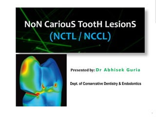
  • 1. 1 NoN CariouS TootH LesionS (NCTL / NCCL) Presented by: D r Ab h isek Gur ia Dept. of Conservative Dentistry & Endodontics
  • 2. 2 Contents  Introduction  Definition  Classification (Grippo) Atrition  Definition  Classification  Proximal surface attrition  Occlusal surface attrition  Etiology and Clinical features  Treatment modalities  The Dahl concept  Schools of thought to increase the V.D.  Endodontic considerations  Treatment strategies for Dentinal hypersensitivity  Nerve desensitization  Anti-inflammatory agents  Covering or plugging dentinal tubules  Restorative materials  Periodontal surgery  Lasers 5/11/2020 Management of Non-carious lesion
  • 3. 3 Management of Non-carious lesion Abrasion, Erosion, Abfraction  Definition  Etiology and pathogenesis  Clinical features  Treatment modalities  Monitoring of lesion progression  Restorative decision making  Perimolysis Fracture line/tooth crack  Etiopathogenesis  Classification  Characteristics  Distribution  Clinical diagnosis  Radiographic examination  Treatment modalities  Management of peg laterals  Management of palatogingival groove 5/11/2020
  • 4. 4 Management of Non-carious lesion  Localized Non Hereditary Enamel Hypoplasia  Localized Non Hereditary Enamel Hypocalcification  Localized Non Hereditary Dentin Hypoplasia  Localized Non-hereditary Dentin Hypocalcification  Amelogenesis Imperfecta  Dentinogenesis Imperfecta  Definition  Etiology  Clinical features  Treatment modalities  Conclusion  References 5/11/2020
  • 5. 5 Introduction Non carious tooth tissue loss (NCTL):  Surface loss due to a disease process other than dental caries. (Pual A Brunton ,Decision making in Operative Dentistry )  25% of tooth destruction does not originate from a carious process.(Marzouk) 5/11/2020 Management of Non-carious lesion
  • 6. 6 Etiology of NCTL 1. Attrition 2. Abrasion 3. Erosion 4. Abfraction 5. Localized Non- Hereditary Enamel Hypocalcification 6. Localized Non- Hereditary Dentin Hypolpasia 7. Localized Non- Hereditary Dentin Hypocalcification 8. Fracture lines 9. Amelogenesis imperfect 10. Dentinogenesis imperfecta 5/11/2020 Management of Non-carious lesion • John O Grippo,Marvin Simmering,JADA 2004 135;1109-1118 • Osborne-Smith KL, Burke FJ, Wilson NH. Int Dent J. 1999 Jun;49(3):139-43. Review
  • 7. 7 Attrition • Mechanical wear of the incisal or occlusal surface as a result of functional or parafunctional movements of mandible (tooth to tooth contacts) (Sturdevant) • Surface tooth structure loss resulting from direct frictional forces between contacting teeth. (Marzouk) • It also includes the proximal surface wear at the contact area because of the physiologic tooth movement 5/11/2020 Management of Non-carious lesion
  • 8. 8 Etiology 1. Bruxism 2. Developmental enamel defect 3. Physiologic 5/11/2020 Management of Non-carious lesion
  • 10. 10 A) Proximal surface attrition (proximal surface facets)  Results from surface tooth structure loss and flattening , widening of the proximal contact areas.  Mesiodistal dimension of the teeth is decreased, leading to drifting , with the possibility of overall reduction in the dental arch. 5/11/2020 Management of Non-carious lesion
  • 11. 11 B) Occluding surface attrition ( OCCLUSAL WEAR)  It is the loss ,flattening, faceting or reverse cusping of the occluding elements.  It leads to loss of vertical dimension of the tooth . A) If the LOSS IS SEVERE & accomplished in a relatively short time 5/11/2020 Management of Non-carious lesion No chance for the alveolar bone to erupt occlusally to compensate for the occlusal tooth loss Loss of VD overclosure during mandibular functional movements strain areas on stomato-gnathic system.
  • 12. 12 Management of Non-carious lesion B) If the loss occurs over a long period- • The alveolar bone can grow occlusally, bringing the teeth to their original occlusal termination. • Vertical dimension loss will be confined to teeth but not imparted to face. • Deficient masticatory capabilities • Cheek biting • Decay 5/11/2020
  • 13. 13 Clinical presentation • Flattened occlusal surfaces. • The degree of wear in both arches is normally equal. • Sometimes there may be presence of peripheral, ragged, sharp enamel edges . • The presence of hypertrophic masseter is a warning sign of the impact of bruxism . • TMJ problems can be elicited especially by the over closure situation 5/11/2020 Management of Non-carious lesion
  • 14. 14 A. When surface attrition is SLOWER & compensated by, intrapulpal deposition of secondary & tertiary dentin, then there will be no pulpal exposure. B. At other times, the attrition is faster than the intrapulpal dentine deposition, leading to direct pulpal exposure. 5/11/2020 Management of Non-carious lesion
  • 15. 15 Treatment modalities • Pulpally involved teeth →endodontic therapy /extraction depending upon their restorability . • Para functional activities ,( bruxism)-- be controlled with protecting occlusal splints. 5/11/2020 Management of Non-carious lesion
  • 16. 16 Management of Non-carious lesion  Myofunctional, TMJ/ any other symptoms in the stomato- gnathic system -----diagnosed and resolved (modifying the occlusal splint).  Occlusal equilibration : should be performed by : 1. Selective grinding of tooth surfaces that includes rounding and smoothening the peripheries of the occlusal tables. 2. By creating adequate overlap between the working inclines to prevent further cheek biting. 5/11/2020
  • 17. 17 Any exposed sensitive dentin should be protected and actual carious lesion be obliterated . Periodontium be examined and any pathology be treated . Restorative modalities can than be initiated. 5/11/2020 Management of Non-carious lesion
  • 18. 18 Management of Non-carious lesion  Restorations are only needed in the following situations:  Noticeable loss of vertical dimension  Or a progressive loss of tooth structure is observed compromising the tooth strength .  Caries ,if present  Defect contributes to a periodontal problem.  Worn tooth contour, (usually proximal ) which is not conducive to the maintenance of periodontium .  A tooth is cracked or endodontically treated. 5/11/2020
  • 19. 19 Procedure • Amount of V.D. lost is estimated . • It gives an estimate up to what should be the height of the worn clinical crowns be increased . Management of Non-carious lesion5/11/2020
  • 20. 20  The additional V.D. that the stomognathic system can accommodate without untoward effects is estimated.  Composite temporary restorations are most frequently used  Permanent restoration should be done in a cast alloy  These restorations should be cemented only temporarily for an extendedperiod of time ,until it is established that no untoward symptoms would occur. 5/11/2020 Management of Non-carious lesion
  • 21. 21  An acrylic splint( as a stabilization splint) may be necessary 5/11/2020 Management of Non-carious lesion
  • 22. 22 Restorative treatment  Tooth wear can be followed and re-evaluated during recall examinations.  Less severe anterior wear can be treated with adhesive composite resin. (Strassler HE, Kihn PW, Yoon R. Conservative treatment of the worn dentition with adhesive composite resin. Contemp Esthet Restor Pract. 1999) 5/11/2020 Management of Non-carious lesion
  • 23. 23 Management of Non-carious lesion When the wear is more severe, a number of treatment modalities are available. 1. Bonded porcelain veneers have been used to treat incisal wear. (Ibsen RL, Ouellet DF. Restoring the worn dentition. J Esthet Dent. 1992;4:96-101.) 1. In some cases, the incisal edges can be restored to the original vertical dimension with direct composite resin. (Strassler HE, Kihn PW, Yoon R. Conservative treatment of the worn dentition with adhesive composite resin. Contemp Esthet Restor Pract. 1999) 5/11/2020
  • 24. 24 Management of Non-carious lesion  Hemmings and coworkers reported on the restoration of severe anterior wear with composite restoration including re- establishment of the occlusal vertical dimension.  They reported a 89.4% success at 30 months. (Hemmings KW, Darbar UR, Vaughan S. Tooth wear treated with direct composite restorations at an increased vertical )dimension: results at 30 months. J Prosthet Dent. 2000;83:287-293. 5/11/2020
  • 25. 25 Management of Non-carious lesion  Adhesive cast metal restorations have also been used to replace missing tooth structure. ( Nohl FS, King PA, Harley KE, et al. Retrospective survey of resin- retained cast- metal palatal veneers for the treatment of anterior palatal tooth wear. Quintessence Int. 1997)  In cases where the occlusion is severely altered by attrition, the only treatment choice may be a reconstruction with crowns and bridges. (Stewart B. Restoration of the severely worn dentition using a systematized approach for a predictable prognosis. Int J Periodontics Restorative Dent. 1998;18:46-57.) 5/11/2020
  • 27. 27 The Dahl concept  Dahl and his coworkers (1975) described the use of a partialbite raising appliance‘ to create inter- occlusal space in an 18-year-old patient with severe localised attrition.  The removable appliance was cast in cobalt-chromium, placed on the palatal aspects of the upper anterior teeth, and worn 24 hours a day. 5/11/2020 Management of Non-carious lesion
  • 28. 28 Management of Non-carious lesion  The creation of inter- occlusal space significantly reduced the amount of tooth preparation required, especially on the already compromised palatal surface.  Teeth were restored with full coverage porcelain bonded crowns 5/11/2020
  • 29. 29 2 schools of thought to increase the V.D.  Addition of increments : gradual increments by progressively adding to the hard splint at 1mm /week, until the patient reaches the increased V.D. for restorative purposes----time consuming  The second approach ----taking the patient immediately to needed increase in V.D.----considerably lesser adjustments are made ,lesser time consuming Management of Non-carious lesion5/11/2020
  • 30. 30 Anterior Bite plane  Used in the reduction of overbite.  Occurs by altering the rate of eruption of posterior teeth relative to the eruption of lower incisors that are in contact with the bite plane.  Overbite reduction by this method --- most successful in actively growing patients 5/11/2020 Management of Non-carious lesion
  • 31. 31 Endodontic considerations In certain cases intentional endodontic therapy has to be performed  Hypererupted teeth  Pulpal involvement For location of calcified canal  Use of magnification  staining the pulp chamber floor with 1%methylene blue dye .  searching for canal bleeding points .  Performing the sodium hypochlorite ―champagne bubble test are helpful in locating calcified.  Long ,thin Ultrasonic tips can also be used 5/11/2020 Management of Non-carious lesion
  • 32. 32 Dentinal hypersensitivity Dentin Hypersensitivity is a condition characterized by short , sharp pain arising from exposed dentin in response to stimuli typically thermal , evaporative , tactile, osmotic or chemical and which cannot be ascribed to any other form of dental defect or pathology . ( Holland et al 1997) 5/11/2020 Management of Non-carious lesion
  • 33. 33 THEORIES 1. The transduction theory 2. Direct neural stimulation theory 3. The hydrodynamic theory
  • 34. 34 Treatment strategies for Dentinal hypersensitivity ( DCNA 53,2009; 47-60) 1. Nerve desensitization  Potassium nitrate 2 .Anti-inflammatory agents  Corticosteroids 3. Covering or plugging dentinal tubules • calcium hydroxide • sodium fluoride 5/11/2020 Management of Non-carious lesion
  • 35. 35 Management of Non-carious lesion • Sodium monoflourophosphate • Stannous fluoride • Oxalates • Strontium chloride  Protien precipitants  formaldehyde  glutaraldehyde • Flouride iontophoresis • Resins and Adhesives 4) Restorative materials 5) Periodontal surgery 6) Lasers 5/11/2020
  • 36. 36 Nerve desensitization  Potassium nitrate  5% concentration in a low abrasive tooth paste was able to desensitize dentin for up to 4 weeks compared to a control paste . (Tarbet et al )  In bio adhesive gels at a concentration of 5% and 10% has also been shown to be effective in reducing dentinal hypersensitivity .(Freschoso SC,Menendez M,2003) Management of Non-carious lesion5/11/2020
  • 37. 37 Anti inflammatory agents: Corticosteroids  glucocorticoids to the cavity preparation may reduce dentinal hypersensitivity by their effect on pain mediators  Lawson and Huff found that paramethasone had a significant desensitizing action.  Furseth and Mjor reported complete obturation of dentinal tubules after corticosteroid application to exposed dentin – reduce dentin permeability.  However there is a little experimental evidence to support or refute the use of such agents 5/11/2020 Management of Non-carious lesion
  • 38. 38 Sodium fluoride  Treatment of exposed root surface with fluoride toothpaste (1.1%) and conc. fluoride solutions(0.2%) is very efficient. (Minkow B,1975;Kerns D G 1991)  Mechanism- precipitated fluoride compound mechanically blocking the exposed dentinal tubules. (Tal et al) 5/11/2020 Management of Non-carious lesion
  • 39. 39 Sodium monoflourophosphate • Tooth pastes containing sodium monoflourophospshates have been shown to be effective in managing dentinal hypersensitivity. (Hernandez F,Mohammed C,1972) • Its mechanism of action is unclear (,Scherman A et al 1992) 5/11/2020 Management of Non-carious lesion
  • 40. 40 Stannous fluoride  Stannous fluoride in aqueous solution or in glycerin gelled with carboxymethylcellulose (Miller JT et al 1969)  Mode of action : induction of high mineral content which creates a calcific barrier blocking the tubular openings on the dentine surface . (Furseth R ,1970) 5/11/2020 Management of Non-carious lesion
  • 41. 41 Fluoride iontophoresis • It is the process of influencing ionic motion by an electric current and has been used as a desensitizing procedure in conjunction with sodium fluoride . (Mc Fall WT,1986)  Studies report that there is a immediate reduction in sensitivity after treatment with iontophoresis, but the symptoms gradually return over the next six months (Kern DA et al 1989 ) 5/11/2020 Management of Non-carious lesion
  • 43. 43 Oxalates  potassium oxalates have both tubule obturation properties and inhibitory effects caused by potassium ions on nerve activity (Pashly DH ,1986)  Oxalate ion reacts with calcium to form insoluble calcium oxalate crystals that bind tightly to dentin and obturate dentinal tubules (TrowbridgeHO,1990)  Three types of oxalates are available : 1. 6% ferric oxalate (Sensodyne Sealant ) 2. 30% di-potassium oxalate( Butler Protect ) 3. 3% monohydrogen monopotassium oxalate 5/11/2020 Management of Non-carious lesion
  • 44. 44 Strontium chloride  strontium ions have the capacity to reduce sensory nerve activity, but less effectively than potassium ions. (MarkowitzK , kim S 1990).  Dentifrices containing 10% strontium chloride (Sensodyne) - widely used as desensitizing agents and were one of the first agents to be marketed for that purpose. 5/11/2020 Management of Non-carious lesion
  • 45. 45 Protein precipitants (Formaldehyde and glutaraldehyde)  Have ability to precipitate salivary proteins in the dentinal tubules.  However this effect has been questioned since various formulations have been found to have little or no effect on dentinal hypersensitivity(Addy M,Mostafa P,1988) 5/11/2020 Management of Non-carious lesion
  • 46. 46 Resins and Adhesives • rationale – they seal the dentinal tubules and hence to preclude the transmission of pain stimuli to the pulpal nerve fibers. • This mode of treatment is performed on localized hypersensitive dentin. • Resin-based materials have been reported to successfully reduce dentinal hypersensitivity.(Kakaboura A ,2005) 5/11/2020 Management of Non-carious lesion
  • 47. 47 Management of Non-carious lesion  Copeland reported successful treatment of dentinal hypersensitivity for up to 18 months in 89% of hypersensitive teeth treated by Scotchbond.  5% glutaraldehyde and 35% hydroxyethyl methacrylate (Gluma Desensitizer) has been reported to be an effective desensitizing agent for up to 9 months. ( Kakaboura A ,2005) 5/11/2020
  • 48. 48 Bioactive glass:  NovaMin - for remineralisation of teeth, treating hypersensitivity.  The active ingredient is called Calcium Sodium Phosphosilicate.  NovaMin is an ionic form of calcium, phosphorus, silica, and sodium which are necessary for bone and tooth mineralization. 5/11/2020 Management of Non-carious lesion
  • 49. 49 Lasers  Nd:YAG laser irradiation in combination with 5% sodium fluoride varnish has higher efficacy in the management of DH than either treatment alone. (Kumar and Mehtas ,2005)  Slutzky-Goldberg (2008) demonstrated that CO2 laser treatment resulted in decreased permeability of dentinal tubules as shown by a dye penetration test. 5/11/2020 Management of Non-carious lesion
  • 50. 50 Restorative materials • The use of restorative materials is generally an invasive solution to the problem of hypersensitivity. • Commonly used materials include composite resins and glass ionomer restorations. 5/11/2020 Management of Non-carious lesion
  • 52. 52 Management of Non-carious lesion  Abnormal tooth surface loss resulting from direct frictional forces between the teeth and external objects or from frictional forces between contacting teeth components in the presence of abrasive medium. (Sturdevant 5 th edition)  It occurs most frequently on the cervical neck of the teeth.  The labial or buccal surfaces. (tooth brush abrasion )  Labial or buccal and lingual surfaces( in case of poorly fitted clasps and artificial dentures ) . 5/11/2020
  • 53. 53 Causes of abrasion : • Traumatic occlusion . • Improper brushing technique . • Occupational (Habits such as holding bobby pins in between the teeth .) • Tobacco chewing /tobacco pipe . • Vigorous use of tooth picks between the adjacent teeth. • Excessive mastication of coarse foods . 5/11/2020 Management of Non-carious lesion
  • 54. 54  Tooth brushabrasion results in a horizontal cervical notches on the buccal surfaces of exposed radicular cementum and dentin .  Notching in right central incisor caused by improper use of bobby pins . 5/11/2020 Management of Non-carious lesion
  • 55. 55 clinical signs and symptoms  The surface of the lesion is extremely smooth and polished and it seldom has any plaque accumulation or caries activity in it .  The surrounding walls tend to make a V shape ,by meeting at an acute angle axially.  Peripheries of the lesion are angularly demarcated from the adjacent tooth surface.  Probing or stimulating the lesion can elicit pain .  Hypersensitivity may be intermittent in character appearing and disappearing at occasional or frequently repeated periods . 5/11/2020 Management of Non-carious lesion
  • 56. 56 Treatment modalities  Diagnose the cause of the presented abrasion.  A detailed history is to be taken considering various factors such as: • Oral hygiene techniques ( use of abrasive tooth cleaning techniques and materials) • Habits- pipe smoking, chewing tobacco, professional habits • Iatrogenic causes,if any.  Avoidance or counteraction of the causes which may lead to its production.  Instituting proper oral hygiene measures.  incorporating correct method of tooth brushing . 5/11/2020 Management of Non-carious lesion
  • 57. 57 Management of Non-carious lesion  Have the habit of chewing tobacco ,toothpick , etc discontinued . If successful in breaking the habit proceed with the restorative treatment as planned.  Correcting or avoiding ill fitting metal clasps and dentures 5/11/2020
  • 58. 58  Abrasive lesions at non-occluding tooth surfaces should be: • Evaluated critically for the need for restoring them. • If the lesions are multiple, shallow( not exceeding 0.5 mm in dentin) and wide → no need to restore them . • If there is involvement of cementum / enamel only → no need to restore . • If lesion is wedge (V) shaped and exceeds 0.5 mm into dentin → restoration is performed . Management of Non-carious lesion
  • 59. 59 Management of Non-carious lesion  If restoration is not indicated for a lesion, then : 1. Edges of the defect should be eradicated to a smooth, non- demarcating pattern relative to adjacent tooth surface. 2. Tooth surface then should be treated by fluoride solution to improve caries resistance 5/11/2020
  • 60. 60 Restoring cervical abrasions  Indicated when 1. Caries ,if present . 2. Sensitivity is present. 3. Lesion is esthetically objectionable . 4. If the defect contributes to a periodontal problem 5. The area to be involved in the design of a removable partial denture. 6. When the depth of defect is found to be close to pulp 7. Or a progressive loss of tooth structure is observed compromising the tooth strength . 5/11/2020 Management of Non-carious lesion
  • 61. 61 Restorative materials : • Glass ionomer restorative material. • Resin modified glass ionomer. • Polyacid-modified resin composites. • Resin composites. 5/11/2020 Management of Non-carious lesion
  • 63. 63 Management of Non-carious lesion  High modulus restorative materials are unable to flex in the cervical regions when the tooth structure is deformed under occlusal load and ,therefore the restorative materials can be displaced from the cavity . (Heymann HO ,Sturdevant Jr ,Baynes S ,JADA,122(2) 41- 57 )  An intermediate material with reduced elastic modulus may function as a stress absorbing layer and improve marginal sealing . (Kemp-Scholte CM ,Davidsson,CL complete marginal seal of class V resin composite restorations affected by increased flexibility .JDR 1990 ;69:1240 -3 ) 5/11/2020
  • 64. 64 Management of Non-carious lesion  As a result materials with low elastic modulus such as : 1. Microfilled composites (Heymann and others ,1991 :Levitch and others ,1994 ) 2. Flowable resins (Unterbink ,Liebenberg ,1999: Li and others 2006 ) 3. Glass ionomer cements (Loguercio and others ,2003:Burgess and others ,2004) Have been used in restoring cervical lesions ,with the aim of absorbing the stresses generated during the polymerization shrinkage of composites and mechanical loading in which the teeth are subjected during function . 5/11/2020
  • 65. 65 Literature  Two-year clinical evaluation of four polyacid- modified resin composites and a resin-modified glass-ionomer cement (Vitremer) in Class Vabrasion/erosion lesions. (Ermiş RB.. Quintessence Int. 2002 Jul- Aug;33(7):542-8) Result • Retention levels at 2 years were 90% for F2000, 90% for Dyract AP, 89% for Compoglass F, 84% for Elan, and 95% for the Vitremer restorations. 5/11/2020 Management of Non-carious lesion
  • 66. 66 Discussion • GIC have an inhibitory effect on secondary caries, and the release of fluoride is considered to be one of the major benefits associated with GIC • Polyacid- modified resin composites may not be recharged again with fluoride as are GIC
  • 67. 67 Comparative analysis of techniques of restoring cervical lesions- (Quintessence Int 1993:24:553- 559.) • Aim - the sandwich technique and the GIC restoration, from the standpoint of marginal leakage. • While GIC may not have as good esthetic properties as resin materials • the sandwich technique does not have universal application, because it requires space : not the technique of choice in shallow cavities.
  • 68. 68 Result 1. The acid-etch technique was effective in reducing marginal leakage along the tooth - composite resin interface in enamel. 2. None of the techniques studied consistently provided a complete seal at the gingival aspect of cervical restorations. 3. Composite resin inserted over a GIC liner demonstrated significantly less leakage than when the liner was not used. 4. The use of the sandwich technique or GIC alone provided the most effective seals in cervical wedge-shaped cavities, compared to the all- composite restoration. 5. There was no significant difference between the marginal leakage of GIC and sandwich technique restoration
  • 69. 69 5-year clinical performance of resin composite versus RMGIC in non-carious cervical lesions. Franco Eb, Benetti A, SK Ishikiriama,SL Santiago ,JRP Lauris METHOD AND MATERIALS: One operator placed 70 restorations (35 RMGIC and 35 resin composite restorations) in 30 patients under rubber dam isolation. CONCLUSIONS : After 5 years of evaluation, the clinical performance of resin modified glass ionomer restorations was superior to resin composite restorations.
  • 71. 71 Erosion • Def- Loss of surface tooth structure by chemical action in the continued presence of demineralizing agents(acids). (Sturdevant- 5th edition) • There is no convincing etiology ,and multiple factors have been theorized for its pathogenesis: Mechanical factors: The action of the muscles of lips and cheeks , and of tooth brush against affected surfaces .
  • 72. 72 Chemical factors : • Ingested acids : citric acids esp. if use in large amounts , can precipitate or initiate erosive lesion • Secreted acids : the acidity of crevicular fluid has been correlated to cervical erosion (Bodecker CF. Local acidity: a cause of dental erosion- abrasion.Ann Dent 1945) • Acid fumes : acid vapours from nitric acid and sulphuric acids, acting in the mouths of workers in the factories ,where these acids are largely used or manufactured ( Miller) • Refused acids : as a result of chronic , frequent regurgitation ,the stomach‘s hydrochloric acid can hit the teeth at specific locations ( atypical pattern of erosion affecting buccal surfaces of lower posterior teeth)
  • 73. 73 Clinical presentation • Extensive loss of buccal and occlusal tooth structure • Raised amalgam restoratins. • Occlusal view of maxillary dentition exhibiting concave dentin depressions surrounded by elevated rims of enamel
  • 74. 74 • Multiple cupped out depressions corresponding to the cusp tips • Extensive loss of enamel and dentin on the Buccal surface of maxillary bicuspids. ( pt had sucked chronically on tamarinds )
  • 75. 75 Palatal surfaces of maxillary dentition in which the exposed dentin exhibits a concave surface and a peripheral white line of enamel • Perimylosis: decalcification of the teeth caused by exposure to gastric acid in patients with chronic vomiting, as may occur in anorexia or bulimia • Loss of lingual enamel and dentin due to acid regurgitation aggravated by circular movements of tongue. • Associated with stress reflux syndrome
  • 76. 76 • A similar appearance is found in patients with eating disorders- • Anorexia - immoderate food restriction and irrational fear of gaining weight, as well as a distorted body self-perception • Bulimia nervosa - consuming a large amount of food in a short amount of time followed by an attempt to rid oneself of the food consumed, typically by vomiting • Rumination - effortless regurgitation of most meals following consumption • Chronic alcoholism produces a similar pattern of erosion, although usually more generalized. • ( ND Robb and BGN Smith, Anorexia and bulimia nervosa (the eating disorders): conditions of interest to the dental practitioner, J Dent (1996)
  • 77. 77 • Any food substance with a critical pH value of less than 5.5 can become a corrodent and demineralize the teeth. ( Stephan RM, JADA 1940) ,( Gray JA, J Dent Res 1962) , (Zero DT. Cariology. Dent Clin North Am 1999) • Holding ,swilling or retaining acidic drinks and foods in the mouth prolongs the acid exposure on the teeth increasing the risk of erosion . (Mossazzez R ,Smith BGN,Barlett DW,Oral Ph and drinking habit during the ingestion of carbonated drink in a group of adolescents with dental erosion ,J Dent 2000)
  • 78. 78 • The corrosive potential of an acidic drink does not depend exclusively on its ph value, but also is strongly influenced by its buffering capacity of the acid and by the frequency and duration of ingestion. (Lussi A. Dental erosion: clinical diagnosis and case histor taking. Eur J Oral Sci 1996 )
  • 79. 79  The other substances that can corrode teeth.  Chewable vitamin C tablets  Aspirin tablets  Aspirin powders  Use of the amphetamine drug ecstasy  Have been associated with corrosion on the occlusal surfaces of posterior teeth.
  • 80. 80 The potential effects of pH and buffering capacity on dental erosion. Owens BM. Gen Dent. 2007 Nov-Dec;55(6):527-31 • This in vitro study evaluated five different soft drinks (Coca- Cola Classic, Diet Coke, Gatorade sports drink, Red Bull high- energy drink, Starbucks Frappucino coffee drink) and tap water (control) in terms of initial pH and buffering capacity. • Initial pH was measured in triplicate for the six beverages. The buffering capacity of each beverage was assessed by measuring the weight (in grams) of 0.10 M sodium hydroxide necessary for titration to pH levels of 5.0, 6.0, 7.0, and 8.3.
  • 81. 81 Mean ph values for each beverage • Coca cola • Diet Coke • Gatorade • Red Bull • Starbucks Frappucino • Tap water 2.49 3.12 2.93 3.24 6.59 7.12
  • 82. 82 • Red Bull had the highest mean buffering capacity (indicating the strongest potential for erosion of enamel), followed by Gatorade, Coca-Cola Classic, Diet Coke, and Starbucks Frappucino.
  • 83. 83 • Beverages with high buffering capacities compete with natural buffering characteristics of saliva and resist ph changes as a result . • Greater the buffering capacity , more time it takes for saliva to restore the pH value ,which causes beverage pH to decline to a sustained level --------- prolonged periods of oral acidity ---- thus increasing the erosive potential .
  • 84. 84 Monitoring tooth wear Most effective way to monitor wear is : • comparing the dated study casts to the clinical conditions of teeth over time . • It can also be used as a part of preventive regime .
  • 85. 85 • Active wear → smooth and unstained ,clean surfaces. → erosion of tooth around the existing restoration . (Restoration is resistant to acid ,remains unchanged ,but the tooth is gradually dissolved leaving the restoration proud) • Inactive wear — stained .
  • 86. 86  Diminish the frequency and severity of acid challenge. • ↓ the amount and frequency of acidic foods or drinks • Acidic drinks should be drunk quickly rather than sipped. • Use of straw reduces erosive potential  Treating the underlying medical disorder or disease. • GERD ,anorexia ,bulimia → refer to a physician /psychologists Protocol for the prevention of progression of erosion (Beatrice K,Edmond L ,J Contemp.Dental practise ,1999)
  • 87. 87  Enhance the defense mechanisms of body: • Saliva provides buffering capacity→ increases with salivary flow rate. • Saliva supersaturated with Ca, P → inhibits demineralization of tooth structure. • Stimulation of salivary flow → sugarless lozenge, candy/gum is recommended
  • 88. 88  Enhance acid resistance, remineralization and rehardening of the tooth surfaces. • Daily use topical flouride at home • Fluoride application in office- 2-4 times a year ,flouride varnish recommended.  Decrease abrasive forces. • Use a soft bristled toothbrush and brush gently.
  • 89. 89 • No brushing should be done immediately after consuming acidic food and drink as teeth will be softened. • Rinsing with water is better than brushing after consuming acidic foods and drinks. (Gandara, B.K; E.L Truelove ,Diagnosis and management of dental erosion. Journal of Contemp.Dental Practice 1999)
  • 90. 90
  • 91. 91 Improve chemical protection • Neutralize acids in mouth ---dissolving sugar free antacid tablets 5 times a day ,particularly after an intrinsic or extrinsic acid challenge. • Dietary components- hard cheese ( provides Ca and PO4), held in  mouth after acidic challenge.
  • 92. 92 Mechanical protection • By application of composites and direct bonding where appropriate – to protect exposed dentin • Occlusal guard /Acrylic splint in the form of stabilization splint necessary to protect dentition from further damage due to erosion . Management of Non-carious lesion
  • 93. 93  Monitor stability • by use of casts /photos to document tooth wear status. • Regular recall examinations to review diet, oral hygiene methods, compliance with medications, topical flouride and splint usage.
  • 94. 94 Restoration • Metallic restorations should be the choice of material ,if restoration indicated . (more resistant to erosion ) • Tooth colored materials may also be used with minimal or no tooth preparation, with the assumption that restoration may require periodic replacement .
  • 96. 96 NoN CariouS TootH LesionS (NCTL / NCCL) Presented by: D r Ab h isek Gur ia Dept. of Conservative Dentistry & Endodontics SHDCH , Hassan Sem - 14 Date of Presentation : 26/02/T20
  • 97. 97 Contents  Introduction  Definition  Classification (Grippo) Atrition  Definition  Classification  Proximal surface attrition  Occlusal surface attrition  Etiology and Clinical features  Treatment modalities  The Dahl concept  Schools of thought to increase the V.D.  Endodontic considerations  Treatment strategies for Dentinal hypersensitivity  Nerve desensitization  Anti-inflammatory agents  Covering or plugging dentinal tubules  Restorative materials  Periodontal surgery  Lasers 5/11/2020 Management of Non-carious lesion
  • 98. 98 Management of Non-carious lesion Abrasion, Erosion, Abfraction  Definition  Etiology and pathogenesis  Clinical features  Treatment modalities  Monitoring of lesion progression  Restorative decision making  Perimolysis Fracture line/tooth crack  Etiopathogenesis  Classification  Characteristics  Distribution  Clinical diagnosis  Radiographic examination  Treatment modalities  Management of palatogingival groove 5/11/2020
  • 99. 99 Management of Non-carious lesion  Localized Non Hereditary Enamel Hypoplasia  Localized Non Hereditary Enamel Hypocalcification  Localized Non Hereditary Dentin Hypoplasia  Localized Non-hereditary Dentin Hypocalcification  Amelogenesis Imperfecta  Dentinogenesis Imperfecta  Definition  Etiology  Clinical features  Treatment modalities  Conclusion  References 5/11/2020
  • 100. 100 Abfraction  Def- The pathological loss of tooth substance caused by the biomechanical loading forces that result in flexure and failure of enamel and dentin at a location away from the loading.(Grippo)  Some authors explain the formation of cervical, wedge shaped defect by the heavy force in eccentric occlusion resulting in flexuring (elastic bending) of the tooth.
  • 101. 101  Loss of tooth surface at the cervical areas caused by tensile and compressive forces during tooth flexure Milosevic, 1998 5/11/2020 Management of Non-carious lesion
  • 103. 103  The size and the development of abfractive lesions are proportional to the direction, magnitude, frequency, duration, and location of the static and cyclic loading forces causing the stress. 5/11/2020 Management of Non-carious lesion
  • 104. 104 Management of Non-carious lesion  Enamel prisms and orientation of dentinal tubules.  The cervical region depicts parallel prisms and tubules, which result in less resistant dental tissue than in the occlusal/incisal regions. 5/11/2020
  • 105. 105  Stress-strain pattern by finite element analysis.  Stress distribution was performed using maximum principal stress, identifying areas with tensile stress (positive) and/or compressive stress (negative). 5/11/2020 Management of Non-carious lesion
  • 106. 106  Microfractures can foster loss of tooth structure from  tooth brush abrasion  from acids in the diet or plaque or both .  The resulting defect has a smooth surface .  Also known as idiopathic erosion. (Lee WC, Eakle WS, J Prosthet Dent 52(3): 374 -380, 1984.)  Stresses that concentrate to produce abfractions in teeth usually are transmitted by occlusal loading forces. ( Whitehead SA, Wilson NHF, Watts DC. J Esthet Dent 2000),(Pintado MR, DeLong R, Ko C, Sakaguchi RL, Douglas WH. Correlation J Prosthet Dent 2000)  Occlusal interferences, premature contacts, habits of bruxism and clenching all may act as stressors.
  • 107. 107 Clinical features  The lesion is typically wedge shaped with sharp line angles  Affects buccal/labial cervical areas of teeth  Deep, narrow V-shaped notch  Commonly affects single teeth with excursive interferences or eccentric occlusal loads
  • 108. 108 Types 1. Hairline Abfractions 2. Striation Abfractions 3. Saucerized Abfractions 4. Semilunar Abfractions 5. Cusp tip invagination Abfractions Grippo. J of Esth Dent. 1991 Management of Non-carious lesion
  • 109. 109 Treatment modalities Passive treatment Active treatment Passive treatment  Monitoring  Prevention The form of the preventive treatment will be dependent on the aetiology 5/11/2020 Management of Non-carious lesion
  • 110. 110 Active treatment  Restorative – for Sensitivity, Aesthetics, Function,  Orthodontic – for Aesthetics, Function  Prosthodontic – for Aesthetics, Function, Space loss in the vertical dimension. 5/11/2020 Management of Non-carious lesion
  • 111. 111 Monitoring abfraction lesions • A novel method of determining the activity of abfraction lesions over time ----Scratch test . ( Kaidonis JA.The tooth wear :view of anthropologists ,Clin Oral Investig 2008) • A no.12 scalpel blade is used to superficially scratch the tooth surface . • Visual observation gives an indication of rate of tooth structure loss • Loss of scratch definition or loss of the scratch altogether signifies active tooth structure loss.
  • 112. 112 Restoration • When clinical consequences (e.g. dentin hypersensitivity ) have developed or likely to be developed . • Aesthetics demands are a concern .
  • 113. 113 Tyas recommended the RMGIC should be the first preference (Tyas MJ,the class V lesion –aetiology ,restoration,Aust. Dental Journal.1995) • In esthetically demanding cases, • RMGIC/GIC liner laminated with resin composite. Vandelwalle and Vigil ( Gen Dent 1997) • Recommended the use of microfilled resin composite(low modulus of elasticity ) as it will flex with tooth and not compromise retention .
  • 114. 114 Occlusal adjustments  Occlusal adjustment may involve (Piotrowski BT JADA 2001 and Ichim IP Dent Mater 2007): • Altering cuspal inclines, • Reducing heavy contacts • Removing premature contacts.  Occlusal splints Aimed at reducing the amount of nocturnal bruxism and non axial tooth loading when constructed properly
  • 115. 115 Fracture lines The cause of fractures may include : i. Physical trauma ii. Occlusal prematurities iii. Repetitive heavy and stressful chewing iv. Resorption weakened teeth v. Iatrogenic dental treatment  It has been suggested that the determination of a fractured tooth is often more of a prediction rather than a definitive diagnosis based on a collective analysis of subjective and objective findings.
  • 116. 116 • Five types of longitudinal fractures have been described (American Association of Endodontists,2008 ): (1) Craze line:  Affect only the enamel,  originate on the occlusal/labial surface  typically from occlusal forces and are asymptomatic .
  • 117. 117 2) Fractured cusp:  occur on the cusps and cervical margins of the root  can have acute pain to mastication and cold. 3) Cracked tooth :  occurs on the crown and may extend into the root ,  develop from damaging occlusal forces or weakened tooth structure
  • 118. 118  (4) Vertical root fracture:  occur and originate only in the roots,  have variable but a lesser degree of signs and symptoms  caused by wedging forces within the roots (i.e. root canal obturation or posts)
  • 119. 119 5) Split tooth:  a fracture through the crown and roots,  developing from damaging occlusal forces or weakened tooth structure,  separating the tooth into two segments, with the tooth typically being painful to mastication
  • 120. 120 • Cracked teeth are thought to occur as a result of parafunctional habits or from weakened tooth structure • The symptoms that develop subsequent to these cracks have been termed as ―cracked tooth syndrome • acute pain that results during the mastication (or release) of small hard food substances and also exacerbates with cold. (Cameron CE,JADA,1964 : American Associationof Endodontists,2008) • However, the signs and symptoms of a cracked tooth may also be consistent with an irreversible pulpitis or necrosis.
  • 121. 121 two main groups of cracked teeth : A) Tooth infarctions: (incomplete tooth fractures extending partially through a tooth ) that includes :  Craze lines  Cuspal fractures  Cracked teeth B) Vertical root fractures : (that occur in endodontically treated teeth )
  • 122. 122 Characteristics of tooth infarctions Problems in diagnosis : • Infarctions typically originate internally and extend peripherally → not likely to be identified by percussion until the fracture extends to involve the periodontal ligament . • The fractures are incomplete, tend to present in a mesial distal orientation and are generally centered on the occlusal table ,radiographs are not very diagnostic .
  • 123. 123 • Also difficult to differentiate masticatory pain /pain from infarction /pain from microleakage associated with restorations . • Infarctions ….not readily visualized without magnification (unless they are at least 20um ) 5/11/2020 Management of Non-carious lesion
  • 124. 124 Distribution  Molars and premolars are the teeth are almost exclusively involved  Teeth with restorations are most likely to develop infarctions
  • 125. 125 Pain characteristics • Occurs when there is release of pressure from biting (rebound pain or relief pain ) • Can be duplicate diagnostically by having the patient bite on a moist cotton roll
  • 126. 126 Etiology • Excessively large and incorrectly designed restorations . • Use of pins for supporting large restorations ,esp. self threading and friction locked ) • Abrasion ,erosion ,caries ,along with age changes in dentin.
  • 127. 127  Biting onto hard objects ,bruxism and clenching , wedging effect of the cusp in the opposing fossa  Use of both high speed handpieces and course diamond burs can lead to infarctions  Acute trauma to the teeth.
  • 128. 128 Clinical examination • Begins with the chief complaint i.e. pain on chewing ,elevated sensitivity to cold food and sweets • Absence of carious etiology …trigger a suspicion of infarction • Visual examination by …….Transillumination and …..Dyes (methylene blue) • Any existing restoration in the tooth should be removed to reveal the infarction lines
  • 129. 129  Removal of the restoration and highlighting with the dyeto detect infarction  Use of optic light source to identify an infarction .  Note that the beam of light does not cross the infarction
  • 130. 130 Biting test : • Biting on rubber wheels, cotton tip applicators ,moist cotton, commercial biting appliances like tooth slooth. • Tooth slooth …….. Differentiates biting pain from restorations with microleakage /pain from infarction……pressure is placed first onto the restoration followed by tooth cusps .
  • 131. 131  teeth with infarctions respond to a lower threshold to cold and EPT as compared to the non cracked teeth.  Cameron suggested the use of thin sharp explorer tip to probe around the cervical circumference of the suspected teeth….the click of the explorer‘s tip and the patient‘s response can provide a clue
  • 132. 132 Radiographic examination  radiographs are not very diagnostic .  Even cone beam volumetric tomography (CBVT) scans cannot consistently visualize these fractures, the coronal- apical progression of fractures cannot always be objectively assessed until the tooth has been extracted.
  • 133. 133 Treatment  Aims :  Preventing the separation of the hard tissue entities ,  Keeping the bacteria's from colonizing the space caused by infarction It is not clear whether all the teeth with infarction require root canal therapy, it depends on the extent of the fracture .
  • 134. 134  Orthodontic band was placed to bind the crown together .  After 3 weeks the tooth was completely asymptomatic and the patient chose to restore it with a crown
  • 135. 135 Localized Non Hereditary Enamel Hypoplasia 5/11/2020 Management of Non-carious lesion DEFINITION CAUSE Clinical presentation Treatment During enamel formation , AMELOBLASTS are injured/irritated ,their metabolic product i.e enamel matrix, would not be properly formed resulting in formation of either hypoplastic or hypomineralized enamel When the teeth erupt,these defects will be apparent in the crown portion of teeth (tooth) which is called as localized non hereditary enamel hypoplasia a) Systemic disorders b) Localized disorders c) Fluorides • Isolated pits to widespread linear defects, • depressions, or loss of a segment in the enamel . In contrast with the caries and erosion and abrasion lesions, Enamel hypoplasia does not Progress a) Systemic disorders Exanthematus diseases NutritionaL Deficiencies (especially vitamins A,C and D) Hypocalcemia Microbial process e.g . (syphilis) These defective areas will have different color from the surrounding enamel If defects are minimum ( narrow lines /isolated pits /shallow depressions) - then selective odontomy/esthetic reshaping can be performed .
  • 136. 136 Management of Non-carious lesion 5/11/2020 CAUSE Clinical presentation Treatment b) Localized disorders- These include periapical infections of the preceding deciduous tooth (Turner‘s hypoplasia ), traumatic intrusion of the preceding deciduous tooth etc If odontomy and esthetic reshaping of the tooth enamel can‘t produce a pleasing functional effect, then Direct tooth colored resinous material (composite material) is inserted with /without tooth preparationc) Fluorides : Metabolizing fluorides in excessive amounts could poison the ameloblasts and disturb their activities to variable degrees, leading too slightly mottled enamel or a completely disfigured crown in its enamel
  • 137. 137 Localized Non Hereditary Enamel Hypocalcification 5/11/2020 Management of Non-carious lesion DEFINITION CAUSE Clinical presentation Treatment Hypomineralized enamel results when normal amount of enamel matrix fails to achieve full mineralization and is a usual consequence of damage to ameloblasts 1.Childhood fever, 2.Trauma / Flourosis During developmental stages of tooth formation 1.Appear chalky 2.soft to indentation. 3.Stainable. 4. If extensive- these Lesions predispose to attrition and abrasion. 5. Enamel chipped if lesion involves the entire surface If diagnosis is made early in tooth‘s life ,while the uncalcified enamel is still intact an attempt at remineralization should be made . Can be done using- A)Fluoride applications B)Fluoride iontophoresis C)strict prevention of plaque accumulation in these areas .
  • 138. 138 Treatment  Vital bleaching  Laminated veneering  Composite  Crowns 5/11/2020 Management of Non-carious lesion
  • 139. 139 Localized Non Hereditary Dentin Hypoplasia DEFINITION CAUSE Clinical presentation Treatment Odontoblasts are the specialized cells ,any disturbance in their function deficient Or complete absence of dentin matrix Deposition Leads to the development of localised non-hereditary Dentin hypoplasia It appears to be a hereditary disease, transmitted as an Autosomal dominant characteristics There would be NO apparent destruction to be diagnosed or treated ,till the time the lesion is covered with enamel Various intermediary bases that can be used are : Zinc oxide eugenol Calcium hydroxide Zinc phosphate Cement Polycarboxylate cement Varnishes Glass ionomer cement During tooth preparation for a restoration , these defects may get exposed 5/11/2020 Management of Non-carious lesion
  • 140. 140 LOCALIZED NON-HEREDITARY DENTIN HYPOCALCIFICATION 5/11/2020 Management of Non-carious lesion DEFINITION CAUSE Clinical presentation Treatment In hypocalcification, there is failure of union of many globules, the dentin will be present in substance ,but would be softer ,more penetrable , and less resilient a) Systemic disorders b) Localized disorders c) Fluorides There would be NO apparent destruction to be diagnosed or treated ,till the time the lesion is covered with enamel Various intermediary bases that can be used are : Zinc oxide eugenol Calcium hydroxide Zinc phosphate Cement Polycarboxylate cement Varnishes Glass ionomer cementDuring tooth preparation for a restoration , these defects may get exposed
  • 141. 141 Amelogenesis Imperfecta  Amelogenesis – enamel formation; imperfecta – improper
  • 142. 142 Amelogenesis Imperfecta  Relatively rare group of inherited disorders characterized by abnormal enamel formation  AI can be inherited as an x-linked, autosomal recessive (AR), or Autosomal dominant (AD) condition.  AI classified into four main types based on the enamel defects
  • 143. 143 Types Type Clinical Appearance Enamel Thickness Radiographic Appearance Inheritance Hypoplastic (Type I) Crowns size varies from small to normal, small teeth may lack proxmial contacts, color varies from normal to opaque white – yellow brown Varies from thin and smooth to normal thickness with grooves, furrows and/or pits Enamel has normal to slightly reduced contrast/ thin Autosomal dominant, recessive, or X- linked Hypomaturation (Type II) Varies from creamy opaque to marked yellow/brown, surface of teeth soft and rough, dental sensitivity Normal thickness with enamel that often chips and abrades easily Enamel has contrast similar to or more than dentin, unerupted crowns have normal morphology Autosomal dominant, recessive, or X- linked
  • 144. 144 Types Type Clinical Appearance Enamel Thickness Radiographic Appearance Inheritance Hypocalcified (Type III) Opaque white to yellow-brown, soft rough enamel surface, dental sensitivity and open bite common, heavy calculus formation common Normal thickness with enamel that often chips and abrades easily Enamel has contrast similar to or less than dentin, unerupted crowns have normal morphology Autosomal dominant, recessive Hypomaturation/ Hypoplasia/ (Type IV) White/Yellow- Brown mottled, teeth can appear small and lack proximal contact Reduced, hypomineralized areas and pits Enamel contrast normal to slightly > dentin, large pulp chambers Autosomal dominant
  • 145. 145 Management of Hypoplastic AI Types  involves the use of bonding procedures to protect the malformed teeth from caries and improve esthetics.  Hypoplastic teeth usually have reasonably well mineralized enamel, pitted, making them suitable for restorative therapies involving bonding to the enamel
  • 146. 146  Orthodontic therapy may be used to partially close the interdental spaces prior to restoration  Composite resin or porcelain veneers can be bonded to the anterior teeth when the incisor shape, size and/or color requires modification.  However, if the enamel is extremely thin and malformed the teeth can require full dental coverage with crowns.
  • 147. 147 Treatment of Hypomaturation and Hypocalcified AI Types  full coverage restorations should be placed.  Esthetic anterior restorations can be made using a variety of techniques.  Open face stainless steel crowns with composite inserts  composite restorations can greatly reduce tooth sensitivity and provide reasonable esthetics.
  • 148. 148  Resin crowns can be placed on permanent incisors soon after they begin to erupt during the mixed dentition (about age 7 – 10 years).  As the gingival margin becomes exposed during continued tooth erupt the resins are easily modified by adding resin to the gingival margin of the tooth.  Ultimately, custom fabricated crowns can be placed on the dentition.
  • 149. 149 Dentinogenesis imperfecta  It is a hereditary mesodermal defect  It may be associated with “Osteogenesis imperfecta”, also a mesodermal defect.  There is a wide range of difference in manifestation of the lesions or defects.  It is classified in 3 types
  • 150. 150 Types  Type I – Dentinogenesis imperfecta with Osteogenesis Imperfecta  Autosomal dominant trait  Clinical appearance varies greatly  Type II - Dentinogenesis imperfecta without Osteogenesis imperfecta  Also called as hereditary opalescent dentin  Autosomal dominant trait  Most common dominantly inherited disorder  Type III - Brandywine type  This is a racial isolate in Maryland  Inherited as autosomal dominant trait
  • 151. 151 Types Type Clinical Appearance Radiographic Appearance Type I Clinical appearance varies greatly Deciduous teeth are mostly affected Partial or precocious obliteration of the pulp chamber Type II Both dentitions are equally effected Partial or precocious obliteration of the pulp chamber Type III Both dentitions are involved Variable Changes similar to type I and II In Brandywine population characteristic “Shell teeth” are present
  • 152. 152 Clinical features  Colour - gray to brownish violet or yellowish brown  translucent or opalescent hue  Enamel may be lost early through fracturing away because of abnormal DEJ  With the early loss of enamel the abnormal dentin undergoes rapid attrition  Teeth are prone for fracture
  • 153. 153 Histological feature  The appearance of enamel is normal  The pulp chamber is usually obliterated by the continuous deposition of dentin.  The dentin is composed of irregular tubules,  Dentinal tubules are larger in diameter and less in number  Some times tubules may be absent  Dentin often shows large areas of uncalcified matrix
  • 154. 154  The water content is greatly increased, as much as 60% above normal  Inorganic content is less than that of normal dentin  Micro-hardness of dentin closely corresponds to that of cementum
  • 155. 155 Radiographic Features  The Type I and Type II diseases are radiographically similar and they often exhibit “bulb shaped” or “bell shaped” crowns of the teeth with abnormally constricted cervical areas.  The roots of the teeth are thin and spiked.  varying degrees of obliteration of the coronal as well as the radicular pulp chamber.  The cementum, Pdl and the alveolar bone radiographically appear normal.
  • 156. 156  The type III - similar to those of the type I and type II,  although in many cases the affected teeth exhibit extremely large pulp chambers surrounded by a thin shell of dentin and enamel.  Because of their typical appearance the affected teeth are often called “shell” teeth.  These teeth frequently exhibit multiple pulp exposure and associated periapical pathology.
  • 157. 157 Management  Any possible success for treatment depends upon early di- agnosis and care.  Only two treatment modalities : selective odontotomy and permanent full veneering.  Metal and ceramic crowns are given.  These teeth are not suitable candidates for playing the role of abutments for any bridge work since the roots are small and they also tend to fracture under frictional stress.  In case of severe generalized attrition, complete denture prosthesis may be necessary.
  • 159. 159 Palatogingival groove (PGG)  Radicular groove (1908): A developmental anomaly that begins near the tooth cingulum and extends from the CEJ in apical direction along the root with a wide range of depths and lengths. (Black) 5/11/2020 Management of Non-carious lesion
  • 160. 160 History  1958, Oehlers first reported radicular invagination of a maxillary lateral incisor in a Chinese female  1968, Lee et al. proposed the term PGG 5/11/2020 Management of Non-carious lesion
  • 161. 161 Other names  Distolingual groove,  Corono-radicular groove,  Radicular lingual groove,  Vertical developmental radicular groove,  Cingulo-radicular groove,  Developmental radicular anomaly,  Interruption groove,  Palatoradicular groove 5/11/2020 Management of Non-carious lesion
  • 162. 162 Etiology a) consequence of an alteration in growth, such as an infolding of the inner enamel epithelium and epithelial sheath of Hertwig, b) variant of dens invaginatus c) alteration of a genetic mechanism d) attempt to form another root. 5/11/2020 Management of Non-carious lesion
  • 163. 163 Clinical charracteristics  Prevalance -2-18%  Commonly lateral incisor  high prevalence in Sino-Americans and low prevalence in Sub- Saharan Africans and Sahul-Pacific people  Clinically, patients with a pathologic lesion related to PGG often complain of dull intermittent or acute pain,  mobility of the teeth, pain on percussion,  Pus discharge, sinus tract formation and gingival swelling 5/11/2020 Management of Non-carious lesion
  • 164. 164 Management of Non-carious lesion  They can also present with no symptoms.  In most cases, the patient had no history of dental caries, trauma, or discoloration of the teeth.  Pulp vitality was retained or lost contingent upon pulpal nerve involvement.  Cases with advanced lesions along deep grooves frequently show no response to thermal or EPT  Funnel shaped hollow grooves with an accumulation of plaque and calculus, along with loss of epithelial attachment, pocket formation, and bleeding on probing.  Observed bilaterally in the oral cavity. 5/11/2020
  • 166. 166 Relationship between PGG and periodontal/pulpal health  Hypothesis 1-  Al-Rasheed et al. studied the association between PGG and periodontal health by investigating the plaque index (PI), bleeding index (BI), and pocket depth (PD)  They reported that teeth with an apical groove showed PI and BI of 100%;  In several case reports, PGG cases presented as a combined type of primary periodontal lesion with secondary involvement of the pulp 5/11/2020 Management of Non-carious lesion
  • 167. 167 Management of Non-carious lesion  Hypothesis 2  some authors have insisted that the pulpal and periodontal lesions are unrelated with different etiologies that simply coincide in time.  the root apex, accessory foramina, lateral canals, and dentinal tubules as possible communication channels between periodontal defects  pulpal tissues, and speculated that bacteria and necrotic debris from a primarily infected root canal could extend into the apical base of the fissure groove with subsequent coronal progression along the groove. 5/11/2020
  • 168. 168  Hypothesis 3  irritants and microorganisms from the periodontal pocket could progress along the hollow, funnel-shaped PGG surface, advancing to the periodontal breakdown and root surface contamination  resulting in retrogenic pulp necrosis even though PGGs do not reach the apex and are not very deep. 5/11/2020 Management of Non-carious lesion
  • 169. 169 Diagnosis  Radiographically, teardrop-like radiolucency can be observed in cases with primary pulpal infection and pear shaped radiolucency can be observed in the coronal aspect with apical periodontal widening 5/11/2020 Management of Non-carious lesion
  • 170. 170  CBCT can provide accurate, sensitive information to assess and plan the treatment 5/11/2020 Management of Non-carious lesion
  • 171. 171 Treatment of PGG 1. Basic principle for treatment a) complete eradication of microbials b) permanent and thorough sealing of the root groove that communicates between the root canal and the periodontium c) periodontal regeneration and complete healing of the periodontium. 5/11/2020 Management of Non-carious lesion
  • 172. 172 Treatment interventions needed in cases of relatively shallow PGG (Kerezoudis et al 2003) 1. surgical removal of granulation tissue and irritants 2. a gingivectomy and apically positioned flap 3. surgical exposure and flattening of the groove by grinding, with or without application of guided tissue regeneration 4. positioning a restoration in the groove 5. orthodontic extrusion of the tooth. 5/11/2020 Management of Non-carious lesion
  • 173. 173 2. Treatment of mild PGG  ‘Saucerization’ to the level of the crestal bone with a rotary cutting and polishing instrument 5/11/2020 Management of Non-carious lesion
  • 174. 174 3. Treatment of complex PGG  granulation tissue removal through a flap,  Saucerization with or without the guided tissue regeneration,  Intentional extraction of a problematic tooth to achieve complete removal of the groove and subsequent reimplantation (intentional replantation), 5/11/2020 Management of Non-carious lesion
  • 175. 175 4. Various regenerative materials currently used to fill the groove  Dragoo et al. indicated that any subgingival restoration material should include the following characteristics: (i) biocompatibility, (ii) a dual-cure set, (iii) adhesiveness, (iv) radiopacity, (v) compactness, (vi) surface hardness, (vii) insolubility in oral fluids, (viii) absence of microleakage, (ix) low coefficient of thermal expansion, and (x) low cure shrinkage.  In addition, convenience of use and rapid setting that does not interfere with hemostasis are important properties in clinical circumstances 5/11/2020 Management of Non-carious lesion
  • 176. 176  Several materials, including amalgam, glass ionomer cement (GIC), composite resin, and calcium silicate based-cement such as mineral trioxide aggregate (MTA), have been selected for PGG treatment. 5/11/2020 Management of Non-carious lesion
  • 177. 177 5. Various regenerative materials currently used to fill intrabony defects  GTR membrane  Bone graft  PRF  enamel matrix derivative (EMD, Emdogain, Straumann, Basel, Switzerland) 5/11/2020 Management of Non-carious lesion
  • 178. 178 Prognosis Depends on several factors:  The location of the groove,  severity of the accompanying periodontal disease,  accessibility to the defect area, and  type of groove (shallow/deep or long/ short) 5/11/2020 Management of Non-carious lesion
  • 179. 179 Management of Non-carious lesion 5/11/2020
  • 180. 180 Conclusion  Depending on the degree of tooth wear, restorative treatment can range from placement of bonded composites in a few isolated areas of erosion, to full mouth reconstruction in the case of the devastated dentition.  Regardless of the type of restorative therapy provided, prevention of the progression of tooth wear should be the basis of management.  This will increase the likelihood of successful, long-term outcomes of the restorative treatment.  The patient should be monitored at regular intervals to determine compliance and success of treatment.
  • 181. 181 References 1. Art and science of operative dentistry- Sturdevant 5 th edition 2. Operative Dentistry- Modern Theory and Practice: Marzouk 3. Shafer‘s Textbook of Oral Pathology- Shafer, Hine, Levy 4. Abfraction : separating fact from fiction --ADJ 2009 5. Fracture necrosis :Diagnosis ,Prognosis assesment and Treatment Recommendations – Louis H Berman,Sergio Kettler 6. Non carious cervical lesions and abfractions :A re – evaluation –JADA 2003 ;134:845—850 7. Role of erosion in tooth wear :aetiology ,prevention and management ---IDJ(2005) 55,277- 284 8. Erosion –Chemical and biological factors of importance –IDJ (2005 ) 55 285-290 9. Removable Orthodontic Appliances—K.G.Isaacson 10. Quintessentials 3 – Decision–Making in Operative Dentistry Brunton, Paul A.

Editor's Notes

  1. Vd in rest, vd in occlusion Freeway space
  2. In this approach, space is created by placing restorations intentionally ‗high‘ – i.e. in supra-occlusion – allowing axial tooth movement that, over time, re-establishes complete occlusion.
  3. Overbite reduction should be evident within first two months of fitting the appliance . It is important to increase the thickness of bite plane slowly with progressive additions of cold cure acrylic as the overbite reduces .
  4. Red line test and white line test
  5. Pica syndrome
  6. Meaning “Breaking away”
  7. (EMD, Emdogain, Straumann, Basel, Switzerland) is to emulate this process and facilitate the role of acellular cementum in the formation and repair mechanisms of the periodontal ligament and alveolar bone.33 Hammarström et al. reported the actual formation of acellular cementum with EMD.60 EMD has also been used to prevent external root resorption and ankylosis onto the bone in cases of intentional replantation