Quick study urogenital system
•
5 likes•1,555 views

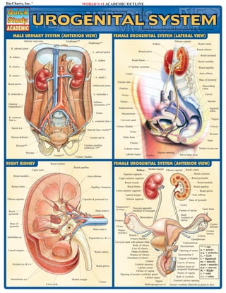
1. The document provides diagrams and labels of the male and female urinary and urogenital systems, including the kidneys, ureters, urinary bladder, and other associated organs. 2. Key structures of the male system shown include the testes, epididymis, vas deferens, seminal vesicles, prostate, and penis. The female system depicts the ovaries, uterus, uterine tubes, vagina, and associated organs and tissues. 3. Also shown are diagrams of the nephron, the functional unit of the kidney, and its role in filtering blood to form urine.

Recommended
Recommended
More Related Content
Featured
Featured (20)
Quick study urogenital system
- 1. BarCharts, Inc. ® WORLD’S #1 ACADEMIC OUTLINE MALE URINARY SYSTEM (ANTERIOR VIEW) FEMALE UROGENITAL SYSTEM (LATERAL VIEW) Inferior vena cava Esophagus** Fibrous capsule Diaphragm** Kidney Renal cortex R. adrenal gland Renal column L. adrenal gland Renal pelvis Renal pyramid R. kidney L. kidney Renal hilum Renal medulla R. renal a. 3rd lumbar vertebrae Renal papillae L. renal a. Ureter Area cribosa R. renal v. L. renal v. Base of pyramid Uterine tube Renal pelvis Abdominal aorta Descending Fimbria colon R. testicular a. L. testicular v. Ovary Cervix Inferior Uterus Sacrum Anterior mesenteric a.** longitudinal l. Endometrium Sigmoid colon Ureter Myometrium R. common iliac a. Cervical canal Vagina L. common iliac v. Urinary bladder Coccyx Sacral n.n. Internal iliac vessels** Urine Rectum Ductus deferens Levator ani m. Pubic bone Anus Clitoris Rectum** Urachus (median umbilical l.)** Labium minus Tendon levator ani Prostate Vaginal opening Urethra** Labium majus Urethra Pelvic bone m.m. Urinary bladder RIGHT KIDNEY Renal column FEMALE UROGENITAL SYSTEM (ANTERIOR VIEW) Renal papillae Upper pole Kidney Medial margin Fibrous capsule Renal cortex Superior segment Renal columns Renal medulla Area cribrosa Upper anterior segment Renal pyramids Renal vessels Renal medulla Renal cortex Papillary foramina Renal hilum Renal papillae Lower anterior segment Renal pelvis Lateral margin Area cribosa Fibrous capsule Capsular & perirenal a.a. Inferior segment Base of pyramid Ureter Suspensory l. of ovary Vesicula appendix Tubal folds Renal Main renal a. chydatid of morgagni Fundus of uterus pyramids Body Mesosalpinx (corpus) of Fimbria uterus Base of Corpus pyramid Ovary albicans Main renal v. Proper Corpus ovarian l. luteum Round l. Follicle Interlobar a.a. Segmental a.a. & v.v. Urinary bladder Epoöphorum Cervical canal with palmate folds Endometrium Body of clitoris Myometrium ** = cut Crus of clitoris Lateral margin Glans of clitoris Opening of ureter a. = artery Renal calices a.a. = arteries Prepuce of clitoris Sacrouterine l. L. = Left Frenulum of clitoris Trigone of bladder l. = ligament Urethra m. = muscle Cervix of uterus Arcuate a.a. & v.v. Urethral opening m.m. = muscles Renal pelvis Inferior fascia of Labium minus n.n. = nerves urogenital diaphragm Orifice of vagina R. = Right Fornix of vagina v. = vein Opening of greater vestibular gland Labium majus Bulb of vestibule v.v. = veins Interlobular a.a. Medial margin External uterine opening Ureter Vagina Lower pole Bulbospongiosus m. Greater vestibule (Bartolin’s) gland & duct
- 2. NEPHRON Afferent glomerular arteriole MALE UROGENITAL SYSTEM (LATERAL VIEW) Fibrous capsule Kidney Fibrous capsule Renal cortex Capsular branches Glomerular Glomerulus Area cribosa Renal column (Bowman’s capsule) Efferent glomerular arteriole Renal pelvis Renal pyramid Renal hilum Renal medulla Stellate venules Distal 3rd lumbar vertebrae Renal papillae convoluted uriniferous Cortex Ureter Base of pyramid tubule Interlobar a. Seminal vesicle Proximal Interlobar v. Urinary bladder convoluted uriniferous Straight Ejaculatory duct Urine tubule arterioles Sigmoid colon Peritubular Arcuate a. & v. Vas deferens capillaries Rectum Spermatic cord Renal (uriniferous) Straight venule Sacrum tubule (nephron) Urethra Bulbourethral Pubis gland Straight segments Outer zone of medulla (Cowper’s) of renal tubules Prostate gland Fat Renal medulla (pyramid) Corpus cavernosum Inner zone of Pelvic bone medulla Corpus spongiosum m.m. Loop of Henle: Anus Descending limb Navicular fossa Vasa recta Ascending limb Epididymus Glans penis Testicular tubules Collecting duct Interlobar a. & v. Prepuce (foreskin) Lobar a. & v. Scrotum Area cribosa External urethral meatus Testis RENAL CORPUSCLE Basement membrane MALE UROGENITAL SYSTEM (ANTERIOR VIEW) Macula densa Superior segment Fibrous capsule Distal convoluted tubule Kidney Renal cortex Basement membrane Upper anterior segment Medial margin Renal column Juxta glomerular Renal vessels cells Renal pyramids Renal hilum Renal medulla Afferent arteriole Lower anterior segment Efferent Renal papillae arterioles Inferior segment Renal calices Blood Lateral margin Base of pyramid flow Urinary bladder (sectioned & Renal pelvis Red blood cells transparent) Ureter Trigone of urinary bladder Endothelium External iliac vessels Opening of ureter Inferior epigastric vessels Glomerulus Urachus** Seminal vesicle Smooth m. Prostatic utricle Ductus deferens Glomerular Prostate (sectioned & capsular transparent) Uvula of bladder space Urethral crest Basement Abdominal m.m. membrane Opening of ejaculatory duct of capillary Inguinal l. seminal colliculus Fenestrations Bulb of penis Opening of prostatic ducts Crus of penis Bulbourethral (Cowper’s) gland Pedicles Opening of bulbourethral duct Pampiniform venous plexus Bowman’s capsule: Plasma Dartos Testicular a. Visceral Cremaster m. Epididymis epithelium Pseudo Internal spermatic fascia Appendix epididymis (podocytes) fenestrations External spermatic fascia Appendix testis Parietal epithelium Scrotal skin Testis (covered by visceral layer Cell nucleus of tunica vaginalis) Basement Corpus cavernosum membrane Corpus spongiosum Parietal layer Endothelial cells Tunica albuginea Trabeculae Urethral glands Intercavernous septum of Glomerular capillary deep (Buck’s ) fascia Filtration slits (slit pores) Corona of glans Valve of navicular fossa Proximal tubule Navicular fossa Primary urine (filtrate) Glans of penis Microvilli Prepuce External urethral meatus U.S.$3.95 Customer Hotline # 1.800.230.9522 NOTE TO STUDENT CREDITS This QUICKSTUDY® reference guide is the single most comprehensive “UROGENITAL SYSTEM” guide ISBN-13: 978-142320759-7 Images ® Vincent Perez ever published. Use it to your advantage in class, during homework and as a memory refresher while preparing perezstudio.com for exams. Reinforce your knowledge of human anatomy with our “UROGENITAL SYSTEM” guide. It is a ISBN-10: 142320759-9 powerful study tool that can be quickly and repeatedly referred to during and well beyond your college years. Layout: Dominic Thompson All rights reserved. No part of this publication may be reproduced or transmitted in any form, or by any means, free downloads & electronic or mechanical, including photocopy, recording, or any information storage and retrieval system, without hundreds of titles at written permission from the publisher. ©2003, 2005 BarCharts, Inc. 0608 quickstudy.com